
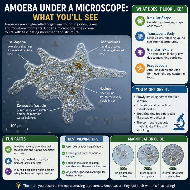
What Does an Amoeba Look Like Under a Microscope?
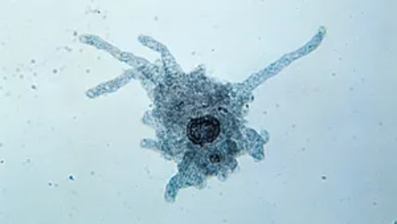
Viewing an amoeba under a microscope reveals a colorless, shapeless organism that constantly flows and shifts as it crawls across your slide. At 100×–400× magnification you’ll see blunt finger-like projections — called pseudopods — slowly extending from one side of the cell while the rest of the cytoplasm streams in behind them, like slow-moving lava. There is no fixed front or back; the cell just oozes in whatever direction its “false feet” are pushing. The granular interior and a clear, gel-like outer edge are both visible at medium magnification. At 400× you can often spot a pulsing contractile vacuole and food inclusions scattered through the cytoplasm.
Amoeba proteus — the species most commonly used in school labs — measures roughly 250–750 µm (0.25–0.75 mm), making it one of the largest single-celled organisms on Earth and easily visible on a standard compound light microscope. No electron microscope, no oil-immersion objective, no special stains required.
Quick-Reference: What’s Visible at Each Magnification

The table below gives a fast overview. The sections that follow explain each level in detail.
| Magnification | Objective | What you can see |
|---|---|---|
| 40× | 4× objective | Grayish irregular speck; slow drift; no internal detail |
| 100× | 10× objective | Overall shape, pseudopods, granular cytoplasm; movement trackable |
| 400× | 40× objective | Nucleus, contractile vacuole, food vacuoles, ectoplasm/endoplasm, cytoplasmic streaming |
To understand how these magnification levels are calculated, see our guide on how to calculate total magnification — multiplying eyepiece power by objective lenses (4×, 10×, 40×) gives you the total.
What You See at Each Magnification (40×, 100×, 400×)
40× — Finding the Organism
At 40× total magnification (4× objective) an amoeba appears as a small, grayish, irregular speck drifting almost imperceptibly. Don’t expect drama. Your job here is just to locate the organism and center it before switching to a higher power. The outlines are fuzzy and the interior looks uniform. Patience is key — scan the slide methodically, especially over debris-rich zones.
100× — The Workhorse View
Switch to the 10× objective and the amoeba’s characteristic shape becomes clear. The pseudopods are obvious now — stubby lobes pushing out from the cell body in one or more directions. The cytoplasm looks distinctly granular compared to the clear surrounding water. You can track the organism’s movement in real time and watch pseudopods extend and withdraw. This is the best magnification for observing how the whole cell navigates its environment. Most students do the bulk of their observation here.

400× — Internal Structures
At 400× (40× objective) the internal landscape becomes genuinely interesting. Look for:
- A defined outer rim of clear, gel-like cytoplasm (ectoplasm)
- A granular, fluid interior (endoplasm) that visibly streams toward the active pseudopod
- The nucleus — a disc-shaped or slightly biconcave structure; it can be difficult to see without staining in a live specimen but is often apparent as a denser region
- The contractile vacuole — a clear, round bubble that slowly fills and then contracts, expelling water
- Food vacuoles — darker, variable-sized inclusions scattered through the cytoplasm
This is also the level at which cytoplasmic streaming becomes most spectacular, which we’ll cover in the movement section below.
The Key Structures You Can Identify
Pseudopods (“false feet”): Blunt, lobose extensions of the cytoplasm used for both locomotion and feeding. They extend, grip, and then the rest of the cell flows into them. The shape of the cell is entirely defined by where pseudopods happen to be reaching at any moment.

Ectoplasm and endoplasm: The cytoplasm has two distinct zones. The outer ectoplasm is clear and gel-like — it’s in a relatively solid state. The inner endoplasm is granular and fluid, and it’s this layer that you can watch stream forward as the cell moves. The transition between these two states — sol to gel and back again — is what drives movement.
Nucleus: Amoeba proteus has a single nucleus. In living specimens it’s often described as disc-shaped or biconcave. Without staining it can be hard to pin down visually, but in a well-focused specimen at 400× it appears as a denser, slightly differentiated region in the endoplasm.
Contractile vacuole: This is one of the most satisfying things to watch. A clear, round vacuole slowly enlarges as it fills with excess water from the cytoplasm, then suddenly contracts and disappears — expelling that water across the cell membrane. This is the amoeba’s osmoregulation system at work. Watching it pulse is a reliable sign your specimen is alive and healthy.

Food vacuoles: Darker, often irregularly shaped inclusions where engulfed material — the bacteria amoebas feed on, algal cells, smaller protists — is being digested. Their number and size vary depending on how recently the organism has fed.
No cell wall: Unlike the plant cells you see when looking at onion cells, an amoeba has only a thin, flexible plasma membrane (plasmalemma) at its surface. This is exactly why it can change shape so freely — there’s no rigid wall constraining it.
How an Amoeba Moves (and Why It’s Slow)
Amoeboid movement is driven by a continuous sol–gel transformation of the actin cytoskeleton. Endoplasm (in the liquid sol state) streams forward into a forming pseudopod, where it converts to the gel-state ectoplasm at the leading edge. Simultaneously, ectoplasm at the trailing end converts back to sol and flows forward — the whole system cycling continuously.
The result, seen through the eyepiece, is a slow oozing motion. We’re talking a few micrometers per second — nothing like the rapid darting of a ciliate. You can watch a pseudopod extend over the course of 10–20 seconds; if you blink, you probably won’t miss it, but you might miss a small retraction.
This is fundamentally different from how a Paramecium moves (fast, spinning, cilia-driven) or how a Euglena moves (direct swimming with a flagellum). Amoeboid motion is about flow, not stroke. Many microscopy beginners mistake a drifting amoeba for a dead one — let it sit under observation for a minute before drawing that conclusion.
At 400× you can watch the granular endoplasm streaming inside the advancing pseudopod like a river of particles pushing toward the tip. This is cytoplasmic streaming, and it’s one of the most visually striking things a beginner can see on a basic school microscope.
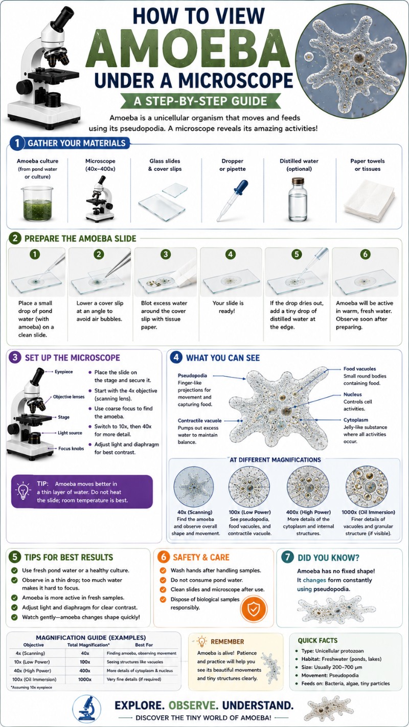
How to Find an Amoeba in Pond Water
Amoebas don’t live in open water — they live in the sediment. The clear water at the top of a pond sample will rarely contain them. You want the gunk at the bottom: decaying leaves, detritus, algal mats, mud. Soil samples from moist garden soil or leaf litter are another excellent source — our guide to viewing soil under a microscope covers what else you’ll find there.
Sampling approach:
- Collect water from the margins of a pond, ditch, or slow stream — scoop some sediment along with the water.
- Let the sample sit in a jar at room temperature for 24–48 hours. Amoebas tend to migrate to the bottom and sides of the container.
- Use a pipette to draw material from the debris layer just above the settled sediment — not from the clear water above.
- Place a small drop on your slide and cover immediately.
Our dedicated guide to collecting and observing pond water covers this process in full, including other organisms you’ll likely encounter at the same time — things like Spirogyra and Volvox that share the same habitat.
How to Prepare and View Your Slide
A standard wet mount works well. Place your drop of sample on a clean glass slide and lower a coverslip at an angle to avoid trapping large air bubbles. If your sample is thick with debris, a depression slide (concave slide) or adding a small piece of broken coverslip as a spacer prevents crushing the organisms. For full wet mount technique, see our step-by-step guide on how to make a wet mount slide.
The biggest challenge with amoebas is contrast. They are nearly transparent and colorless — under bright illumination they practically disappear into the background. The most important technique adjustments:
- Lower the condenser: Bringing the condenser down reduces the cone of light and increases contrast on transparent specimens. This is the single most effective trick for seeing amoebas.
- Close the iris diaphragm: Partially closing the iris diaphragm works similarly — less light, more contrast at the edges of the cell.
- Phase contrast or dark-field: If your microscope has these options, use them. Dark-field microscopy makes transparent cells glow brightly against a black background — amoebas look spectacular under dark-field.
- Work quickly and keep the light low: Bright light warms and dries the slide. Amoebas under harsh illumination will slow down and stop moving within minutes. Use only the light you need.
Start scanning at 40× in a methodical grid pattern. Amoebas move slowly, so they’re easy to overlook if you’re moving the stage too quickly. When you find a likely candidate, increase to 100×, then to 400× for the full structural view. Comprehensive tips on preparing your microscope slides properly will also improve your success rate across all organisms.
Amoeba vs. Other Protists (Paramecium and Euglena)
Three protists frequently appear together in pond-water samples and in biology curricula. Here’s how to tell them apart at a glance:
| Feature | Amoeba | Paramecium | Euglena |
|---|---|---|---|
| Shape | Shapeless, ever-changing | Slipper-shaped, fixed | Elongated, tapered; somewhat flexible |
| Movement | Slow oozing via pseudopods | Fast spinning; cilia all over surface | Directed swimming; single anterior flagellum |
| Speed | Very slow (µm/sec) | Fast (can cross slide in seconds) | Moderate; can also be stationary |
| Distinguishing structures | Pseudopods, contractile vacuole, food vacuoles | Oral groove, two contractile vacuoles, macronucleus | Eyespot (stigma), chloroplasts (green color), flagellum |
| Color | Colorless/gray | Colorless/gray (cilia visible at edges) | Green (from chlorophyll) |
In practice: if it’s green, it’s likely Euglena (or another photosynthetic protist). If it’s zipping around the slide, it’s probably a ciliate like Paramecium. If it’s slowly oozing and constantly changing shape, you’ve got an amoeba.
Amoeba Species: A. proteus vs. Chaos carolinensis
Amoeba proteus is the standard teaching species. It reaches 250–750 µm at most, has a single nucleus, and shows the textbook pseudopod behavior described throughout this article. Biological supply companies sell cultures specifically for classroom use, and it’s the most likely amoeba in a standard school wet mount.
Chaos carolinensis — sometimes called the giant amoeba — is a different organism entirely. It can reach 1–5 mm, making it occasionally visible to the naked eye as a small moving speck. More striking: it is multinucleate, carrying potentially hundreds of nuclei in a single cell. Under the microscope it looks dramatically larger than A. proteus, and at higher magnification you can sometimes see multiple nuclei scattered through the cytoplasm. It is far less common in classroom cultures but sometimes appears in pond-water samples from areas with abundant organic matter.
If you’re into exploring unusual microscopic life, the hunt for different amoeba species pairs well with hunting for tardigrades in the same moss and pond-water habitats.
Is It Safe? A Quick Note on Naegleria
The freshwater amoebas you’ll encounter in typical pond water or purchased lab cultures are completely harmless. They don’t infect humans and pose no threat from observation or normal handling.
You may have heard of Naegleria fowleri, the so-called “brain-eating amoeba” that occasionally appears in news stories. This is a separate organism from Amoeba proteus and is not present in standard school cultures. N. fowleri infections are extremely rare and occur specifically through water entering the nose during activities like swimming in warm freshwater lakes — not through skin contact, not through looking at pond water under a microscope, and not through ordinary lab handling according to the CDC.
Sensible hygiene is all that’s needed: wash your hands after handling pond water samples. That’s the beginning and end of the safety concern for the vast majority of amoeba observation.
For more on the biology of Amoeba proteus specifically, the Britannica entry on Amoeba proteus is a solid reference. Florida State University’s Molecular Expressions gallery has actual video footage of live amoeba movement that pairs well with firsthand observation.
Frequently Asked Questions
What does an amoeba look like under a microscope?
An amoeba appears as a colorless, irregularly shaped organism with blunt finger-like projections (pseudopods) extending from its body. The interior looks granular and you can often see a clear outer rim, darker internal inclusions, and a pulsing contractile vacuole. Its shape changes continuously as it moves. At 100× the overall form is clear; at 400× internal structures become visible.
What magnification do you need to see an amoeba?
You can spot an amoeba at 40× total magnification, but 100× (10× objective on a standard compound microscope) is the most practical viewing magnification. At 100× you can see its shape and movement clearly. For internal structures — nucleus, contractile vacuole, food vacuoles — step up to 400× (40× objective).
Can you see an amoeba with a regular light microscope?
Yes, easily. Amoeba proteus is 250–750 µm — far larger than bacteria or viruses. A standard compound light microscope used in schools and home labs is entirely sufficient. No electron microscope, no special oil-immersion objective, and no staining is needed to observe a living amoeba.
How do you find an amoeba in pond water?
Sample from the bottom sediment and detritus of a pond or ditch — not from the clear water at the top. Let your sample settle for 24–48 hours, then pipette material from the debris layer just above the sediment. Place a small drop on a slide, add a coverslip, and scan at 40× starting in areas with visible particles and organic matter.
How does an amoeba move under the microscope?
Amoebas move by extending pseudopods — blunt cytoplasmic projections — in the direction of travel, then flowing the rest of the cell into them. The granular endoplasm streams visibly toward the leading edge. This process is slow (a few micrometers per second), so movement looks like a gradual ooze rather than swimming. At 400× you can watch the streaming cytoplasm inside each pseudopod.
What is the difference between an amoeba and a paramecium?
An amoeba is shapeless and moves slowly by pseudopods; a Paramecium is fixed slipper-shaped and moves fast using cilia that cover its entire surface. Paramecia also have an oral groove and two contractile vacuoles. In practice: if the organism is zipping around the slide spinning, it’s a Paramecium; if it’s slowly changing shape and oozing, it’s an amoeba.
Why is my amoeba not moving or hard to see?
The most common causes: (1) The organism is there but nearly invisible because your illumination is too bright — reduce light by lowering the condenser and partially closing the iris diaphragm. (2) The slide has dried out or overheated from the light source — work quickly and dim the lamp. (3) You’re looking in the wrong zone — scan areas with debris and particles, not clear water. (4) Amoebas are genuinely slow; give the slide a full minute at 100× before declaring nothing is there.
Are amoebas dangerous to handle?
Freshwater amoebas from ponds or lab cultures are harmless to humans. The dangerous species Naegleria fowleri is a separate organism entirely and is not present in standard cultures or typical pond-water samples. Normal hygiene — washing hands after handling pond water — is the only precaution needed.
Conclusion
Viewing an amoeba under a microscope is one of the most satisfying introductions to single-celled life. At 100× you get the big picture — a shapeless blob that contradicts everything we assume a “creature” should look like. At 400× that blob becomes a complex working cell: pseudopods pushing forward, cytoplasm streaming in behind them, a contractile vacuole pulsing away, food vacuoles scattered through the granular interior. The key techniques — sampling from sediment, lowering the condenser for contrast, and scanning patiently at low power first — make the difference between a frustrating blank slide and a cell that genuinely moves while you watch.
Have you spotted an amoeba in your own pond-water samples? Did you manage to catch cytoplasmic streaming at 400×, or find something that might be the giant Chaos carolinensis? Share what you found in the comments below — we’d love to hear about it.
