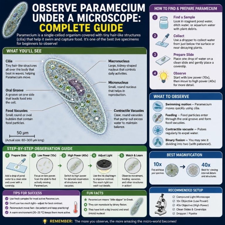
What Does a Paramecium Look Like Under a Microscope?
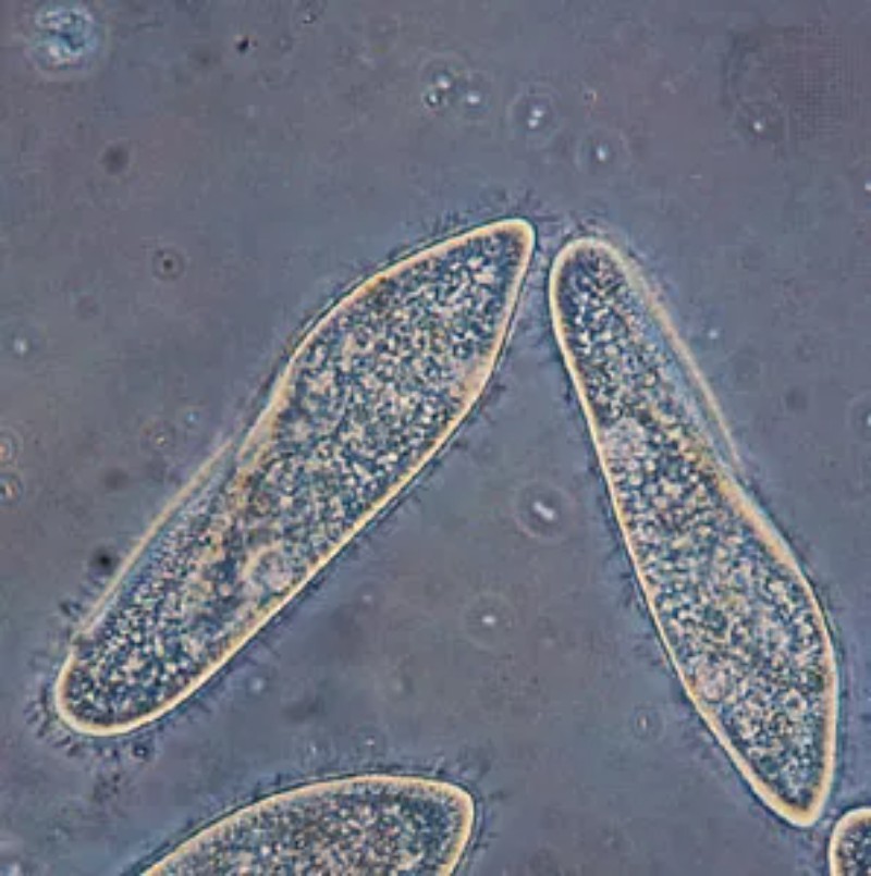
A paramecium under a microscope appears as a translucent, slipper-shaped single cell measuring roughly 50–350 micrometers (µm) long. Thousands of hair-like cilia cover its surface, beating in coordinated waves that drive it in a spiraling, rotating swim. At 100× you can make out the oval body, the diagonal oral groove, and two star-shaped contractile vacuoles that pulse rhythmically. Push to 400× and the cilia come alive as a shimmering fringe, food vacuoles appear as dark spheres circling inside the cell, and the oral groove snaps into sharp detail.
What Is a Paramecium?

Paramecium is a genus of single-celled ciliate protists belonging to the phylum Ciliophora. These organisms live in freshwater and are among the most studied microorganisms in biology — partly because they are abundant, partly because so much of their internal life is visible under an ordinary compound light microscope without any special staining. They are eukaryotes (their DNA is enclosed in a nucleus), which makes them fundamentally different from bacteria, even though both are microscopic.
Paramecia feed primarily on bacteria, yeasts, and small organic particles, making them key players in microbial food webs. The name “paramecium” comes from the Greek for “oblong,” and the common nickname “slipper animalcule” captures their shape perfectly.
Paramecium vs. Bacteria vs. Other Protists
Beginners often confuse paramecia with bacteria or assume they are animals. Here is the quick distinction:
- Bacteria are prokaryotes — no membrane-bound nucleus, no organelles, and far smaller (1–10 µm). A paramecium is roughly 20–100× larger than a bacterium and eats bacteria for food. Read more about what bacteria look like under a microscope to see how different they are.
- Paramecium vs. Euglena: Euglena uses one or two long flagella; paramecia use thousands of short cilia. Euglena is often green (it photosynthesizes); paramecia are colorless.
- Paramecium vs. Volvox: Volvox forms a hollow sphere of hundreds of cells; a paramecium is always a single, free-living cell.
Where to Find Paramecium
Paramecia are found in freshwater environments rich in decaying organic matter and bacteria — ponds, ditches, slow-moving streams, birdbath water, and any container of stagnant water left outdoors. They concentrate near the surface scum, around submerged decaying vegetation, and at the mud-water interface. Observing pond water under a microscope is the fastest route to finding them in the wild.

When collecting, draw your sample from the top centimeter of the water, near plant material. Transfer it to a clear jar and hold it up to light — the largest paramecia (~0.3 mm) appear as faint white specks moving with purpose against a dark background. You cannot distinguish any structure with the naked eye, but the movement confirms life.
How to Make a Hay Infusion to Culture Paramecium
If pond water is not available, a hay infusion is the classic reliable method:
- Bring a handful of dried hay or dried grass to a boil in dechlorinated tap water or pond water for 5–10 minutes.
- Let the infusion cool completely, then pour it into a wide-mouth jar.
- Add a small amount of pond water or soil as a seed culture (paramecia are in most outdoor soil and water).
- Store the jar at room temperature in indirect light — never direct sunlight, which can overheat it.
- A bacterial bloom develops within 2–3 days; paramecia peak around day 5–10 and can be harvested with a pipette from the surface film.
The boiled hay releases nutrients that feed the bacteria, which in turn feed the paramecia. This is one of the simplest and most reliable protozoan cultures you can set up at home or in a classroom.

How to Prepare a Paramecium Wet Mount
Paramecia must be observed alive in a wet mount — they die quickly on dry slides and are not interesting once fixed. Follow these steps for a good wet mount slide:
- Use a clean glass slide and a #1 coverslip. Remove any dust with lens paper.
- Draw a small sample with a pipette from near the surface of your culture — this is where paramecia concentrate.
- Place one drop (about 0.05 mL) at the center of the slide. Avoid a flood — too much water and the cells swim out of the focal plane constantly.
- If using a slowing agent (see below), add one small drop of methyl cellulose solution to the water drop now and mix gently.
- Lower the coverslip at a 45° angle from one edge to the other to avoid trapping air bubbles.
- Blot any overflow at the edges with lens paper, but do not press the coverslip.
- Place on the stage and start at 40× (4× objective) to locate moving cells before increasing magnification.
For more detailed slide technique, see our guide on preparing microscope slides.
How to Slow Paramecium Down for Observation
Paramecia swim fast — a typical cell covers 1–2 mm per second. At 400×, a cell crosses your field of view in under a second. You need to slow them down without killing them:

- Methyl cellulose / Protoslo / Detain: These viscous commercial solutions are the best option. Add a small drop to your water drop before covering. The thick medium slows cells to a crawl without harming them. Protoslo is available from biological supply companies.
- Cotton or lens-paper fibers: Tease a small amount of raw cotton or lens paper into the drop. The fibers create a physical maze that traps cells long enough to study. This is a free, effective classroom technique.
- Gentle coverslip pressure: Press lightly on the coverslip with a pencil eraser to reduce the water layer height. This restricts vertical movement. Caution: too much pressure ruptures cells.
- Reduce the iris diaphragm: Closing the condenser diaphragm to about 70% increases contrast on these transparent cells and helps your eye track them.
- Dark field: If your microscope supports it, dark field microscopy makes paramecia glow against a black background, which is spectacular for watching ciliary motion.
What Magnification to Use for Paramecium
Understanding your microscope objective lenses (4×, 10×, 40×, 100×) is essential here. Here is a practical guide to what you will see at each total magnification:
| Total Magnification | Objective Used | What Becomes Visible | Best For |
|---|---|---|---|
| 40× | 4× | Moving specks; rough slipper outline on larger species | Finding and tracking cells |
| 100× | 10× | Body shape, oral groove, contractile vacuole positions, general cytoplasmic granularity | Watching behavior, feeding, avoidance reaction |
| 400× | 40× | Cilia fringe, contractile vacuole pulsing and structure, food vacuoles, oral groove detail, nucleus (with stain) | Studying organelles — use a slowing agent first |
| 1000× | 100× oil | Fine nuclear detail, cilia individually | Stained fixed specimens only — live cells move too fast and are too thick |
Recommendation: Start at 40× to find your cells, switch to 100× to watch behavior, then move to 400× with a slowing agent for organelle detail. You rarely need oil immersion for living paramecia. Understanding how to calculate total magnification will help you keep track of which objective gives you which power.

Paramecium Organelles You Can See Under the Microscope
The beauty of paramecia is how much internal structure a standard compound light microscope can reveal in a living, unstained cell. Here is what to look for and where to find it.
Cilia and How Paramecium Moves
Paramecia are covered in thousands of short cilia — hair-like projections arranged in rows across the entire cell surface. These cilia beat in coordinated metachronal waves, meaning each cilium beats slightly after the one behind it, like a stadium wave. The result is a characteristic forward spiral swim: the cell rotates along its long axis as it moves forward, rather than gliding straight.
When a paramecium runs into an obstacle, it performs an avoidance reaction: it reverses the direction of ciliary beat, backs up, pivots, and swims forward in a new direction. You can watch this happen repeatedly in real time at 100× — it is one of the most satisfying things to observe in basic microscopy.
Individual cilia appear as a shimmering fringe at 400×, especially if you have reduced the iris diaphragm for contrast. At lower powers they are invisible as individual structures but visible collectively as movement on the cell’s edge.
Oral Groove and Feeding
The oral groove (also called the buccal cavity) is a diagonal, funnel-like indentation on one side of the cell that leads to the cytostome (“cell mouth”). Cilia lining the oral groove sweep bacteria and particles inward. You can see this groove clearly at 100× as a diagonal slash across the cell body.
Once food enters the cytostome, it is packaged into food vacuoles — small membrane-bound spheres that bud off and circulate through the cytoplasm in a process called cyclosis. At 400× these vacuoles appear as dark, granular spheres tumbling slowly around the cell interior. Watching cyclosis in a live cell is one of those moments that makes microbiology feel real.
Contractile Vacuoles and Osmoregulation
Paramecia live in fresh water, which is hypotonic (lower solute concentration than the cell’s cytoplasm) — water constantly floods in by osmosis. To avoid bursting, they use contractile vacuoles to bail water out continuously. Most paramecia have two: one near the anterior end and one near the posterior end.
Each contractile vacuole has a star or rosette shape with radiating canals that collect water from the cytoplasm. The vacuole fills, then contracts rapidly and collapses, expelling water through a pore in the pellicle. At 400× you can watch these vacuoles pulse in a steady rhythm — typically every 6–25 seconds depending on temperature and species. This pulsing is one of the most mesmerizing sights in protozoan microscopy.
Note for beginners: Two pulsing vacuoles is completely normal. Do not assume one is a burst organelle or damage — both vacuoles are supposed to be there.
Macronucleus and Micronucleus
Paramecia have a dual nuclear system:
- Macronucleus: Large, kidney- or oval-shaped, controls day-to-day cell metabolism. In a living unstained cell it is difficult to see clearly — it blends with the surrounding cytoplasm. A brief exposure to methylene blue or methyl green stain makes it pop at 400×.
- Micronucleus: Small, located near the macronucleus, involved in sexual reproduction (conjugation). Usually near-invisible in living wet mounts even at 400× without staining. P. aurelia typically carries two micronuclei; P. multimicronucleatum carries four to seven or more.
The outer surface of the cell is bounded by the pellicle — a flexible but firm protein layer that maintains the cell’s fixed shape even as the cilia beat. This is why paramecia do not deform like amoebae.
How to Identify Paramecium Species
Three species dominate classroom and hobbyist cultures. They differ in size, shape, and micronucleus count:
| Feature | P. caudatum | P. aurelia | P. multimicronucleatum |
|---|---|---|---|
| Size | 170–330 µm | 120–180 µm | up to ~350 µm |
| Shape | Strongly tapered (“tailed”) posterior | More cigar-like, blunt at both ends | Large, robust, more cylindrical |
| Micronuclei | 1 | 2 | 4–7+ |
| Best ID feature | Distinct pointed tail; largest in typical cultures | Smaller size; blunter ends; 2 micronuclei (needs stain) | Very large cells; multiple micronuclei (staining required) |
Without staining you can reliably distinguish P. caudatum from the other two by its pointed posterior. Separating P. aurelia from P. multimicronucleatum in a living unstained cell is difficult — size and micronucleus count require staining. Most commercial cultures sold as “paramecium” contain primarily P. caudatum.
For context on how these species compare to other freshwater microorganisms you might encounter, see our guide on Spirogyra under the microscope — a filamentous alga often found in the same pond water samples.
Reproduction You Might See: Binary Fission and Conjugation
Paramecia reproduce asexually by binary fission, and the manner of division is distinctive: they split transversely — across the short axis — rather than lengthwise. This means you will see a cell that appears to be pinching in half at its midsection. During division, the macronucleus elongates and the cell visually elongates before splitting. In an active culture you can catch this by scanning at 100× — dividing cells are noticeably elongated and have a visible constriction.
Conjugation is the sexual process: two compatible paramecia align at their oral surfaces and exchange micronuclear material over 6–12 hours. Conjugating pairs look like two cells stuck together side by side along their ventral surfaces. This is relatively rare to catch in cultures, but it occurs more often in aging or stressed cultures where the population needs genetic renewal. To maximize your chances, observe a culture that has been growing for 3–4 weeks without refreshment.
Common Mistakes Beginners Make
- Too much light: Paramecia are nearly transparent. Cranking up the condenser to maximum bleaches out internal detail. Close the iris diaphragm to 60–70% for best contrast without staining.
- No slowing agent: Jumping to 400× without methyl cellulose or fibers results in a frustrating blur. Slow them first, then magnify.
- Too much water in the mount: A thick water layer means cells are constantly swimming in and out of focus in the z-axis. Use one small drop, not a full dropper.
- Sampling from the wrong part of the jar: Paramecia concentrate at the surface and around debris. Sampling from the middle of a clear water column gives you very few cells.
- Expecting the naked eye to see a “cell”: You can detect the largest paramecia as moving specks, but you cannot see a cell. Anyone who tells you otherwise is seeing debris or large algae.
- Panicking over two contractile vacuoles: Two pulsing vacuoles is normal anatomy, not damage.
- Pressing too hard on the coverslip: Heavy pressure ruptures cells and creates artifacts. If you need to restrict movement, apply gentle, even pressure — just enough to slightly flatten the water layer.
For a broader look at finding and working with microorganisms in natural water samples, our guide on how to find tardigrades covers a lot of the same collection and mounting skills.
For background on how magnification interacts with what you can actually resolve, see magnification vs resolution — a concept that matters when you push to 400× and wonder why the image looks soft.
Paramecia are also a good gateway to understanding the broader world of single-celled life. Britannica’s overview of Paramecium is a solid reference for the classification and evolutionary context if you want to go deeper on the biology. For the microscopy side, Nikon MicroscopyU’s guide to live cell imaging explains the principles of contrast and illumination that apply directly to watching living paramecia. If you want to explore the peer-reviewed science, the NCBI-hosted research on ciliate biology covers the molecular detail behind what you are observing. And for a classroom-friendly introduction to how protists fit into the tree of life, Khan Academy’s guide to building an evolutionary tree is free and well-structured.

Frequently Asked Questions
What does a paramecium look like under a microscope?
A paramecium appears as a translucent, slipper-shaped cell covered in fine, beating cilia. It swims in a rotating spiral pattern. At 100× you can see the oral groove and contractile vacuoles; at 400× you can see the cilia fringe, food vacuoles circulating inside the cell, and the pulsing of the contractile vacuoles.
What magnification do you need to see a paramecium?
You can spot a paramecium as a moving speck at 40× total magnification. For useful detail — body shape, oral groove, and contractile vacuoles — you need 100×. For organelle detail including cilia and pulsing vacuoles, use 400×. Oil immersion (1000×) is generally not needed for living specimens.
Where can I find paramecium for a microscope?
Paramecia are abundant in freshwater ponds, ditches, and stagnant water near decaying vegetation. Collect from the surface film near plant material. Alternatively, make a hay infusion: boil dried hay in water, let it cool, seed it with a small amount of pond water or soil, and a thriving culture develops in 5–10 days.
How do you slow down paramecium under a microscope?
The most effective method is adding a drop of methyl cellulose, Protoslo, or Detain to your water sample — these viscous solutions slow cells without harming them. You can also tease a tiny amount of cotton or lens-paper fibers into the drop to physically trap cells, or apply very gentle pressure to the coverslip to restrict vertical movement.
What organelles can you see in a paramecium under a light microscope?
In a living, unstained cell at 100–400×, you can typically see: the oral groove, two contractile vacuoles (and watch them pulse), food vacuoles circulating through the cytoplasm, the cilia fringe, and the general outline of the macronucleus (easier with methylene blue stain). The micronucleus is usually too small to see without staining.
How does a paramecium move under the microscope?
Paramecia swim in a forward spiral — rotating along their long axis while moving forward — driven by thousands of cilia beating in coordinated metachronal waves. When they encounter an obstacle, they perform an avoidance reaction: reversing ciliary beat, backing up, pivoting, and swimming forward in a new direction.
Is a paramecium visible to the naked eye?
The largest paramecia (~300–350 µm) are just barely detectable as tiny moving white specks in a clear container held up to light against a dark background. However, no cellular structure is visible — you see only the movement. Magnification of at least 40× is required to see any form or detail.
Conclusion
Paramecium is one of the best starting points in microscopy — large enough to find easily, complex enough to keep you looking for hours. With a basic compound microscope, some pond water or a simple hay infusion, and a drop of methyl cellulose, you can watch cilia beating, contractile vacuoles pulsing, food vacuoles circling the cytoplasm, and — if you are lucky — catch a cell in the middle of dividing. Each magnification level reveals a new layer of biology that a textbook can describe but a microscope makes real.
Have you tried observing paramecia yourself? Whether you cultured your own hay infusion or pulled a sample straight from a local pond, we would love to hear what you found — drop your observations, questions, or photos in the comments below.
