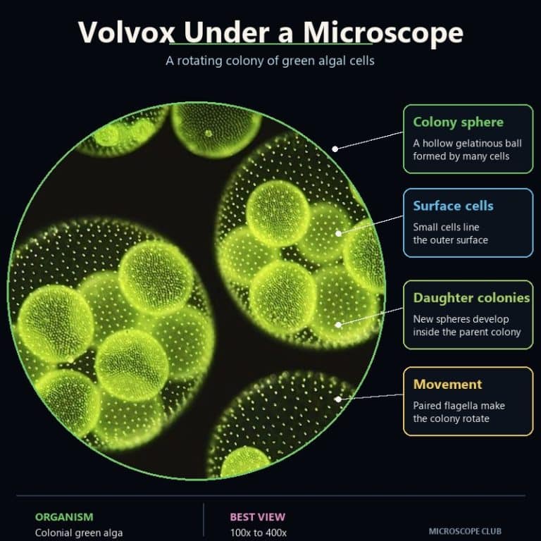
Volvox is one of the few microscopic organisms you can sometimes spot with the naked eye — a tiny green sphere rolling through a drop of pond water, spinning as smoothly as a planet in slow orbit. Put it under a compound microscope and within seconds you will see daughter colonies packed inside the parent like green globes within a globe, flagella flickering at the surface, the whole structure tumbling with unhurried purpose. First-time observers regularly gasp. Here is everything you need to collect, prepare, and study Volvox — from pond to eyepiece.
What Is Volvox?
Volvox is a genus of colonial green alga in the family Volvocaceae (not “Vovlocacae” — the misspelling appears often, including in older posts on this site). A single Volvox colony is a hollow sphere: hundreds to tens of thousands of microscopic cells embedded in a clear gelatinous extracellular matrix, all connected by fine cytoplasmic strands that let the colony act as a coordinated unit.
Two cell types do completely different jobs. Somatic cells — the smaller, numerous ones — are motile but reproductively sterile. Each carries two whip-like flagella that beat in unison with neighboring cells, driving the whole colony in a rolling, spinning motion. Gonidia are the large, non-motile cells that sit deeper in the sphere; they cannot swim but they can divide rapidly to form entirely new daughter colonies. The strict division between body cells and reproductive cells mirrors the germ–soma split found in complex animals — which is exactly why Volvox attracts so much attention from evolutionary biologists.
A fully grown Volvox colony can reach 500 µm in diameter, large enough to appear as a tiny green speck to the naked eye in a jar of pond water held up to the light.
When and Where Was Volvox First Discovered?
Volvox was first observed by Antonie van Leeuwenhoek in 1675, who described the rolling spheres in a letter to the Royal Society of London as “a breathtaking sight.” He noted tiny globules “swimming very nimbly with an as great velocity almost directly backward” — a remarkably accurate description of the colony’s coordinated phototactic retreat from strong light.
Swedish naturalist Carl Linnaeus later gave Volvox the informal nickname “fierce roller” in reference to its characteristic tumbling motion. He classified it under the Zoophyta within class Vermes, though modern taxonomy now places it firmly among the green algae (Chlorophyta).
The Structure of a Volvox Colony


Each component of the colony has a specific role. Understanding them before you look through the eyepiece makes the observation far more meaningful.
- Flagella — Each somatic cell has two flagella that extend outward from the colony surface. Beat coordination across thousands of cells produces the rolling motion you see at 100x. The anterior (north) hemisphere has slightly longer flagella, which steers the colony toward light.
- Eyespot (stigma) — A small cluster of orange-red carotenoid pigment at the anterior side of each somatic cell. Eyespots are most prominent at the colony’s north pole. At 400x you can sometimes resolve individual eyespots as rust-colored dots when the colony rotates into view. They detect light direction and coordinate flagellar response.
- Contractile vacuole — A pumping organelle in each cell that expels excess water, maintaining cell turgor in freshwater where osmotic pressure would otherwise burst the cell.
- Nucleus — Volvox is eukaryotic; each cell has a distinct nucleus containing the cell’s genetic material. The nucleus is involved in both the asexual division of gonidia and, in sexual species, gamete formation.
- Chloroplast — A large cup-shaped chloroplast fills most of each somatic cell, giving the colony its bright green color. Chloroplasts perform photosynthesis, which is why Volvox needs light and why it actively swims toward it.
- Gelatinous extracellular matrix — A thick jelly-like layer surrounds the colony and holds the cells in position. It gives Volvox its characteristic glossy appearance under the microscope and its slight resilience when the colony rolls against the coverslip.
- Gonidia — The large reproductive cells sit in the posterior (south) hemisphere. They appear as darker, slightly larger spheres within the colony wall. Each gonidium divides repeatedly to produce a complete daughter colony while still inside the parent.
- Cytoplasmic strands — Fine threads of cytoplasm connect adjacent somatic cells, allowing chemical signals to coordinate flagellar beating and, in sexual species, pheromone responses across the entire colony.
Common Species of Volvox
Around 20 species of Volvox are traditionally recognized, though modern taxonomy continues to revise this figure as molecular data challenges older morphological classifications. The four species most likely to turn up in a hobbyist’s pond sample are:
- Volvox aureus — One of the most widely studied species. Forms spherical colonies with cells arranged in double rows; a large colony may contain up to 50,000 cells. Common in eutrophic ponds in temperate regions.
- Volvox carteri — The primary laboratory model species, with a fully sequenced genome. A typical colony contains roughly ~2,000 somatic cells plus ~16 gonidia — far fewer than V. aureus. Grows in water temperatures from 13 to 30 °C.
- Volvox globator — Larger colonies, often 20,000–45,000 cells. Prefers slightly warmer water (21–24 °C). Frequently seen when Volvox blooms in summer.
- Volvox barberi — Found in freshwater ponds and slow-moving water. Colonies up to ~4,000 cells. Less commonly encountered in field samples.
Volvox carteri is the species most intensively studied in the laboratory because its germ–soma division is clean enough to use as a model for the unicellular-to-multicellular evolutionary transition (see below).

How Volvox Survives and Reproduces

Volvox thrives in nutrient-rich, still freshwater — ponds, ditches, rice paddies, slow streams. It blooms most abundantly in spring and summer, when warmer temperatures and longer daylight hours fuel rapid asexual division. During a bloom, a single jar of pond scum can contain thousands of colonies.
Asexual reproduction is the default. Each gonidium divides repeatedly by a process called palintomy — producing a miniature colony, still inside the parent, that inverts itself (turning inside-out) to orient its flagella outward. These daughter colonies are the dark spheres you see floating inside the parent at 100x. When the daughter colonies mature they rupture the parent’s gelatinous wall and swim free, leaving the parental body behind.
Sexual reproduction is triggered by environmental stress, most notably heat shock. In Volvox carteri, elevated temperature causes somatic cells to produce and release a ~32-kDa glycoprotein sex-inducing pheromone — one of the most potent biological effectors known, active at concentrations as low as 10⁻¹⁶ mol/L. This pheromone reprograms gonidia in nearby colonies to produce gametes rather than daughter colonies, initiating sexual reproduction. Wounding can trigger the same response. The biological advantage is timing: sexual reproduction produces a resistant zygote (zygospore) that can survive drought or extreme heat, so triggering it in response to heat stress gives Volvox a survival option ahead of hostile conditions.
Why Volvox Matters in Evolutionary Biology
Volvox sits at a pivotal position in the history of life. Its closest relatives, such as the single-celled alga Chlamydomonas, are fully unicellular. Volvox itself has crossed the threshold into genuine multicellularity — somatic cells have irreversibly surrendered their reproductive capacity to serve the colony, a sacrifice that mirrors what happened hundreds of millions of years ago in the ancestors of animals and plants.
The germ–soma division of labor in V. carteri is studied intensively because only a small number of regulatory genes differentiate the two cell types. Turning those genes on or off in lab experiments can collapse a multicellular Volvox back toward unicellular behavior — making it one of the clearest windows we have into how complex life evolved from simple cells. No other organism of comparable simplicity models this transition so directly.
Is Volvox Harmful to Humans?
Volvox itself is not hazardous. The organisms carry no toxins and are far too large and slow to penetrate tissue. However, dense blooms of Volvox — and the cyanobacteria and other algae that often bloom alongside it in eutrophic water — can deplete dissolved oxygen, block sunlight from submerged plants, and create dead zones that kill fish and invertebrates. Algal blooms also raise water-treatment costs when the affected water enters municipal supply systems. The risk is to the ecosystem, not to humans directly.
How to Collect and Prepare Volvox for Viewing
Collecting a sample
Visit a pond, ditch, or slow-moving stream in spring or summer, ideally after a warm spell. Volvox concentrates at the surface in calm, sunlit areas — look for a faint green sheen or slight turbidity. Scoop surface water with a jar or pipette, or squeeze a handful of submerged pond weed directly into your sample container. You do not need a net; Volvox is large enough that a plain jar works fine. Back-lit against a window, a good sample shows tiny green dots moving lazily upward toward the light.
Making the wet mount
To prepare your microscope slide, place a single drop of pond water on a clean glass slide. Lower the coverslip slowly at roughly a 45° angle from one side of the drop, letting it fall gently onto the water — this displaces air and avoids the bubbles that beginner observers almost always introduce. Volvox colonies are large and slow; no chemical retardant is needed to slow them down. If colonies keep drifting out of the field of view, tease a single fiber from a piece of lint or lens tissue and lay it across the slide before covering — it creates a micro-obstacle that catches colonies without harming them.
Work quickly. Volvox in a sealed wet mount can survive 20–30 minutes before the oxygen runs out and the flagella stop. If you want to extend your session, use a coverslip with petroleum jelly at the corners to create a sealed ring with trapped air, or use a well slide.
What You See at Each Magnification

This is the section most observation guides skip — the step-by-step of what your eyes should expect as you increase magnification. Use a compound microscope with a 400x total magnification capability; oil immersion (1000x) is not appropriate for Volvox. Oil immersion is designed for bacteria and sub-micron detail. Volvox at up to 500 µm is best seen with dry objectives at 40x–400x total.
| Magnification | What resolves |
|---|---|
| 40x total | The whole colony fits easily in view — a bright green sphere, slightly fuzzy at the edges. You can see the rolling, tumbling motion in real time. Multiple colonies often appear in the same field. Daughter colonies may already be visible as slightly darker blobs inside larger parents. |
| 100x total | Coordinated colony rotation becomes dramatic — the sphere spins on a consistent axis, sometimes pausing and reversing. Daughter colonies inside the parent are now clearly distinct, darker green spheres in the posterior half. The gelatinous wall catches light differently from the hollow interior. This is the best magnification for the “globe within a globe” effect. |
| 400x total | Individual somatic cells appear on the colony surface as small hexagonal or round units. Paired flagella are visible as fine flickering hairs at the edge of the colony. Eyespots resolve on well-oriented cells as tiny orange-red dots. The gonidium cells are large and dark relative to somatic cells. Focus becomes shallow — you are viewing one focal plane at a time through the sphere’s depth. |
Step-by-step observation procedure
- Place your prepared wet mount on the stage and start at 40x total magnification. Focus roughly on the center of the drop.
- Scan slowly until you find a green sphere rolling across the field. Center it.
- Close your iris diaphragm slightly — reducing light increases contrast on the transparent sphere and makes the gelatinous wall and internal colonies pop. This is more effective than staining for initial observation. Darkfield or phase contrast microscopy are even more effective if your microscope supports them: Volvox is unstained and nearly transparent, exactly the specimen type these contrast methods are designed for.
- Switch to 100x total. Use only the fine focus from this point — coarse focus at higher magnification can crack the coverslip. Re-center the colony and watch the rotation cycle for 30–60 seconds before moving on.
- Switch to 400x total. The colony now fills much of the field. Adjust fine focus up and down through the sphere’s depth. The surface cells resolve in the focal plane you choose; the opposite hemisphere will be soft. Rotate focus slowly to step through layers.
- If a colony drifts, do not chase it at 400x — drop back to 40x, re-locate, and step back up.
Common beginner mistakes
- Confusing Volvox with Pandorina or Eudorina — both are green colonial algae that form smaller spherical colonies. Pandorina colonies are solid (not hollow) and typically 16–32 cells; Eudorina colonies are open spheres of 32–64 cells. If the sphere lacks visible daughter colonies and seems smaller than a grain of table salt, you may be looking at one of these relatives rather than Volvox.
- Sample dying before viewing — if your pond water has been sitting in a warm car for several hours, the Volvox may already be dead. Fresh sample, viewed within an hour or two of collection, gives the best results. Dead colonies collapse slightly and lose the clean spherical outline; flagella stop within minutes of death.
- Too much light — a fully open iris diaphragm makes the transparent sphere nearly invisible against the bright background. Close the iris until the colony outline sharpens. If detail washes out, reduce light further before adjusting magnification.
- Chasing a moving colony at 400x — at high magnification the field of view is tiny. Moving the slide to catch a drifting colony results in rapid loss and frustration. Intercept the colony at 40x, predict its path, move the stage ahead of it, and let it swim into frame.
Frequently Asked Questions
Is Volvox a plant, animal, or protist?
Volvox is a protist — specifically a green alga in the phylum Chlorophyta. It is photosynthetic like a plant, motile like an animal, but belongs to neither kingdom. In older classifications it appeared in both botany and zoology texts, which is why Linnaeus placed it in the animal-adjacent Zoophyta.
What are the green balls inside Volvox?
Daughter colonies — fully formed new Volvox colonies that developed from gonidia inside the parent sphere. They sit in the posterior hemisphere and are the most visually dramatic feature of the organism. At 100x they appear as smaller, darker green spheres suspended within the larger parent. When mature they rupture the parental wall and swim free.
Do you need to stain Volvox to see it?
No. Volvox is large enough to observe unstained. Iodine solution will kill the colony but can highlight the gelatinous matrix and starch granules in the chloroplasts if you want a static structural view. For live observation, skip staining and use contrast methods (closed iris, phase contrast, or darkfield) instead.
How long does Volvox live?
A single asexual colony typically survives a few days to a week before the gonidial daughter colonies rupture the parent and continue the lineage. Under stress — heat, drought, nutrient depletion — the sexual cycle produces resistant zygospores that can remain dormant in pond sediment for months or years.
Conclusion
Volvox is the rare microscopic organism that rewards observers at every magnification: a rolling green sphere at 40x, a world within a world at 100x, and an intricate community of specialized cells at 400x. It also sits at the center of one of biology’s most important questions — how life crossed the threshold from single cells to coordinated multicellular organisms. To get started, collect a fresh pond water sample in spring or summer, make a quick wet mount with a single drop, close the iris diaphragm to boost contrast, and work your way from 40x up to 400x. The daughter colonies inside the parent are unmistakable once you have seen them once; after that, you will recognize Volvox in any sample in seconds.
READ MORE:
- Observing Mold Under Microscope
- Observing Yeast Under Microscope
- Observing Onion Cells Under Microscope
Originally posted 2022-07-03 04:36:00.