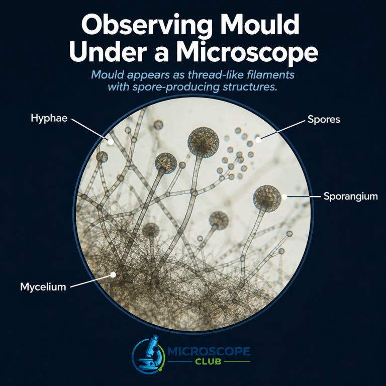
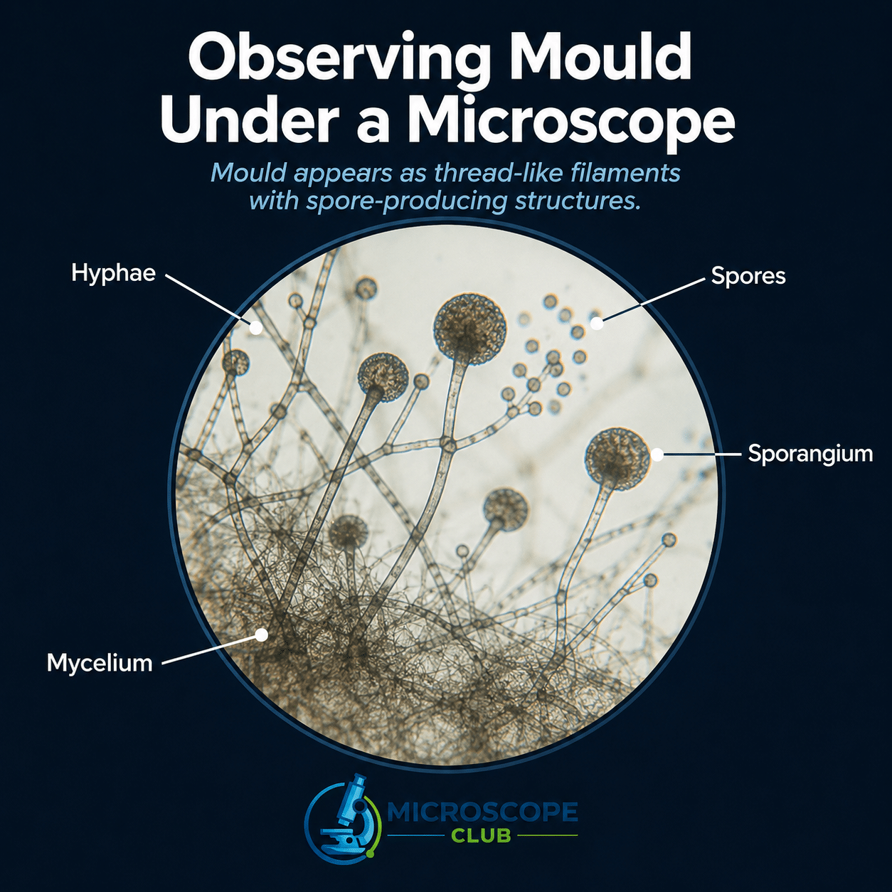
Mould under a microscope is a tangle of branching, translucent threads called hyphae that weave into a mat called the mycelium, studded with spore-bearing structures whose shape — round sac, brush-like cluster, or sprinkler head — tells you exactly which genus you’re looking at. That fuzzy patch on your bread or bathroom tile looks featureless to the naked eye; at 400× on a standard compound microscope it becomes a precision identification exercise. Genus-level ID is achievable at home; species-level ID requires culture morphology or molecular methods.
The Microscopic Architecture of Mould: Thread, Spore, and Structure
Mould is not a plant. Its cell walls are built from chitin — the same polymer found in insect exoskeletons — not cellulose. It feeds saprotrophically: secreting digestive enzymes into its substrate and absorbing the broken-down nutrients rather than photosynthesising. That means mould hyphae are colourless to pale brown under the scope, never green. For a grounded overview of mould biology, Encyclopaedia Britannica’s mould entry is a reliable starting point.
At 40× (scanning power) the mycelium appears as a tangled mass of faint lines. Larger structures — sporangia in bread mould — show as dark dots. Step to 100× and individual hyphae resolve into clear tubes; you can start distinguishing septate from aseptate types. At 400× (high-dry) you can read septa, conidiophore shape, and spore arrangement — the level needed for genus ID. Total magnification equals eyepiece power (almost always 10×) multiplied by the objective: 10× × 40× = 400×.
Key Structures: Hyphae, Septa, and Reproductive Bodies
Hyphae and Mycelium (Septate vs Aseptate)
A hypha is a hollow tube — the fundamental unit of mould. Width varies significantly by type:
- Septate hyphae — Aspergillus, Penicillium, Cladosporium, and Stachybotrys all produce these: thin-walled tubes 3–6 µm wide, divided at intervals by cross-walls (septa) visible at 400× as fine dark lines. The septa don’t completely seal each compartment — cytoplasm flows through a central pore — but they’re unmistakeable once you know to look.
- Aseptate (coenocytic) hyphae — Rhizopus and Mucor (the classic bread moulds, order Mucorales) have no septa. These hyphae run as a single continuous multinucleate cell and are markedly wider: typically 10–20 µm, up to ~25 µm, ribbon-like and irregular, branching near-90°. That wide, pale ribbon is itself an ID cue: if you can’t find a septum at 400× and the hypha looks like a deflated garden hose, you’re likely looking at a Mucoral.
Khan Academy’s fungi module covers hyphal biology accessibly if you want more background.
One thing beginners don’t expect: hyphae collapse under the coverslip if you press too hard, flattening into irregular ribbons that look nothing like the textbook image. Apply the coverslip by lowering one edge first and letting capillary action pull it down — that alone prevents most collapses.
Spores, Sporangia, and Conidiophores
Mould reproduces via spores — single cells released in millions. Two production strategies:

- Conidia — asexually produced spores borne externally on a specialised aerial stalk called a conidiophore. The conidiophore’s architecture is the single most useful genus cue at 400×.
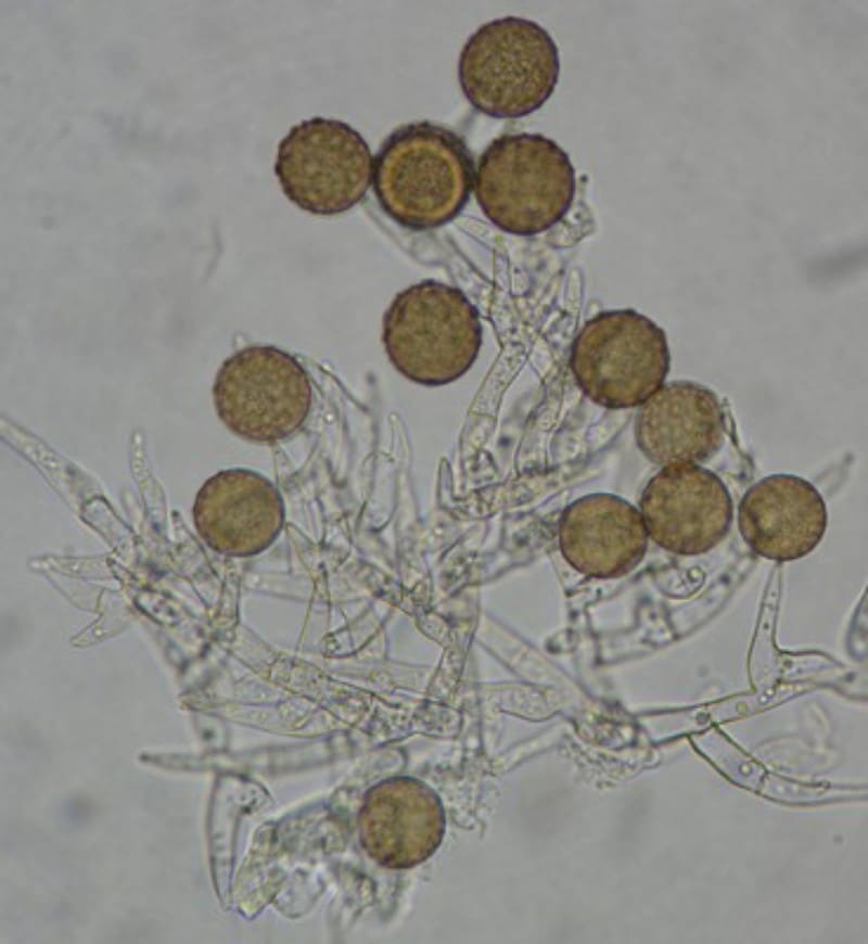
- Sporangiospores — produced inside a round, enclosed sac called a sporangium. When the sporangium ruptures it releases hundreds of spores. This is the hallmark of Rhizopus and Mucor.
Two conidiophore shapes to lock in:
- Aspergillus: the conidiophore tip swells into a round vesicle covered in flask-shaped phialides, from which chains of conidia radiate outward. The structure looks exactly like a sprinkler head — which is how the genus got its name (from the aspergillum, a liturgical water sprinkler). In Aspergillus niger specifically, the vesicle is biseriate — two rows of cells (metulae + phialides) cover the entire vesicle surface, giving a dense, full-coverage radiate head. The conidiophore itself is long, smooth, hyaline, darkening only at the apex; vesicle diameter 30–75 µm. See ScienceDirect’s Aspergillus overview for deeper genus detail.
- Penicillium: the conidiophore branches into a brush-like cluster of phialides called a penicillus (Latin: “little brush”), with chains of conidia hanging from each tip. Yes — this is the genus that gave us penicillin.
In Rhizopus you’ll also see the columella — a dome-shaped cap of sterile tissue sitting at the base of the burst sporangium — and rhizoids, root-like anchoring hyphae gripping the substrate. The columella is unmistakeable once you spot it.
Genus Comparison: What Each Common Mould Looks Like at 400×
| Genus / Common Name | Hyphae (type + width) | Reproductive Structure | Colour (colony / spores) | Magnification to ID | Notes / Risk |
|---|---|---|---|---|---|
| Rhizopus (black bread mould) | Aseptate, 10–25 µm, ribbon-like, ~90° branching | Round sporangium on tall stalk; dome columella; rhizoids | Colony grey/black; sporangia dark | 40–100× | Very common; generally low risk; specific species (R. arrhizus, R. microsporus) can cause mucormycosis in immunocompromised patients and those with uncontrolled diabetes — see StatPearls: Mucormycosis |
| Penicillium | Septate, 3–6 µm | Brush-like penicillus; chains of round conidia | Blue-green colony; conidia grey-green | 100–400× | Source of penicillin; some species (P. expansum, P. citrinum) produce mycotoxins; common on citrus and aged food |
| Aspergillus (general) | Septate, 3–6 µm | Vesicle/sprinkler-head with radiating chains of conidia | Green, yellow, or black depending on species; young colonies start white/cream | 100–400× | Several species pathogenic; A. fumigatus causes invasive aspergillosis primarily in immunocompromised individuals and those with pre-existing lung disease — not healthy adults |
| Aspergillus niger (black mould on onions/lemons) | Septate, 3–6 µm; long smooth hyaline conidiophore | Large biseriate sprinkler head (full-coverage vesicle); rough dark brown/black conidia | Starts white/yellow, turns jet-black as conidia mature; reverse pale | 100–400× | Most common “black mould” found in kitchens; NOT Stachybotrys — distinguished by biseriate full-coverage vesicle at 400×; low risk to healthy adults |
| Cladosporium | Septate, 3–6 µm; distinctive shield-shaped cells | Branching chains of dark conidia; “shield cells” (cladospores) | Olive-green to brown/black | 400× | Most common outdoor mould globally; significant allergen; low toxicity |
| Stachybotrys (“toxic black mould”) | Septate, 3–6 µm | Slimy clusters of ellipsoidal conidia on phialides (7–12 × 4–6 µm); NOT dry chains | Starts colourless/pale, darkens to brown/black with maturity; wet/slimy appearance | 400× | Produces trichothecene mycotoxins; requires ~94% surface RH + cellulose substrate (water-damaged drywall, gypsum board); handle only with professional PPE |
Quick genus key at 400×: Wide ribbon hypha + no septa + sporangium + dome columella → Rhizopus. Brush-like head → Penicillium. Sprinkler-head vesicle → Aspergillus (full-coverage dark biseriate head → A. niger). Branching chains of shield-shaped dark conidia → Cladosporium. Slimy black clumps (not dry chains) → suspect Stachybotrys — handle accordingly.

Colour changes with age — don’t rely on colour alone. Young mould colonies are often white or cream before pigmentation develops. A young Aspergillus or Stachybotrys colony can look pale on the slide; colour is a secondary cue at best, and the weakest one. Structure is what counts.
For comparison, yeast under the microscope — another fungus — looks completely different: scattered oval single cells that bud, with no hyphae at all.
What Magnification Do You Need to See Mould?
You don’t need an expensive research microscope. A standard student compound light microscope with 40×, 100×, and 400× objectives covers everything below.
- 40× (scanning): See the mycelium as a tangle of faint lines. Rhizopus sporangia appear as dark round dots. Good for overall layout — and for ruling out debris.
- 100×: Hyphae resolve clearly as tubes. Sporangia and conidiophores begin to take shape. You can distinguish septate from aseptate hyphae at this level. A useful first stop for any slide.
- 400× (high-dry): Individual conidia, phialide arrangement, septa, and spore chains all become readable. This is your genus identification level — most of your time will be here.
- 1000× (oil immersion): Rarely needed for mould. Useful only for detailed spore surface ornamentation. Requires immersion oil and careful technique; most hobbyists won’t need this.
Understanding depth of field at higher magnification matters here — as magnification increases, the focal plane narrows sharply, so always start at 40× and work up, constantly adjusting the fine focus rather than locking it. The light microscope vs electron microscope comparison is also worth knowing: you won’t see molecular-level detail under light, but all the structures that matter for genus ID are well within range.
How to Prepare a Mould Slide at Home (Step by Step)
Read the safety section below before starting. The tape-lift method is the safest home technique — it minimises airborne spores compared to scraping or blowing on the sample.
New to slide preparation? The guide to making a wet mount slide covers the fundamentals you’ll draw on here, and the general how to prepare microscope slides guide is worth reading through first.
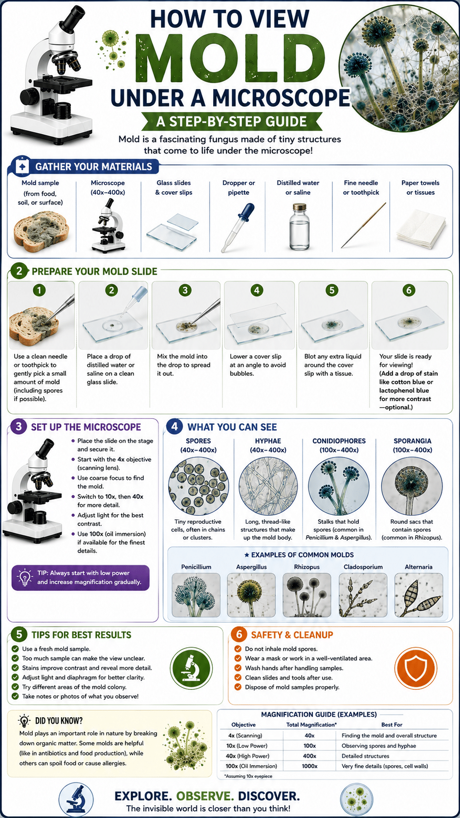
- Source your mould. A small patch on old bread, citrus, or cheese works well. Work in a well-ventilated area. Do not deliberately grow large colonies indoors in open containers — keep any growth sealed in a bag or jar. Bread mould under the microscope makes a good first project because Rhizopus is easy to ID even at 100×.
- The tape lift. Cut a 1–2 cm piece of clear sticky tape. Gently press the sticky side onto the mould surface and peel off. This lifts a thin layer of spores and hyphae without launching them into the air.
- Prepare your stain. Place a small drop of lactophenol cotton blue on a clean glass slide. This binds to chitin in fungal cell walls, stains them blue, and simultaneously kills and fixes the spores. It provides the best contrast of any home-accessible fungal stain. No lactophenol? Methylene blue or dilute food colouring shows structure with less contrast. Iodine (Lugol’s solution) works poorly on fungi — skip it.
- Apply the tape. Lay the tape sticky-side-down onto the drop of stain.
- Add the coverslip. Lower it at a 45° angle from one edge, letting capillary action draw it down. If large bubbles appear, gently press the coverslip edge to push liquid into the gap — don’t press flat, or you’ll collapse the hyphae.
- Start at 40×. Find a clear area first, then work up through 100× to 400×. Use the fine focus knob continuously — mould structures exist at different depths and you need to scan through the focal plane, not lock it.
The first time I did this with a bread mould tape-lift, the initial coverslip had a large bubble sitting right over the most interesting area — classic mistake. I reseated it by touching a corner with a toothpick and the bubble migrated to the edge. At 40× the dark sporangia looked like scattered pepper; stepping up to 100× revealed the tall sporangiophore stalks, and at 400× the columella was unmistakeable — a perfect dome sitting in the mouth of the burst sporangium. Nothing in a textbook quite prepares you for how three-dimensional it looks when you scan the focal plane.
Comparing mould to onion cells under the microscope is a useful exercise — the geometric regularity of onion cell walls makes the irregular, branching architecture of mould hyphae even more striking by contrast.
How to Tell Mould from Slide Artefacts (Dust, Lint, and Bubbles)
A beginner’s first question at 40× is often: “Is that mould, or just debris?” Here’s how to read what’s on the slide:
- Lint fibres: Uniform width end-to-end, flat, often coloured, and far longer than any hypha you’ll see. They don’t branch and carry no spore structures. Hyphae taper, branch, and — at 400× — show parallel walls with internal contents.
- Air bubbles: Thick black refractive ring around a clear centre; shift or move when you press the coverslip edge gently. No mistaking one for a hypha at 100×.
- Dust particles: Irregular shape, non-branching, non-uniform. May look faintly opaque but show no internal structure, no parallel walls, no reproductive bodies.
- Mould hyphae: Branch, have clearly parallel walls, and — at 400× — carry recognisable spore structures (sporangia, conidiophores, chains of conidia). Even at 40× a mycelial mat has a characteristic woven texture no artefact reproduces.
If you’re unsure, step to 400× and look for septa (dark cross-lines inside the tube) or any spore-bearing structure. Debris has neither.
Is It Safe to Look at Mould Under a Microscope?
For most healthy adults handling a small patch of common household mould briefly, the risk is low — but not zero, and a few precautions make it negligible. The US Centers for Disease Control and Prevention provides authoritative guidance on mould health risks.
- Ventilation: Work near an open window or in a well-ventilated space. Don’t blow on the sample.
- Don’t inhale: Position the sample away from your face. Use the tape-lift method — scraping sends spores airborne.
- Fix the sample quickly: Lactophenol cotton blue kills and immobilises spores on contact. Once under the coverslip, the sample is contained.
- Wash hands thoroughly after handling any mould sample or slide materials.
- Dispose carefully: Seal used slides and mould-contaminated materials in a zip-lock bag before binning.
- High-risk individuals should not handle live mould: Anyone with asthma, fungal allergies, or a compromised immune system (chemotherapy, immunosuppressants, uncontrolled diabetes) should avoid mould samples entirely. A. fumigatus can cause invasive aspergillosis in immunocompromised patients and those with pre-existing lung disease — healthy adults face low risk, but the same people who shouldn’t be in a mouldy basement shouldn’t be handling tape-lift samples either — see CDC: Aspergillosis. Stachybotrys (“toxic black mould”) should only be sampled by professionals with appropriate PPE — never from water-damaged building materials at home.
For students and children, the My First Lab Duo-Scope is robust and easy to clean — adult supervision is appropriate for any mould activity. Dark-field microscopy is worth knowing too: it provides high contrast for low-pigment specimens and makes translucent hyphae pop without staining.
Mould Spores vs Pollen and Other Look-Alikes
- Mould vs pollen: Pollen grains are typically larger (10–100 µm for most species vs 2–10 µm for mould conidia), with sculptured patterned walls — ridges, spines, apertures visible at 400×. Mould conidia are smoother and attached in chains or clusters to a conidiophore. Pollen sits in isolation with no connecting structure.
- Mould vs yeast: Yeast cells are oval or round, single-celled, and show budding (a small daughter cell attached to the parent). No hyphae. A yeast culture looks like scattered ovals; mould looks like threads. Some fungi are dimorphic — they can switch between forms — but the thermally-dimorphic types (like Histoplasma) that go yeast-form at body temperature are specialised pathogens you won’t encounter on kitchen bread. Common household moulds (Rhizopus, Penicillium, Aspergillus, Cladosporium, Stachybotrys) are always filamentous at room temperature.
- Mould vs algae: Filamentous green algae like Spirogyra can look superficially similar — but their cells contain clearly visible bright green chloroplasts. Mould hyphae are colourless to pale brown, never green. If you’re comparing across sample types, pond water samples show this contrast well — algae and filamentous fungi can both appear in the same slide, and the chloroplast tells you which is which instantly.
- “Black mould” ≠ Stachybotrys by default: Both Aspergillus niger and Cladosporium produce dark/black colonies. Without a scope you cannot distinguish either from Stachybotrys. At 400×: A. niger has the distinctive full-coverage biseriate sprinkler head; Cladosporium has branching chains of shield-shaped conidia; Stachybotrys has slimy clumped phialides and no dry chains. Colour is not diagnostic. A young A. niger colony may still be white — colony age matters as much as colour.
- Mould vs bacteria: Bacteria are far simpler and smaller — single cells in the 1–10 µm range, with no hyphal network, no reproductive structures, and no internal complexity visible at standard light microscopy. Even at 400× a bacterial smear shows nothing resembling mould architecture.
Frequently Asked Questions
What if the structures look blurry or I can’t get the mould in focus?
Blurriness is almost always a focus-depth problem. Mould structures sit at different depths within the slide — hyphae at one level, conidiophores and spores at another. Use the fine focus knob continuously and scan slowly through the focal plane rather than locking it in place. If contrast is poor and structures are hard to distinguish, the stain is usually the cause: lactophenol cotton blue gives the best contrast for fungal walls — switching to it from methylene blue or plain water makes hyphae, septa, and conidiophore shapes significantly clearer. Always start at 40× to find a clear area before stepping up to 100× and 400×.
How long do prepared mould slides last, and can you keep them?
A simple lactophenol cotton blue wet mount will dry out within hours to a few days as liquid evaporates from under the coverslip. To keep one as a reference, seal the four coverslip edges with clear nail varnish (or mounting medium such as Permount) once settled — a sealed lactophenol slide lasts months to a few years. For a truly permanent mount, PVA-based products like BlueMount embed the specimen and stain together.
Where do you buy lactophenol cotton blue, and how much does it cost?
Laboratory suppliers including Hardy Diagnostics, Sigma-Aldrich, Thomas Scientific, and Carolina sell it in 10–15 mL dropper bottles for around £5–10 / US$15–25. It contains phenol — toxic and corrosive — so some suppliers (Carolina, for example) ship only to schools and businesses, not residential addresses. Safer alternatives with similar fungal-wall staining: lactoglycerol cotton blue (glycerol replaces the phenol) or Congo red.
Where can I safely find mould to look at, and what should I avoid?
The safest sources are small patches on old bread, citrus peel, or cheese — common, low-risk genera (Rhizopus, Penicillium) you can handle briefly with basic precautions. Avoid dark mould on water-damaged drywall, ceiling tiles, or wallpaper: that environment favours Stachybotrys, which requires ~94% surface humidity and cellulose-rich material (gypsum board, fibreboard, straw) to establish — and should only be handled by professionals. Sample from food in your kitchen, not from damp building materials.
How long does the whole process take, and can it be done in one sitting?
If you already have a mouldy sample, the tape-lift, staining, and viewing steps take 10–20 minutes start to finish — easily a single sitting. The time-consuming part is growing your own: a bread slice in a sealed plastic bag in a warm, dark spot shows visible mould in 3–5 days, with a well-developed colony after 7–10 days. Keep the bag sealed throughout and open it only briefly to take the tape-lift.
How do I know it’s mould and not lint or a bubble on the slide?
Lint fibres are uniform-width, flat, often coloured, and far longer than any hypha — they don’t branch. Air bubbles show a thick black refractive ring and move when you press the coverslip. Dust is irregular and structureless. Mould hyphae branch, have clearly parallel walls, and at 400× carry recognisable spore structures. When in doubt, step to 400× and look for septa inside the tube — lint and debris have none.
Conclusion
Mould is far more structured and visually rich than its fuzzy surface appearance suggests. Under a microscope the mycelial network, the shape of conidiophores, the presence or absence of septa, and the arrangement of spores give you a reliable window into which genus you’re dealing with — and what that means for health and home. The key tools are simple: the genus comparison table, 400× magnification, and the ability to distinguish a real hypha from a lint fibre or bubble. Start with old bread, use lactophenol cotton blue, and work up from 40× — the sprinkler heads and brush-like clusters of a household mould colony are among the most satisfying structures you’ll find on a student scope.
Originally posted 2026-06-13 03:10:23.