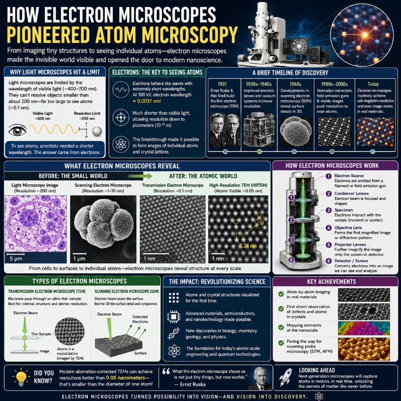
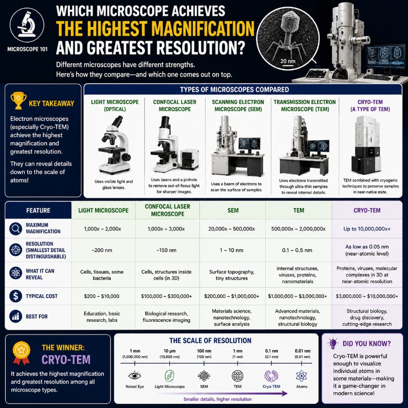
How Electron Microscopes Pioneered Atom Microscopy

Photo of How Electron Microscopes Pioneered Atom Microscopy

For most of human history, the atom was a philosophical concept — matter’s smallest theorized unit, invisible by definition. That changed in 1931, when a pair of German scientists aimed a beam of electrons at a thin specimen and produced an image no light microscope could ever rival. Today, we don’t just see atoms; we …

Read more

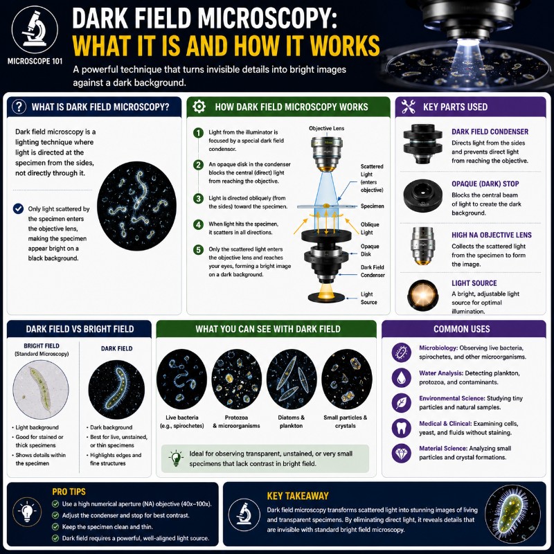
Dark Field Microscopy: What it is And How it Works

Photo of Dark Field Microscopy: What it is And How it Works

Dark field microscopy illuminates a specimen with a hollow cone of oblique light so that only the light scattered or diffracted by the specimen reaches the objective — leaving everything else in pitch-black. That simple optical trick turns colourless, unstained, and nearly transparent specimens into bright glowing structures you simply cannot see under conventional bright …

Read more

Bright field Microscope: Facts and FAQs

Photo of Bright field Microscope: Facts and FAQs

You might be wondering what a brightfield microscope is, but chances are, you have already seen one- more specifically, a compound light microscope. The thing is, brightfield microscopes are really common and can be found in just about any lab and science classroom. These microscopes operate through a basic form of optical microscopy, and are …

Read more

Super-Resolution Microscopy: STED, STORM, PALM & SIM Explained

Photo of Super-Resolution Microscopy_ STED, STORM, PALM & SIM Explained

Super-resolution microscopy is the family of optical techniques that breaks the ~200 nm diffraction limit of conventional light microscopy, resolving biological structures down to 10–50 nm — while still using light and, in many cases, still imaging living cells. It earned the 2014 Nobel Prize in Chemistry and has since revealed biological architecture that was …

Read more

Polarizing Microscope Uses: 8 Real Applications (and Why They Work)

Photo of Polarizing Microscope Uses: 8 Real Applications (and Why They Work)

A polarizing microscope is a specialized compound microscope fitted with two crossed polarizing filters and a rotating stage, purpose-built to examine birefringent materials — substances that split light into two rays — which glow with vivid interference colors against a pitch-black background that a standard brightfield microscope simply cannot produce. Understanding polarizing microscope uses matters …

Read more

Phase Contrast Microscope: How It Turns Invisible Cells Visible

Photo of Phase Contrast Microscope: How It Turns Invisible Cells Visible

A phase contrast microscope makes living, unstained, transparent specimens visible by converting invisible differences in light’s phase into visible differences in brightness — something a standard bright-field microscope simply cannot do. It achieves this through two matched optical components: an annular diaphragm in the condenser and a phase plate in the objective, which together exploit …

Read more

Fluorescence vs Confocal Microscope: 7 Key Differences

Photo of Fluorescence vs Confocal Microscope: 7 Key Differences

Fluorescence vs confocal microscope is one of the most common points of confusion in biological imaging — and the answer surprises most people: a confocal microscope is a fluorescence microscope. The difference is a single component, a tiny pinhole aperture, that blocks out-of-focus light and lets confocal produce sharp optical sections through thick samples. Widefield …

Read more

Observing Yogurt Under a Microscope: Bacteria You’ll See

Photo of Observing Yogurt Under a Microscope_ Bacteria You'll See

Observing yogurt under a microscope reveals two types of live bacteria — rod-shaped Lactobacillus and bead-like chains of Streptococcus — but you need about 1000x magnification with oil immersion and a simple stain to see them clearly. At 400x they appear as faint dark specks, enough to confirm they’re there; at 1000x you can distinguish …

Read more

How to View Soil Under a Microscope (Step-by-Step)
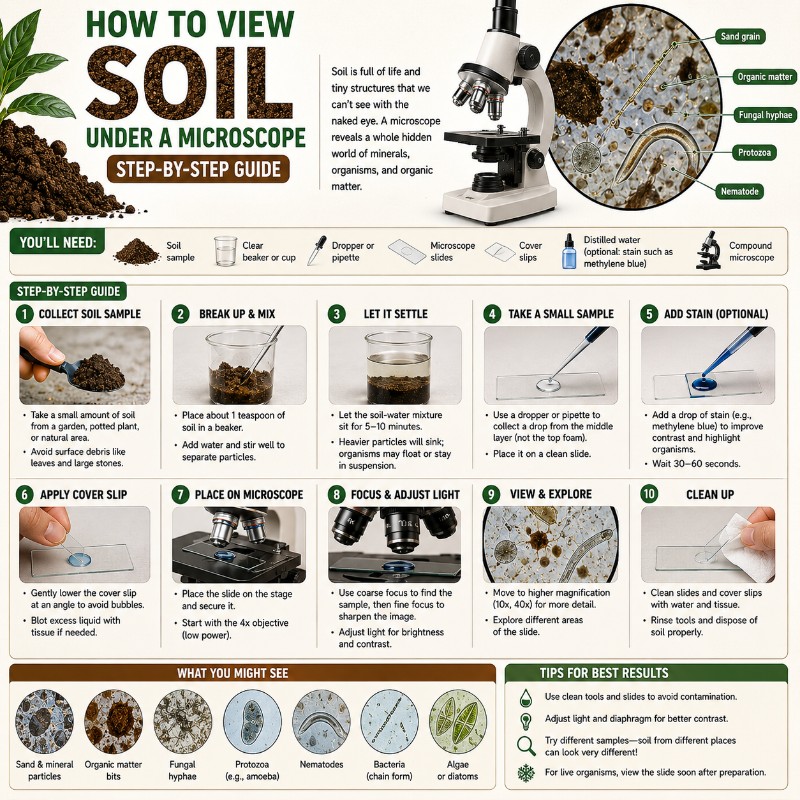
Photo of How to View Soil Under a Microscope (Step-by-Step)

Learning how to view soil under a microscope comes down to five steps: collect a fresh sample from the top 2–4 inches of ground, disperse a small amount in distilled water, pipette a drop of the cloudy liquid onto a slide, add a coverslip, and scan from low to high magnification. Done right, you’ll find …
Read more