Trichomes Under a Microscope: A Plant Hair Field Guide

Non-glandular, glandular and stinging plant trichomes compared under a microscope

Trichomes under a microscope reveal a plant’s leaves and stems as far stranger surfaces than the naked eye suggests. Expect forests of glassy spikes, translucent balloon-headed glands, and even a tiny hypodermic needle. A trichome is simply a fine outgrowth of the plant epidermis — a plant hair — and it falls into one of …

Read more

Salmonella Under a Microscope: Pink Rods, No ID

Gram-negative pink rod-shaped bacteria under a microscope; appearance alone cannot confirm Salmonella

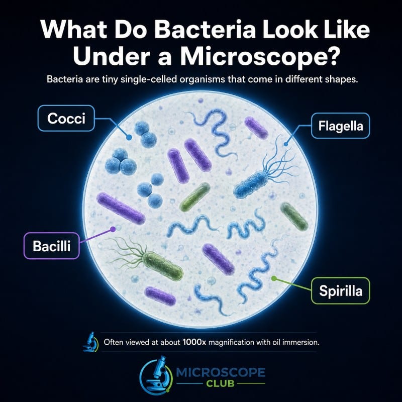
Salmonella under a microscope appears as tiny, straight, pink rods after a Gram stain — small enough that you need 1000x oil immersion just to resolve their outline. A light microscope can show you that shape, size, and color, but it cannot tell you the rods are Salmonella specifically. That confirmation only comes from a …

Read more

Skin Cells Under a Microscope: What You Really See

Surface skin cells and a stained epidermis cross-section compared under a microscope

Skin cells under a microscope look nothing like the tidy textbook diagram most people expect: a simple home scrape shows a single sheet of flat, colorless, dead cells with no visible layering at all. The famous five-layer structure of the epidermis — from the tough outer stratum corneum down to the actively dividing stratum basale …

Read more

Observing Cancer Cells Under The Microscope

One of the more useful and essential uses of microscopy is in identifying, analyzing, and treating certain diseases, ranging anywhere from bacterial and viral infections, to something a lot more serious and fatal, such as cancer. There are a lot of distinctive differences between cancer cells and normal cells when it comes to appearance, characteristics, …

Read more

Observing Ants Under The Microscope

Ants reward every magnification step with something new — a whole body in motion at 10x, individual hairs and mandible teeth at 40x, and individual ommatidia in the compound eye at 400x on a prepared slide. Knowing what to expect at each level, and how to prepare your specimen correctly, is the difference between a …

Read more

Observing Salt Under The Microscope

Salt under the microscope reveals something most people don’t expect: a perfectly geometric crystal, its flat faces and right-angle edges so precise they look machined rather than grown. The first time you focus on a recrystallized grain at 40x, the cubic shape is unmistakable — no soft curves, no randomness, just clean right angles repeating …

Read more

Muscle Tissue Under a Microscope: 3 Types Compared

Skeletal, cardiac and smooth muscle tissue compared under a microscope, showing striations, nuclei and cell shape

Muscle tissue under a microscope reveals its identity through four telltale features: striations, nuclei count and position, cell shape, and intercalated discs. Skeletal muscle is long, cylindrical, striated, and packed with nuclei pushed to the cell’s edge. Cardiac muscle is branched and striated too, but its cells carry dark connecting lines called intercalated discs. Smooth …

Read more

Observing Sand Under The Microscope

Sand grains under a microscope reveal more than most people expect — not just shape and color, but the entire travel history of a grain: whether it was hurled by desert winds, tumbled down a river, or built grain-by-grain from the skeletons of sea creatures. A $40 stereo scope and a pinch from your local …

Read more

Observing Yeast Under The Microscope

Observing Yeast Under The Microscope

Yeast under a microscope reveals a world far more dynamic than the plain beige powder from a baking packet suggests. At 400x magnification you can watch living cells bud off daughter cells in real time — and a single drop of methylene blue tells you in seconds which cells are alive and which are dead. …

Read more

Electron Microscope Images That Show The Power of Electron Microscopes

Photo of Electron Microscope Images That Show The Power of Electron Microscopes

Electron microscope images reveal a world that visible light can never reach — individual atoms, flash-frozen viral proteins, and the claw of a mite clinging to a honeybee’s back. This guide explains how electron microscopes produce those images, what the color and scale bars actually mean, and how to read every picture in the gallery …

Read more