Observing sperm cells under a microscope is another interesting experiment to conduct. Analyzing sperm can give information about the reproductive function of a male patient. Observing sperm cells is also part of a routine semen analysis and is done to identify issues with the male reproductive cells. An observer can come up with normal sperm count, motility, and morphology. But before heading towards medical complexities, let’s find out more about their structure and microscopy.
What are Sperm Cells?
Sperm cells are produced in the testicular organ of male human beings and animals. It is gametes or sex cells. Similar to female gamete or oocyte, sperm cells have 23 chromosomes resulting from a process called meiosis.
Gametes are cells involved in sexual mode or reproduction for both humans and animals; wherein there is an interaction of the sex cells.
General Morphology:
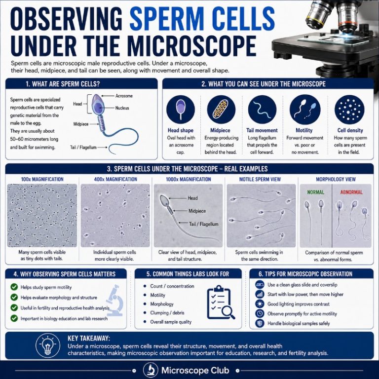
The sperm cells typically consist of the following parts:
- Distinct head
- Midpiece (body)
- Tail
Structure and Functions:
Before proceeding to the functions and structure of the cells, it is equally essential to understand how they are produced. The process is called spermatogenesis.
What is Spermatogenesis?
In male beings, the testosterone level in the blood is closely monitored by the hypothalamus. A dip in the hormone levels indicates low testicular activity. Thus, the hypothalamus releases the gonadotropin-releasing hormone of the GnRH.
This hormone will flow towards the pituitary gland and stimulate the production of the follicle-stimulating hormone (FSH) and the luteinizing hormone (LH).

The luteinizing hormone flows from the pituitary gland and stimulates the Leydig in the testicles to produce testosterone. On the other hand, the follicle-stimulating hormone plays a vital role in concentrating the LH in the seminiferous tubule to start the formation of the sperm.
The primary spermatocytes (haploid) are produced in the inner walls of the seminiferous tubules by a group of cells known as a spermatogonial germ. Spermatogonia, or immature sperm cells, are created through meiosis in the process of spermiogenesis. The spermatids are then formed due to second meiotic division and develop into mature male cells.
The actual process of sperm development takes around 74 days.

Spermatogenesis is made up of two distinct phases. The first (meiosis) eliminates half of the chromosomes, whereas the second (spermiogenesis) modifies size and form as the sperm mature to their standard form.
The Sperm Structure

Sperm cells come in various shapes and sizes, with some having heads, bodies, and tails while others do not.
Due to various anomalies, they may vary in form and size, but other differences can be seen on any portion of the cell (head, body, tail).
The following are the physical characteristics of a normal sperm:
- The head of a typical sperm has a smooth surface and resembles that of an egg.
- The sperm’s head is 2.5 to 3.5 micrometers in diameter and 4.0 to 5.5 micrometers in length (um=micrometers). It gives a 1.50 to 1.70 ratio of height to width.
- They have a well-developed acrosome covering 40 to 70 percent of the oval-shaped head.
- The body is narrow and approximately the same length as the head.
- A finer tail section with a diameter of around 45 micrometers.
A sperm cell comprises three components: a head, a body (midsection), and a tail. Each part has its own set of molecules and more minor details that enable the entire sperm to function correctly.
The Sperm Head
The standard sperm head is smooth and oval. Due to the broad base and tapering apex, it has a similar appearance to an egg.
The head is the essential component because it contains the nucleus (genetic material with 23 chromosomes) needed to produce a new organism.
Aside from the nucleus, the head consists of several components.
The Acrosome and Acrosomal Cap
The acrosomal region is made up of the acrosome and the acrosomal cap. The acrosome results from the Golgi complex and includes several components, including the acrosin enzyme in the acrosomal matrix, formed during spermiogenesis.
Other components in the acrosome include mannose, hexosamine, and galactose. Additionally, the acrosome contains such polysaccharides as mannose, hexosamine, and galactose.
The space between the interior plasma membrane and the nuclear membrane is the acrosome. The inner and outer acrosomal membranes form the acrosome (acrosomal membrane), which borders the plasma membrane, while the internal acrosomal barrier lies against the nuclear membrane.
The acrosomal filament is a complex structure that consists of the major structural proteins termed fibrous layer proteins, intermediate filament proteins, and components of the microtubular system.
The acrosome is involved in a variety of essential processes during fertilization. For example, it is utilized in recognizing the oocyte (egg) to be fertilized with many of its associated chemicals.
The sperm cell is attracted to the egg jelly’s diffusible molecules when it comes in touch with them, causing the cell to swim towards the eggs. Chemotaxis is the ability of a cell to respond to specific chemical substances.
The cell swims towards the egg (high molecule concentration zone) and makes physical contact when it detects a high quantity. As a result of physical touch, the acrosome reaction occurs.
- Sperm chemotaxis is the ability of sperm to sense chemical cues and move toward them. As a result, this is an essential mechanism that ensures that a conspecific egg (that is, within the same species) is fertilized.
- The target gamete is recognized by its primary ligands (proteins) located close to the acrosome.
The Sperm Acrosome Reaction
An acrosome reaction is an important event when the sperm comes into touch with the oocyte membrane at various locations.
Sperm contact with zona pellucida on the oocyte’s plasma membrane, for example, leads to an acrosome reaction in certain animals. It is a calcium-dependent process that causes the outer acrosome membrane to peel off. It reveals enzymes within the acrosome.
The reaction between eggshell and calcium adenosine triphosphatase results in the release of acrosin (e.g., acrosin) and proacrosin. Acrosin/proacrosin is involved in the lysis of the thick membrane that covers the ovum’s (eggs) exterior.
The acrosome is a structure inside the testis that protects sperm from damage during fertilization. The enzyme (acrosin) is maintained in the acrosome in an inactive form called zymogen. The pH level within the acrosome is lower, keeping the enzyme inaccessible.
The enzyme is transformed into acrosin, an active form that can act on the ovum membrane when it comes into touch with the glycoproteins of the ova membrane (zona pellucida). It allows the sperm cell to enter and penetrate the egg for fertilization.
The Nucleus
Typically known as the head’s nucleus, the haploid nucleus is found at one end of spermatozoa and contains 23 chromosomes. It is covered with an acrosome, which protects the nucleus while navigating through the female reproductive system.
The sperm cell fuses with the egg to form a zygote with 46 chromosomes. The 46 DNA in the new organism is derived from the combined gametes of both parents (egg and sperm).
The Sperm Midpiece
The midpiece is the middle portion of the sperm cell, between the head and tail. The midpiece, like the head, accounts for roughly 10% of the overall sperm length. The midpiece contains densely packed mitochondria that supply energy for swimming.
Mitochondria are believed to have an anti-inflammatory effect and a role in programmed cell death known as apoptosis.
The Centriole
The centriole, located in the midpiece of the sperm cell, is involved in flagellar functions. Each centriole comprises nine sets of microtubules that form part of the cytoskeleton known as an axoneme. A bundle of microtubules (axonemal) contains a single flagellum.
The purpose of these is to allow for the movement and fusion of the pronuclear with the female genome. In addition, during cell division, the centrioles are involved in making the mitotic apparatus, which separates chromosomes and at the same time serves as the template for all future centrioles.
The Sperm Tail
The sperm tail, or flagellum, is the longest part of the sperm cell and makes up roughly eighty percent of its length. The tail is composed of a single axoneme that contains microtubules. The microtubules are arranged in a spiral pattern, which provides strength and stability to the tail.
The flagellum propels the sperm cell through the female reproductive tract and into the egg. The movement of the flagellum also creates a current that draws nutrients and oxygen to the mitochondria in the midpiece, providing energy for swimming.
The tail consists of numerous components, each with its distinct purpose.
- Connecting piece
- Midpiece
- Principal piece (axial filament)
- End piece
The primary and end pieces of the flagella contribute to the waveform that allows for motion.
Sperm Motility

Motility is one of the main characteristics of a well-developed sperm cell. In mammals, two types of physiological motility have been identified, and they are the following:
Activated motility: This is the sort that can be found in the early phases of motility (in the epididymis and freshly ejaculated sperm). In movement, the flagella of the sperm beat gently from one side to another as the cell travels along what appears to be a straight line.
Hyperactivated motility (hyperactivation): The second form of physiological motility is non-activated motility. This type of movement takes place in the female reproductive canal (site of fertilization) instead of activated motility, which occurs in the male reproductive system.
Furthermore, the flagellum shows a symmetrical, lower-amplitude waveform in individuals with hyperactivated motility. More power is required for movement because of the asymmetrical frequency of motion in hyperactivated motility.
The Axoneme and the Molecular Mechanism of Motility
The tail’s axoneme is the flagellum’s core strand (flagellum). It’s a significant flagellum component and is commonly known as the motility motor. From the connecting piece of the tail to the end piece, the axoneme is made up of microtubule doublets (inner and outer axonemal dynein) and a central pair (9+2 structure).
The microtubules within the flagellum are connected via nexin bonds (nine microtubule doublets). Furthermore, they are linked to the central pair through radial spokes. These projections (radial spokes) also have the essential function of aligning the microtubules around the center pair.
Dynein in the microtubules, on the other hand, causes the microtubule to slide with its surrounding microtubules, which promotes motility—the axonemal moves toward the flagellum base due to energy derived from the mitochondria (ATP).
The resistance to movement, which causes the flagellum to bend, is due to the connection between the microtubules and connecting pieces behind the head. This motion causes the flagellum to form a whip-like bend.
Some reasons for the movement are:
- Dynein detaches from an adjacent microtubule, causing the microtubules to break down.
- One side of the axoneme is where processes take place.
Adaptations of Sperm Cells
The sperm cell has evolved in different mechanisms to make its way through the female reproductive system and fertilize an egg.
Here are some common adaptations noted
- Streamlined body: The sperm has a simplified form that allows it to travel to the target egg cell swiftly. The head, for example, has a tapering apex that aids in the cell’s movement through the female reproductive system.
- Tightly packed mitochondria: The midpiece of sperm has about 70 mitochondria, which provide the energy (ATP) required for movement. It provides adequate power for propulsion while the cell travels towards the ovum. Once a male sperm head enters an egg, the mitochondrial membranes are shed.
- Primary amines: The sperm contains a variety of amines such as cadaverine and spermine, among others. These amines are responsible for the semen’s alkaline (slightly fundamental) nature, protecting the sperm. Because the vaginal canal is acidic, these compounds protect the DNA from degradation and assist in successful fertilization.
- Acrosome: The acrosome plays an essential part in chemotaxis by identifying the target female gamete and contains lysosomal enzymes that break down the egg’s thick membrane. As a result, it aids in fertilization.
Microscopy of the Sperm Cell

Microscopy is one of the methods utilized in the study. It is possible to observe the morphology, population, and movement of male cells with a basic wet mount procedure under a microscope.
The Wet Mount Procedure
Requirements
- A phase-contrast microscope (or a differential interference contrast microscope) is a compound microscope.
- Thin tissue culture slide (pre-warmed)
- Sperm sample
- Warm extender or buffered saline
- Coverslip
Procedure
- In a mixing chamber or bottle, dilute the sample with warm buffered saline or an extender.
- Using a pipette, add around 20 ul of the solution to the microscope slide (a pre-warmed glass is beneficial for the sample).
- Cover the sample with a cover slide by gently folding the coverslip at an angle to eliminate air bubbles.
- Starting with low power, observe the slide under the microscope.
RECOMMENDED COMPOUND MICROSCOPE
No products found.
It’s possible to see the overall morphology and sperm motility by employing this method. This technique is best for viewing live sperm cells and sperm motility while they’re in action.
Under a phase contrast or differential interference contrast microscope, sperm cells may be easily recognized.
Staining Procedure
The pros are that it’s less likely to harm the cells when it comes to staining. Staining allows for more significant differentiation, enabling you to look at different areas of a sperm cell compared to a wet mount (which is less harmful). However, this has the downside of destroying sperm cells.
Procedure
- Prepare a smear with a cotton swab onto a clear glass slide, then clean the area using rubbing alcohol or isopropyl alcohol.
- Remove the smear by dipping the slide into a fixative for 5 minutes.
- Dry the smear for about 15 minutes on a heating plate set to 37°C.
- Soak the slide in tap water for about a minute, then dip it into stain A (Spermac A) to remove any stains.
- Submerge the slide for a few seconds in a sink or pail, then one minute in stain B (Spermac B).
- For about one minute, submerge the slide in water, then strain C (Spermac C)
- Wipe the slide clean with a soft cloth dampened in tap water and leave it to dry for approximately 12 hours.
- Under high power, mount the slide and inspect it under oil immersion.
Staining makes it easier to see all parts of the sperm cell. It is also possible to identify any irregularities in the sperm here. Sperm cells appear red, while the tail, centerpiece, and acrosome are green.
Summary
So, what is the point of learning about sperm cells? Understanding more about their function and structure can help you in understanding your reproductive health. If you’re interested in having a semen analysis done to see how well your reproductive system is functioning, it may be helpful for you to learn more about these cells under a microscope.
RELATED POST:
- How do bacteria look under a microscope
- What does the grass look under a microscope
- What does cancer look like under a microscope
- What does onion skin cell look like under a microscope
- What do blood cells look like under a microscope
Originally posted 2022-06-21 07:01:30.
Last update on 2026-07-08 / Affiliate links / Images from Amazon Product Advertising API