Gram Positive vs Gram Negative Bacteria: 9 Key Differences

Compare Gram-positive and Gram-negative bacteria by cell wall structure, staining result, outer membrane, examples and microscope appearance.
Discover the fundamentals of microscopes, from understanding different microscope types to learning how to use one effectively. Our Microscope Basics category provides comprehensive guides on the essential components, functions, and operation of microscopes for enthusiasts and beginners alike.
Compare Gram-positive and Gram-negative bacteria by cell wall structure, staining result, outer membrane, examples and microscope appearance.

See how cilia and flagella differ in length, number, movement and function while sharing the same microtubule-based axoneme.

Compare prokaryotic and eukaryotic cells by nucleus, DNA, size, organelles and reproduction, with examples and microscope viewing tips.

Explore the phospholipid bilayer, fluid mosaic model, membrane transport and observable effects of osmosis in plant and animal cells.
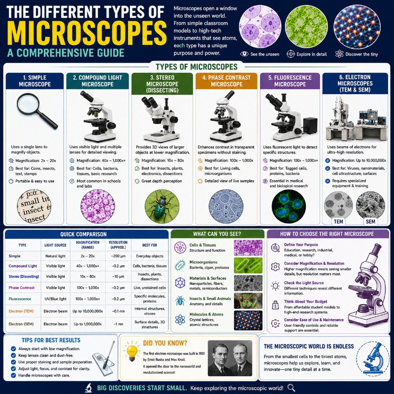
Most people picture a compound microscope the moment the word comes up — the upright tube and a rack of objectives that defined every middle school science class. But that single image barely scratches the surface: there are dozens of microscope types in regular use today, from handheld USB devices a kid can run off …
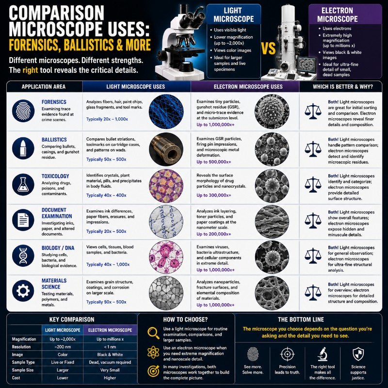
A comparison microscope is used to view two specimens simultaneously in a single split field of view — most famously in forensic science, where examiners place a bullet from a crime scene on one stage and a test bullet fired from a suspect weapon on the other, then judge whether the microscopic scratch patterns match. …
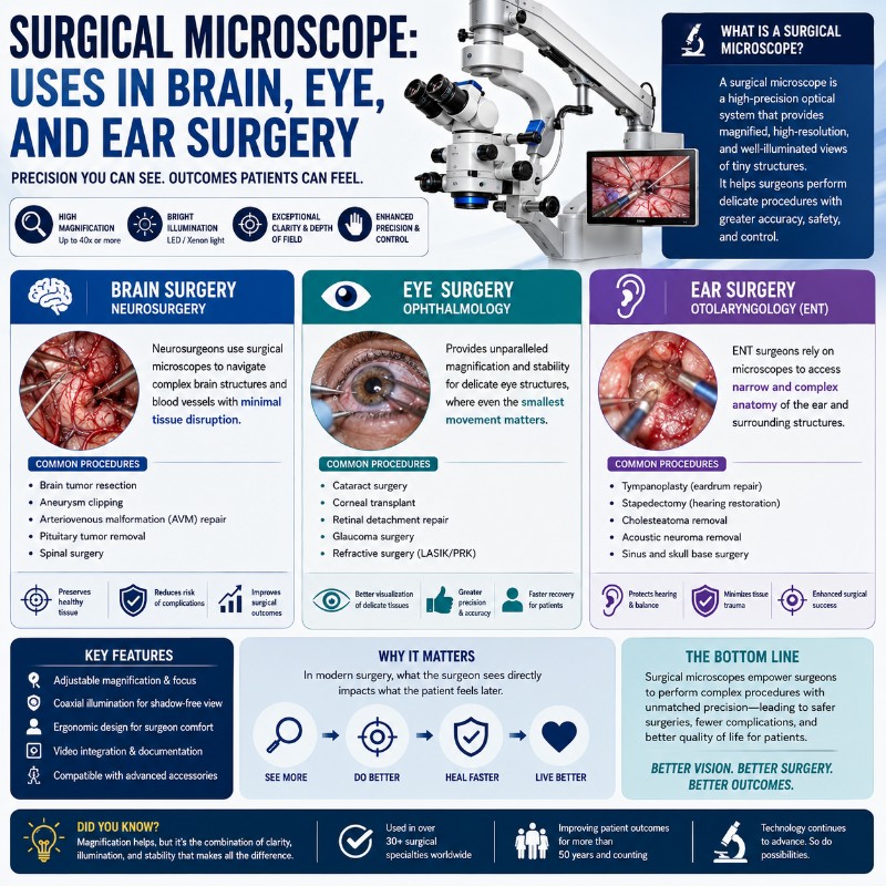
A surgical microscope — also called an operating microscope — is a binocular stereo microscope mounted on a counterbalanced stand that gives surgeons a magnified, brilliantly lit, three-dimensional view of the operative field. Unlike the high-powered compound scopes used in a biology lab, it operates at modest magnification (roughly 4× to 40×) with a working …
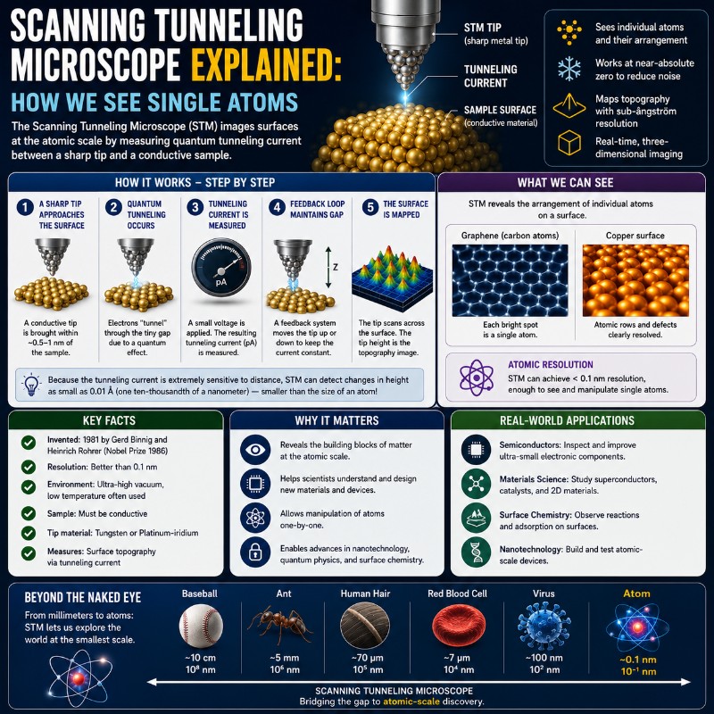
A scanning tunneling microscope (STM) is a device that images surfaces at the scale of individual atoms by measuring a tiny quantum-mechanical current that flows between a sharp metal tip and a conductive sample without the two ever touching. Invented in 1981 at IBM Zurich by Gerd Binnig and Heinrich Rohrer — work that earned …
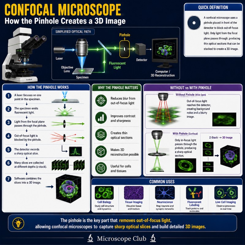
A confocal microscope is a type of optical microscope that uses a tiny pinhole aperture to physically block out-of-focus light, producing razor-sharp “optical slices” through a specimen that can be stacked into a full 3D reconstruction. Unlike a compound light microscope, which captures every plane at once in a blurry superimposition, a confocal microscope builds …
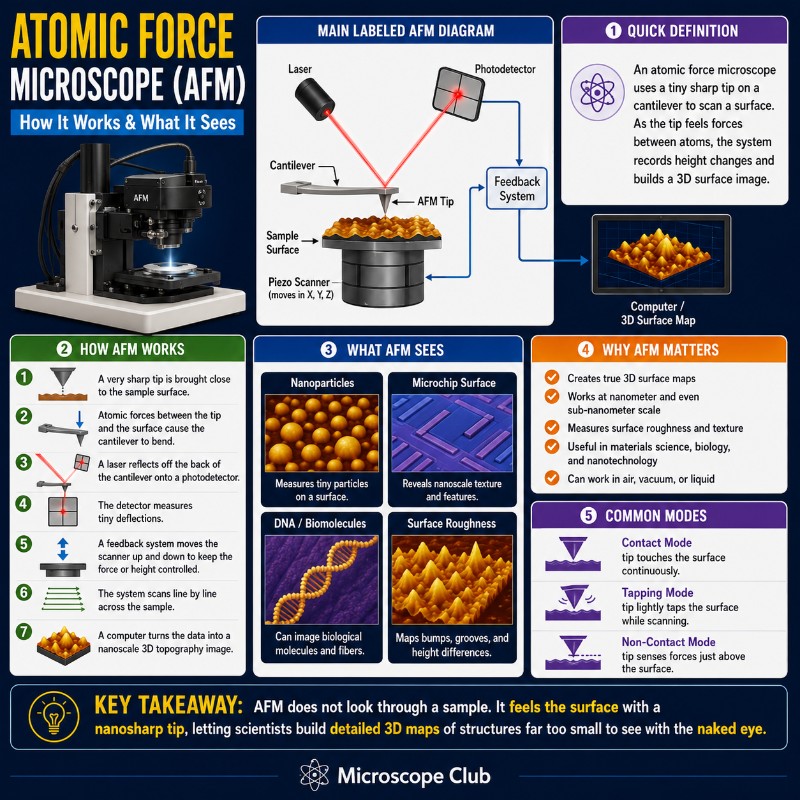
An atomic force microscope (AFM) is a scanning probe instrument that maps surfaces in three dimensions. It does this by dragging — or tapping — an ultra-sharp nanoscale tip across the sample and measuring the tiny forces between the tip and the material beneath. Unlike optical or electron microscopes, it uses no lenses and no …