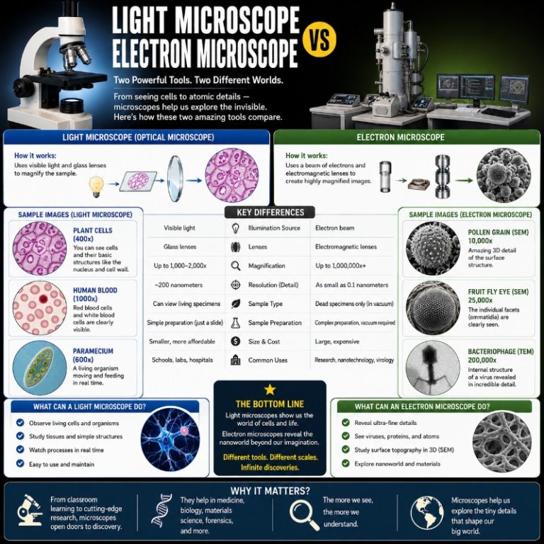
Light microscopes and electron microscopes are both designed to reveal what the naked eye cannot see — but they operate on entirely different physics, deliver very different images, and suit very different jobs. Understanding the gap between them is the first step to knowing which tool you actually need.
Quick comparison: light microscope vs electron microscope
| Attribute | Light microscope | Electron microscope |
|---|---|---|
| Radiation source | Visible light (~400–700 nm wavelength) | Accelerated electron beam (~0.004 nm at 100 keV) |
| Max useful magnification | ~1,000–1,500× | Up to ~1,000,000× (TEM); ~500,000× (SEM) |
| Resolution | ~200 nm | TEM ~0.05 nm · SEM ~0.5–20 nm |
| Image color | True color | Greyscale (false-color added in post-processing) |
| Living specimens | Yes | No (vacuum environment; cryo-EM = frozen, not live) |
| Sample preparation | Minimal — mount and stain in minutes | Extensive — fixation, dehydration, embedding or coating; hours to days |
| Size & cost | Benchtop, portable; tens to a few thousand dollars | Room-sized (or benchtop SEM); ~$100,000 to several million dollars |
How each microscope illuminates a specimen
Light microscope

A compound light microscope works through bright-field microscopy: a lamp below the stage fires a focused beam of visible light up through the specimen and a series of glass lenses, and the magnified image arrives at your eye — or a camera — through the eyepiece. Because the light carries actual color information, what you see through the eyepiece is in true color. A prepared blood smear, for instance, shows red erythrocytes and the deep-purple stained nuclei of white cells at a glance. The contrast you see depends on how much of that light the specimen absorbs; thin, unstained sections can look nearly invisible until you add a dye.
The hard ceiling on light microscopy is physical, not engineering: the Abbe diffraction limit. Resolution is approximately λ / (2 × NA), where λ is the wavelength of light (~550 nm for green) and NA is the numerical aperture of the objective. The math works out to roughly 200 nm — two points closer than that blur together and can never be resolved, no matter how good the lens. Superresolution techniques like STORM and PALM push past this with clever fluorophore chemistry, but they sit in a category of their own.
Electron microscope

Electron microscopes replace the light beam with a tightly focused stream of accelerated electrons. Electrons have a de Broglie wavelength of roughly 0.004 nm when accelerated at 100 keV — about 100,000 times shorter than visible light. Because resolving power scales with wavelength, that enormous difference is why electron microscopes can reveal details that are simply invisible under any light microscope (Biology LibreTexts).
The tradeoff is that electrons cannot travel through air without scattering, so the entire beam path, sample chamber, and detector must be held at a high vacuum. That vacuum requirement is what prevents imaging living, hydrated cells under most EM configurations.

Types of electron microscopes: TEM vs SEM
Electron microscopes come in two main classes, and “what is the difference between TEM and SEM?” is the first follow-on question most readers have.
Transmission electron microscope (TEM) — The electron beam passes through an ultrathin section of the sample (typically 50–100 nm thick). Because electrons must transmit through the material, TEM produces a 2D cross-sectional image showing internal ultrastructure. Resolution reaches approximately 0.05 nm with aberration correction, which is sub-Ångström and capable of imaging individual atoms in crystalline materials (Thermo Fisher Scientific). This is the instrument used to visualize ribosomes, mitochondrial cristae, and viral capsid proteins in atomic detail. See our full transmission vs scanning electron microscope comparison for more.
Scanning electron microscope (SEM) — The electron beam is scanned across the surface of the specimen, and detectors collect low-energy secondary electrons emitted as the beam hits each point. The result is a three-dimensional-looking surface topography image, like a landscape photograph of the sample surface. SEM resolution is typically 0.5–20 nm — far lower than TEM, but still orders of magnitude beyond light microscopy. SEM is the instrument that produces those dramatic images of insect compound eyes or the surface of a metal fracture. Visit our scanning electron microscope guide for a deep dive.

Two more types worth knowing: STEM (scanning transmission EM) combines the scanning approach with transmission imaging for atomic-scale chemical mapping; FIB-SEM pairs a focused ion beam that mills the sample with an SEM for 3D reconstruction of sub-cellular volumes.
Magnification: how much can each enlarge?
Magnification for a light microscope is calculated simply: total magnification = eyepiece magnification × objective magnification. A standard 10× eyepiece paired with a 40× objective gives 400×. Swap in a 100× oil-immersion objective and you reach 1,000× — near the practical ceiling for a light microscope. You can push to ~1,500× with specialized optics and immersion oil, but beyond ~1,000× most of the additional enlargement is “empty magnification”: you are not resolving any new detail, just making the blur bigger (Nanoscience Instruments). For a broader look at magnification records, see our guide to the highest magnification microscope.
Electron microscopes are in an entirely different league. TEM routinely achieves one million times magnification or beyond; SEM typically covers 1× to 500,000×. The exact ceiling depends on the instrument and the sample, but both types surpass any light microscope by factors of hundreds to thousands.
Resolution: what these microscopes can actually distinguish
Magnification without resolution is meaningless — a blurry image enlarged ten million times is still a blurry image. Resolution is the real measure of what a microscope can distinguish:
- Light microscope: ~200 nm — enough to resolve organelles like the nucleus, mitochondria, and chloroplasts, but not the internal structure of a ribosome or viral protein.
- SEM: ~0.5–20 nm (best instruments reach ~0.4 nm) — resolves the fine surface morphology of cell membranes, nanoparticles, and material surfaces (Measurlabs).
- TEM: ~0.05 nm (50 pm) with aberration correction — sub-Ångström resolution capable of imaging atomic columns in crystalline samples and near-atomic detail in frozen proteins.
To put those numbers in context: a hydrogen atom is about 0.1 nm across. TEM can, in the right configuration on a suitable sample, resolve features close to that scale. Light microscopy is separated from that by a factor of roughly 1,000 to 4,000.

Sample preparation: the practical cost of each technique
This is where the two approaches diverge most dramatically in day-to-day lab life.
Light microscope: Prepare a slide in minutes. For a quick look at pond water microorganisms, a drop on a slide and a coverslip is enough. For fixed tissue, you add a brief staining step — hematoxylin and eosin, for instance. Even a moderately stained slide takes under an hour. Living cells in culture can be placed directly on the stage and imaged in their growth medium.
Electron microscope: Sample preparation for EM is a multi-step, often multi-day process, and the steps differ by instrument type:
- TEM prep: chemical fixation (glutaraldehyde + osmium tetroxide) → dehydration in an alcohol series → resin embedding → ultrathin sectioning (~70–90 nm) with a diamond knife on an ultramicrotome → heavy-metal staining (uranyl acetate, lead citrate) to add contrast. A biological TEM sample typically takes 1–3 days. The thin-section requirement is also a common point of confusion: the ~70–90 nm figure is the section thickness electrons must pass through, not a limit on how large the specimen can be — you slice a large specimen into ultrathin sections.
- SEM prep: fixation → critical-point drying (to prevent collapse from surface tension) → conductive sputter-coating with gold, gold-palladium, or carbon for non-conductive samples so charge does not build up on the surface and blur the image. Note: gold coating is an SEM-specific step, not universal EM preparation.
Color: why electron microscope images look the way they do
Light microscopes produce true-color images — the color is intrinsic to the wavelengths of light the specimen absorbs or transmits. The red of a stained red blood cell is real red. Electron microscopes produce greyscale images only: electrons carry no color information, and detectors simply measure electron intensity at each point. Every vivid colored EM image you have seen — the neon blue neuron, the magenta coronavirus — had color added in post-processing by a digital artist or scientist choosing which structures to highlight. The raw data is always grey.
Living specimens and cryo-EM

Light microscopes can observe living organisms in real time. You can watch an amoeba extend pseudopods across a slide, observe bacteria tumbling through water, or image a live cell dividing under fluorescence. This is one of the most significant practical advantages of light microscopy.
Conventional electron microscopes cannot image living cells — the high vacuum kills hydrated biological specimens instantly, and the intense electron beam would damage them regardless. This was considered a fundamental limitation of EM until cryo-electron microscopy (cryo-EM) was developed. Cryo-EM flash-freezes samples in vitreous (glass-like) ice so fast that water molecules have no time to form crystals, preserving biomolecules in their near-native, hydrated shape inside the vacuum. The technique earned Jacques Dubochet, Joachim Frank, and Richard Henderson the 2017 Nobel Prize in Chemistry and has since delivered atomic-resolution structures of membrane proteins, viruses, and ribosomes that were impossible to solve any other way (NobelPrize.org). Cryo-EM does not image living, moving cells — but it does capture their molecular architecture in unprecedented detail.
Size, cost, and where these instruments live
A standard benchtop compound light microscope stands about a foot tall, weighs a few kilograms, and costs anywhere from a few hundred to a few thousand dollars. You can set one on a kitchen counter, pack it in a case for fieldwork, or store it in a cabinet. That portability is why light microscopes are standard equipment in school science labs worldwide.
Traditional research-grade electron microscopes are the opposite: room-filling instruments that require purpose-built facilities. A high-resolution TEM can stand two to three meters tall, weigh several tonnes, and cost $1 million or more to purchase and install. Vibration is the enemy — even the footsteps of someone walking past can blur a sub-nanometer image — so EM suites are typically placed on ground floors or basements on thick vibration-isolation slabs, or mounted on active anti-vibration tables. Electron microscopes are often placed on lower floors or in basements, and the primary reason is vibration isolation from building movement and traffic, not magnetic shielding. Magnetic and acoustic shielding matter for the highest-resolution TEM installations, but vibration is the baseline requirement.
That said, benchtop SEMs have changed this picture significantly. Modern desktop SEMs from manufacturers such as Thermo Fisher, Hitachi, and JEOL sit on a lab bench, run off standard power, require minimal facility prep, and cost roughly $100,000–$250,000. They cannot match the resolution of a full-sized research SEM, but they bring SEM imaging within reach of industrial quality-control labs, universities, and contract testing facilities that cannot build a dedicated EM suite.
What can you see with each microscope?
Light microscopes excel at viewing:
- Living cells and microorganisms — bacteria, protists, algae, blood cells
- Tissue cross-sections (histology) — muscle fiber arrangement, tumor margins
- Cell organelles visible at ~200 nm+ — nucleus, mitochondria, chloroplasts
- Chromosomes during cell division
- Whole small organisms — nematodes, small insects, plant material
Electron microscopes can reveal:
- Viral particles and protein complexes (TEM)
- Atomic columns in crystal lattices (aberration-corrected TEM/STEM)
- Cell membrane and organelle ultrastructure in 3D (SEM)
- Surface topography of materials — fractures, coatings, nanoparticles (SEM)
- Individual heavy-metal atoms on suitable samples (STEM)
- Near-atomic resolution of frozen biomolecules (cryo-EM)
The one thing a conventional electron microscope cannot do that a light microscope can: show you a living, moving specimen. That remains a fundamental distinction between the two technologies.
Which should you use? A decision guide
Use a light microscope when:
- You need to observe living or hydrated specimens in real time
- True color matters (histology, clinical diagnosis, microorganism ID)
- You need results quickly — hours or less from sample to image
- Budget is limited or portability is required
- ~200 nm resolution is sufficient for your question
Use an SEM when:
- You need high-resolution 3D surface morphology of a non-living or fixed sample
- Elemental composition at the surface matters (most SEMs accept energy-dispersive X-ray spectroscopy, EDX)
- The sample can tolerate vacuum and conductive coating
- ~1–20 nm surface resolution is the target
Use a TEM when:
- Internal ultrastructure at sub-nanometer detail is needed
- Atomic-scale imaging of crystal defects, grain boundaries, or nanoparticles is the goal
- You can invest in multi-day sample preparation
- You have access to a dedicated TEM facility
Wondering what electron microscopes cost in practice? Our electron microscope cost guide breaks down pricing from benchtop SEM to full research TEM. For a full overview of instrument types, see our guide to the different types of microscopes.
Frequently asked questions
What is the main difference between a light microscope and an electron microscope?
A light microscope uses photons of visible light to illuminate a specimen; an electron microscope uses a beam of accelerated electrons. Because electrons have a wavelength roughly 100,000 times shorter than visible light, electron microscopes achieve far higher resolution and magnification — but require a vacuum environment, extensive sample preparation, and cannot image living specimens.
Which has higher resolution — a light or electron microscope?
Electron microscopes are significantly higher-resolving. The best light microscopes are limited to ~200 nm by the Abbe diffraction limit. SEM achieves ~0.5–20 nm; TEM reaches ~0.05 nm (50 pm) with aberration correction — roughly 1,000 to 4,000 times better than light.
Can an electron microscope see living cells?
No, not in a conventional electron microscope. The high vacuum required kills live, hydrated specimens. Cryo-EM can image biomolecules in near-native conformation by flash-freezing them in vitreous ice, but it still does not capture cells in a living, active state — it preserves a snapshot of their structure.
Why can’t electron microscopes show color?
Electrons carry no color information — they are simply particles with a charge and kinetic energy. Detectors measure electron intensity, not wavelength, so the raw output is always a greyscale map of electron signal. Any color in an EM image was added digitally in post-processing to highlight different structures or make images more intuitive for a non-specialist audience.
What is the difference between TEM and SEM?
In a TEM, the electron beam passes through an ultrathin sample section, producing a 2D image of internal structure with the highest available resolution. In an SEM, the beam is scanned across the sample surface, and secondary electrons are collected to build a 3D-looking surface image. TEM resolution (~0.05 nm) is higher than SEM (~0.5–20 nm), but SEM handles larger, bulkier samples more easily.
Do electron microscopes need a vacuum?
Yes. Electrons scatter off air molecules almost instantly, so the entire optical column, sample chamber, and detectors must be maintained at high vacuum. Achieving and maintaining that vacuum is one reason electron microscopes are large, expensive, and require a power supply and pumping system running continuously.
What is the maximum magnification of a light microscope vs an electron microscope?
A standard compound light microscope reaches ~1,000–1,500× useful magnification (higher magnification produces empty enlargement without new detail). Electron microscopes reach up to ~1,000,000× or more (TEM) and ~500,000× (SEM) — hundreds to thousands of times greater.
Conclusion
Light microscopes and electron microscopes are complementary tools, not competing ones — the right choice depends entirely on what question you are trying to answer. Light microscopy wins when you need living cells, true color, speed, or portability, and its 200 nm resolution ceiling is sufficient for the vast majority of biological and educational work. Electron microscopy wins when surface ultrastructure or internal architecture at the nanometer or sub-nanometer scale is the target, at the cost of complex sample preparation, vacuum infrastructure, and specimens that can no longer be alive. If you are starting out, a compound light microscope is the natural first step; if your research has outgrown what visible light can resolve, an SEM is usually the accessible entry point into electron microscopy before committing to a full TEM installation. Either way, understanding the physics behind each instrument — wavelength, resolution, and sample state — will guide you to the right tool for the job.
