Microscope field of view is the diameter — in millimeters or micrometers — of the circular specimen area you can actually see through the eyepiece at any given magnification. Get this number wrong and you’ll spend ten minutes hunting for a 0.55 mm object on a slide that you could have found in seconds by knowing where to look.
What field of view means in a microscope

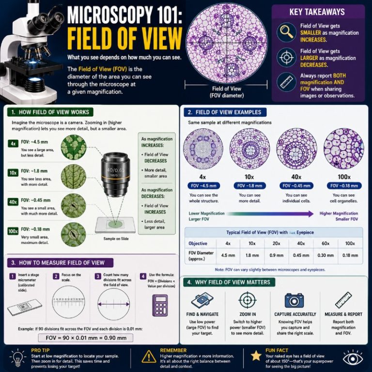
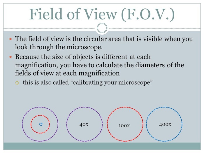
When you look through a compound light microscope, you see a bright circle of light — that circle is your field of view. It is not an angle; it is a diameter measured at the specimen plane, expressed in mm or µm. The area you can view shrinks as magnification rises, which is why you always start a search at low power and zoom in once you’ve found your subject.
For context: the human binocular visual field spans roughly 200° horizontally, with a central ~120° zone where both eyes overlap and true stereoscopic vision occurs. A microscope’s “field of view” is an analogous concept scaled to the specimen — how wide a slice of reality the instrument lets you examine at once.
Field number: the key number on your eyepiece

Look at the barrel of your eyepiece. You’ll see two numbers stamped on it — something like 10× / 22. The first (ending in ×) is the eyepiece magnification. The second is the field number (FN): the diameter in millimeters of the intermediate image circle the eyepiece is physically capable of transmitting to your eye. Everything outside that circle is blocked by the field diaphragm.
Field number is a property of the eyepiece alone. It does not change when you swap objectives. What changes is how much of that FN circle gets “used up” by the objective’s magnification — which is exactly what the FOV formula captures.
FN vs. apparent field of view (AFOV)
Beginners often see “AFOV” on eyepiece spec sheets and confuse it with field number. They are different things:

- Field number (FN) — a linear measurement (mm) at the intermediate image plane. This is what you plug into the formula.
- Apparent field of view (AFOV) — the angular width (degrees) the eyepiece presents to your eye. A 10× eyepiece with FN22 has an AFOV of roughly 22°; a wide-field 10× with the same FN but different optics may show a wider angle. AFOV does not directly predict specimen coverage.
When calculating how much of your slide you can see, always use FN — not AFOV.
Modern standard widefield (WF) 10× eyepieces ship with FN18, FN20, or FN22; FN22 is now the de facto standard for quality student and research scopes. Ultra-wide eyepieces reach FN26.5–28 mm. The older 16 mm figure you still see cited is an outdated budget spec. (Olympus/Evident on field number)
The FOV formula — and the mistake most beginners make

The formula is:
FOV (mm) = Field Number ÷ Objective Magnification
Notice what’s not in that formula: eyepiece magnification. You divide by the objective power only, not by the total magnification (objective × eyepiece). (Nikon MicroscopyU — FOV formula)
The most common beginner error is dividing FN by total magnification (e.g. 200× instead of 20×), which gives a field diameter ten times smaller than reality. The eyepiece magnification controls how large the intermediate image looks to your eye — it does not shrink the specimen area the objective captures.
Worked example
Eyepiece: 10× with FN22. Objective: 40×.

FOV = 22 mm ÷ 40 = 0.55 mm (550 µm)
Swap to a 10× objective: FOV = 22 ÷ 10 = 2.2 mm (2200 µm) — four times wider, so you can orient yourself on the slide before zooming in.
When there’s a tube factor (auxiliary lens)
Some microscopes — particularly older research scopes or those fitted with a 1.25× or 1.5× intermediate magnification changer — have an extra lens between the objective and eyepiece. That lens multiplies the effective objective magnification:
FOV (mm) = FN ÷ (Objective Magnification × Tube Factor)

Example: FN22, 40× objective, 1.5× tube = 22 ÷ (40 × 1.5) = 22 ÷ 60 = 0.37 mm (370 µm). If you ignore the tube factor, you’ll think you’re seeing more slide than you actually are.
FOV at every common objective — quick-reference table
Fixed eyepiece: 10× WF, FN22 (the modern standard). All values calculated as 22 ÷ objective magnification.
| Objective | Total magnification (×10 eyepiece) | FOV diameter |
|---|---|---|
| 4× | 40× | 5.5 mm (5500 µm) |
| 10× | 100× | 2.2 mm (2200 µm) |
| 20× | 200× | 1.1 mm (1100 µm) |
| 40× | 400× | 0.55 mm (550 µm) |
| 100× | 1000× | 0.22 mm (220 µm) |
At 4× you can see nearly the entire wing of a small insect in one view. By the time you reach 100× oil immersion, you’re looking at a window 220 µm wide — narrower than the diameter of three human hairs side by side. That dramatic collapse is what makes low-power orientation so important, and it’s also why how magnification limits what you can see is such a critical concept before you crank up the power.
Why magnification shrinks your field of view
Here’s the physical reason, not just the formula: the objective lens projects a magnified intermediate image of the specimen onto the plane where the eyepiece field diaphragm sits. Higher objective magnification stretches the image — so the same fixed-diameter diaphragm opening now covers a smaller original area on the slide. You’re zooming into a portion of what the diaphragm could theoretically show at lower power.
This is why the relationship is inversely proportional and exactly linear: double the objective magnification, halve the FOV diameter, quarter the visible area. There’s no workaround within the optics — the only fix is a physically wider diaphragm (higher FN eyepiece) or a different optical design.
How to measure your own field of view (when FN isn’t printed)
Some older or budget eyepieces don’t have the field number stamped on them. Here’s how to find your actual FOV with tools you already have.
Method 1 — Stage micrometer (most accurate)
A stage micrometer is a glass slide with a precision-ruled scale (typically 1 mm divided into 100 divisions of 10 µm each). Place it on the stage the same way you would when preparing a slide for viewing.
- Place the stage micrometer on the stage and focus at the objective power you want to measure.
- Move the micrometer so one end of the scale aligns with the left edge of your field of view circle.
- Count how many divisions fit across the full diameter to the right edge.
- Multiply by the division value (10 µm per division if using a standard 0.01 mm micrometer).
Example: 55 divisions × 10 µm = 550 µm = 0.55 mm. This matches FN22 ÷ 40× exactly — and confirms your eyepiece is a modern FN22 model.
Method 2 — Clear plastic ruler at low power (quick estimate)
- Place a clear mm ruler flat on the stage under the 4× objective.
- Focus until you can read the graduations clearly.
- Count how many millimeters span the diameter of the lit circle.
At 4× with FN22 you should see approximately 5–5.5 mm. This method is only accurate enough for the 4× objective because ruler lines are too coarse to count precisely at higher power — use a stage micrometer for 10× and above.
Camera and digital microscope field of view
When you attach a camera to a microscope, the camera sensor is almost always smaller than the eyepiece field. The sensor captures a crop of the eyepiece image, not the full circle. The formula changes:
Camera FOV ≈ Sensor diagonal (mm) ÷ C-mount adapter magnification
For example: a 1/2.3″ sensor has a diagonal of ~7.7 mm. With a standard 0.5× C-mount adapter: 7.7 ÷ 0.5 = 15.4 mm effective FOV — significantly narrower than the eyepiece’s full FN22 circle. If your photomicrographs always show a narrower view than what you see by eye, the C-mount adapter’s reduction factor is why.
USB and HDMI digital microscopes with integrated cameras have their own sensor-based FOV that is independent of eyepiece FN entirely. The manufacturer should list the FOV at each magnification step; if they don’t, measure it with the clear-ruler method above. Stereo microscopes follow a different FOV calculation path entirely — see our stereo microscope guide for the specifics.
Wider FOV without losing magnification: plan objectives and wide-field eyepieces
The vague “more sophisticated lenses” reference in older articles on this topic usually means two specific technologies:
- Plan and plan-apochromat objectives — designed to project a flat, in-focus image all the way to the edge of the field (not just the center). They don’t increase the FN, but they make the outer portion of the FOV actually usable. Standard achromat objectives can show sharp focus at the center while the edges blur — so even if your FN22 says the FOV is 0.55 mm at 40×, a cheap objective may only give you a usable ~0.35 mm in the center. (Leica on resolved field number)
- Wide-field (WF) and ultra-wide (UW) eyepieces — physically larger field diaphragms with FN20–28 instead of the older FN18 or 16. A WF10×/22 eyepiece on the same objective gives you 22 ÷ 40 = 0.55 mm; replace it with a legacy 10×/16 and you get only 16 ÷ 40 = 0.40 mm — nearly 90% more area with the FN22.
FOV vs. depth of field — not the same thing
Field of view is a horizontal measurement — how wide a slice of the slide you can see. Depth of field in a microscope is a vertical measurement — how thick a layer of the specimen stays in focus at once. Both shrink as magnification rises, but they shrink for different optical reasons and are controlled by different lens properties (numerical aperture for DOF; field diaphragm size and objective mag for FOV).
A common beginner confusion: at 100× oil immersion, both the FOV and depth of field are tiny, so you can only see a tiny, paper-thin slice of the specimen at any moment. This is why focus racking (moving the fine focus wheel slowly through z-planes) is part of every high-power examination.
Electron microscope field of view
For completeness: a transmission electron microscope (TEM) uses electrons rather than light, but it still has a field of view — the area of specimen imaged at a given magnification. At moderate TEM magnification the FOV spans tens to hundreds of micrometres. At maximum magnification (imaging individual atoms) the FOV narrows to the nanometre range, not picometres. TEM resolution reaches sub-Ångström (~0.05–0.2 nm), but resolution and field of view are separate specs.
Frequently asked questions
What is the field number on a microscope eyepiece?
The field number (FN) is the diameter in millimeters of the intermediate image circle the eyepiece transmits. It is stamped on the eyepiece barrel next to the magnification — for example, “10× / 22” means 10× magnification and FN22.
What is the field of view of a 40× objective?
With a standard FN22 eyepiece: FOV = 22 ÷ 40 = 0.55 mm (550 µm). With an older FN18 eyepiece it drops to 0.45 mm. Always check your eyepiece’s FN for an exact value.
What is the field of view of a 10× or 4× objective?
FN22 eyepiece: 10× objective → 2.2 mm; 4× objective → 5.5 mm. See the full table above for all common powers.
Conclusion
Microscope field of view is determined by two numbers you can read directly off your equipment: the field number (FN) stamped on the eyepiece barrel, and the objective magnification. Divide FN by objective power — not total magnification — and you have the diameter of the circular specimen area in millimeters. That single formula, combined with the reference table above, tells you exactly how much of your slide is visible before you even look through the eyepiece. If your eyepiece has no FN printed on it, a stage micrometer gives you the answer in under a minute. Understanding FOV alongside depth of field and microscope resolution gives you real control over what you see — and faster, less frustrating sessions at the bench.
