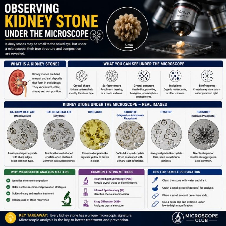
Kidney stones under the microscope reveal something most patients never see: jagged crystalline structures whose shape, color, and optical behavior tell clinicians exactly what went wrong in the kidney and how to prevent it happening again. Understanding what each stone type looks like — and which microscope technique makes those details visible — turns a painful lump of mineral into diagnostic information.
Medical disclaimer: The information here is for educational purposes. Stone analysis is performed by accredited clinical laboratories. Symptoms of kidney stones (especially fever with flank pain) require prompt medical attention — do not self-diagnose. Consult a physician or urologist for any concerns about your kidney health.
What a Kidney Stone Is (and Why Composition Matters)
A kidney stone is a solid mass of crystallized minerals that forms when urine becomes too concentrated. Stones range from a grain of sand to a golf ball in size. Small stones usually pass on their own; larger ones lodge in the ureter and can cause obstructive uropathy — pressure-driven kidney damage — or, if bacteria are present, urosepsis, a life-threatening systemic infection. That is why knowing a stone’s composition matters: the crystal type points to the metabolic or infectious cause, and choosing the right prevention strategy depends on getting that identification right.
Stone formation begins when urine contains too much of certain minerals — calcium, oxalate, phosphate, uric acid — and too little water to keep them dissolved. The minerals nucleate into crystals, which aggregate into a stone over months to years. The stone’s surface layers often form under different conditions than its core, so a lab that only analyzes the surface may miss the true initiating cause. [StatPearls NBK442014]

The Four Types of Kidney Stone and Their Crystal Appearance
Kidney stones fall into four main types, each with a distinct crystal morphology visible under the right scope. Calcium stones account for 75–85% of all cases PMC8229448; uric acid stones make up roughly 8–10%; struvite stones 3–8%; and cystine stones around 1–2%. The table below maps what each type looks like through the lens.
| Stone type | Crystal shape | Color | Birefringence (polarized light) | Typical urine pH | Best scope for ID |
|---|---|---|---|---|---|
| Calcium oxalate monohydrate (COM) | Dumbbell / ovoid | Off-white to tan | Strongly birefringent (bright under crossed polarizers) | Variable (5.5–6.5) | Polarizing compound scope |
| Calcium oxalate dihydrate (COD) | Envelope / bipyramidal octahedron | Colorless to pale yellow | Weakly birefringent | Variable | Polarizing compound scope |
| Calcium phosphate (brushite / apatite) | Thin rosettes of sharp needles / plates | White to chalky | Moderate | Alkaline (>6.5) | Polarizing compound scope |
| Uric acid | Rhomboid / diamond / barrel | Yellow-orange to brick-red | Birefringent; ID confirmed with urine pH | Acidic (<5.5) | Polarizing compound scope + pH |
| Struvite (triple phosphate) | Coffin-lid (rectangular with beveled corners) | White/grey; layered cross-section | Weakly birefringent | Alkaline (>7.2) | Stereo scope (surface); compound (section) |
| Cystine | Hexagonal plates | Amber / honey-yellow; translucent | Weakly birefringent | Variable (often <7) | Polarizing compound scope |
Calcium Stones — the Most Common Type
Calcium oxalate monohydrate (COM) is the most common sub-type, responsible for roughly 67% of all kidney stones on its own. Under polarized light it is one of the most striking crystals in clinical microscopy: crossed polarizers make it blaze white against a black background because it is strongly birefringent. The dumbbell shape is distinctive once you know what to look for, but beginners often mistake COM crystals for cell debris or mucus threads because the background is cluttered. The fix is to focus the polarizer — debris goes dark; COM crystals light up. Calcium oxalate dihydrate (COD), by contrast, forms classic envelope-shaped bipyramidal crystals and is less birefringent. Calcium phosphate presents as rosettes of very thin, sharp needle-like projections — brushite under the scope looks almost flower-like, delicate rather than jagged.
Uric Acid Stones
Uric acid stones account for roughly 8–10% of cases — not the most common type, despite what some sources claim [StatPearls NBK442014]. They form when urine is persistently acidic (pH below 5.5), which is common in gout, obesity, type 2 diabetes, and in patients eating a very high-protein diet. Under the microscope they appear as rhomboid or diamond-shaped crystals ranging from yellow to brick-orange. The color is one of the first visual cues: whole uric acid stones often have an orange or reddish tinge that makes them visually distinct from calcium stones. Uric acid is birefringent, so polarized light helps, but pH confirmation is essential for certain ID — calcium oxalate monohydrate can look similar at some orientations.

Struvite Stones
Struvite stones — made of magnesium ammonium phosphate — form exclusively in the setting of urinary tract infection with urease-producing bacteria such as Proteus mirabilis or Klebsiella. Urease splits urea into ammonia, raising urine pH above 7.2 and driving the precipitation of struvite. These are the stones that can grow enormous: by filling the entire renal pelvis they become staghorn calculi, antler-shaped casts of the collecting system Nature Reviews Urology. Under the stereo microscope, struvite stones are layered and often pale grey-white with a rougher, cratered surface. The coffin-lid crystal shape under the compound scope is unmistakable — rectangular with the corners cut off, like a trapezoid — and is characteristic enough that experienced lab technicians identify it on morphology alone.
Cystine Stones
Cystine stones are rare (1–2% of cases) and arise from a genetic defect — cystinuria — that prevents the kidney from properly reabsorbing the amino acid cystine. They tend to be smooth, amber to honey-colored, and partially translucent, which makes them look almost gemstone-like compared to the chalky calcium varieties. Their hallmark under the microscope is the hexagonal crystal: flat, six-sided plates that are instantly identifiable. Cystine stones are compact and resistant to lithotripsy, which is one reason correct identification matters — it changes the treatment path.

How to View a Kidney Stone Under a Stereo Microscope
A stereo (dissecting) microscope is the right starting point for observing a whole kidney stone. It uses reflected and oblique light at low magnification — typically 7–45×. Unlike a compound microscope, it does not require thin sections, immersion oil, or coverslips; you are looking at the surface of a solid object, not a slide preparation.

You will need:
- Stereo microscope (7–45× range)
- Well plate or black card as a stage background (white stones show better on black)
- Forceps to handle the stone
- Scalpel or razor blade if halving the stone (lab use only)
- Distilled water and soft brush for cleaning
- Camera or phone adapter for documentation
Step-by-step procedure:
- Clean the specimen. Rinse the stone gently with distilled water and use a soft brush to remove any attached tissue. Pat dry with a lint-free cloth — do not use immersion oil; it is not used with stereo microscopes.
- Place the stone on your stage background. For pale stones, a black card background improves contrast. Use forceps, not fingers, to avoid contaminating the surface.
- Set the lowest magnification (7–10×) first. Dial in the lighting — top-reflected light shows surface texture; transmitted oblique light from beneath shows translucency and color in thinner stones.
- Observe the surface. Note overall color, surface texture (smooth, rough, spiky, layered), and luster. Calcium oxalate stones often look crystalline and slightly sparkle; uric acid stones look waxy-orange; struvite can look chalky and stratified.
- Increase magnification incrementally. At 20–45× you will start to see crystal faces, layering, and pitting. Common beginner mistake: jumping straight to maximum magnification loses context — always work up from low power.
- Record observations. Note size, shape, dominant color, surface texture, any visible layering. If the stone has been halved (a clinical procedure), examine the core versus the surface — different crystal zones indicate different formative phases.
- Document with a photo before moving to higher-power or destructive analysis.
For more on slide and specimen preparation techniques, see our guide on how to prepare microscope slides.
Polarized Light Microscopy — How Labs Actually Identify Crystal Type
Surface morphology under a stereo scope tells you a lot, but the definitive clinical identification of kidney stone crystals uses a polarizing microscope on a ground thin section. Here is why that matters: calcium oxalate monohydrate is strongly birefringent — it rotates the plane of polarized light — so under crossed polarizers (two filters at 90°) it appears brilliantly bright on a black background. Uric acid is birefringent too, but less so, and its rhomboid morphology plus acidic urine pH provide the confirming signature. Struvite’s coffin-lid shape is so distinctive it barely needs the polarizer. Cystine’s hexagons are identifiable by shape and the relatively weak birefringence.
The clinical workflow for a passed or surgically retrieved stone goes further than just polarized light. Labs typically do:
- Gross inspection + stereo microscopy — color, size, surface texture, layering.
- Sectioning — the stone is cut in half to expose the core, because core composition can differ from the surface and reveals the initiating event.
- Polarized light on a thin section — crystal morphology + birefringence identifies ~80–90% of common stones.
- FTIR or Raman spectroscopy — infrared spectroscopy provides definitive compositional analysis and detects mixed-type stones (a stone with a calcium phosphate core and a calcium oxalate shell, for instance) that polarized light alone might misread [PMC3309560].
- SEM (scanning electron microscopy) — used for surface ultrastructure and to examine stones with unusual morphology. A scanning electron microscope can resolve individual crystal faces at nanometer scale, revealing growth layers and incorporated organic material. See our electron microscope images gallery for examples of mineral crystals at this resolution.
Combined morphology, birefringence, and urine pH correctly identify approximately 98% of “typical” stone crystals in routine urinalysis [StatPearls NBK606103].
Causes, Risk Factors, and Recurrence
Kidney stones form when urine is persistently oversaturated with stone-forming minerals. The key risk factors vary by stone type, which is another reason accurate microscopic identification matters — the treatment for calcium oxalate stones (increase fluid intake, reduce dietary oxalate, sometimes thiazide diuretics) is different from uric acid stones (alkalinize urine with potassium citrate, reduce dietary purines) and completely different from struvite (treat the underlying UTI, surgical clearance of large staghorn stones).
General risk factors across stone types include [StatPearls NBK442014]:
- Dehydration — concentrated urine is the single most modifiable risk factor for all stone types.
- Diet — high-sodium, high-protein, or high-oxalate intake (spinach, nuts, chocolate) for calcium oxalate; high-purine diet (red meat, shellfish) for uric acid.
- Obesity and metabolic syndrome — strongly linked to uric acid stone formation via insulin resistance reducing urine pH.
- Chronic UTI with urease organisms — the only reliable cause of struvite.
- Genetic predisposition — cystinuria (autosomal recessive), primary hyperoxaluria, distal renal tubular acidosis.
- Previous stone — the single strongest predictor of a future stone; recurrence rates reach 50% within 5 years without targeted prevention.
The symptoms of an active stone include flank or lower-back pain (often severe and colicky), nausea and vomiting, blood in urine, pain on urination, and increased urge to urinate. Fever and chills alongside flank pain signal a possible infected obstruction (pyonephrosis) — seek urgent medical care immediately rather than waiting for the stone to pass, as this can progress to urosepsis StatPearls.
Symptoms of Kidney Stones
A stone can remain silent in the kidney for years. Once it begins to move or grows large enough to obstruct, symptoms appear:
- Severe colicky pain in the flank, lower back, or radiating to the groin
- Blood in urine (hematuria)
- Nausea and vomiting
- Pain or burning during urination
- Frequent or urgent need to urinate
- Cloudy or foul-smelling urine
- Fever and chills — seek urgent care if these accompany flank pain
Smaller stones may pass without any pain at all — “silent stones” exit in the urine unnoticed.

Kidney Stone Under Microscope — FAQ
Can you see a kidney stone without a microscope?
Stones larger than about 3–4 mm are visible to the naked eye. Very small stones — and the crystals that make up their structure — require at least a stereo microscope at low power. Individual crystal faces and sub-millimeter structural detail need a compound microscope at 100–400×.
Will a kidney stone float or sink in water?
It depends on the density of the dominant mineral. Calcium-containing stones are denser than water and sink. Pure uric acid stones have a density close to water and may barely sink or appear to float momentarily; mixed-composition stones always sink.
Why are kidney stones so jagged?
Jaggedness reflects crystal growth pattern and composition. Calcium oxalate monohydrate grows in spiky, elongated habits and creates sharp, irregular surfaces. Calcium phosphate brushite forms rosettes of needle-like crystals — even more jagged at a microscopic level than it appears whole. Uric acid and cystine stones, by contrast, tend to grow in more compact, rounded habits and feel smoother to the touch.
What is the most common type of kidney stone?
Calcium oxalate is the most common, accounting for roughly 67% of stones on its own; calcium stones overall (oxalate + phosphate) make up 75–85% of all cases. Uric acid is second at 8–10%. Struvite is 3–8%, and cystine is 1–2% [PMC8229448].
Why is stone analysis done in a lab rather than at home?
Clinical stone analysis requires a thin-section preparation, crossed polarizers calibrated precisely at 90°, and often FTIR or Raman spectroscopy to resolve mixed-composition stones and confirm crystalline identity. Home or classroom microscopy can show gross morphology and color, but will miss the compositional nuance that drives treatment decisions. All diagnostic stone analysis should go to an accredited clinical laboratory.
MUST-READ: Light Microscope vs Electron Microscope
Conclusion
Kidney stones under the microscope are a masterclass in how crystalline structure encodes biological history: the coffin-lid shape of struvite signals a bacterial infection; the bright birefringence of calcium oxalate monohydrate under crossed polarizers points to a metabolic imbalance; the hexagonal plates of cystine reveal a genetic condition. The key fact to anchor on: calcium stones are the most common type — not uric acid — and getting that right is the foundation for choosing the correct prevention strategy. If you pass a stone, strain your urine through a fine mesh or coffee filter, save it, and ask your urologist to send it for FTIR analysis: that single step is the highest-yield thing you can do to prevent the next one.
RELATED POST:
Originally posted 2022-06-26 01:27:08.