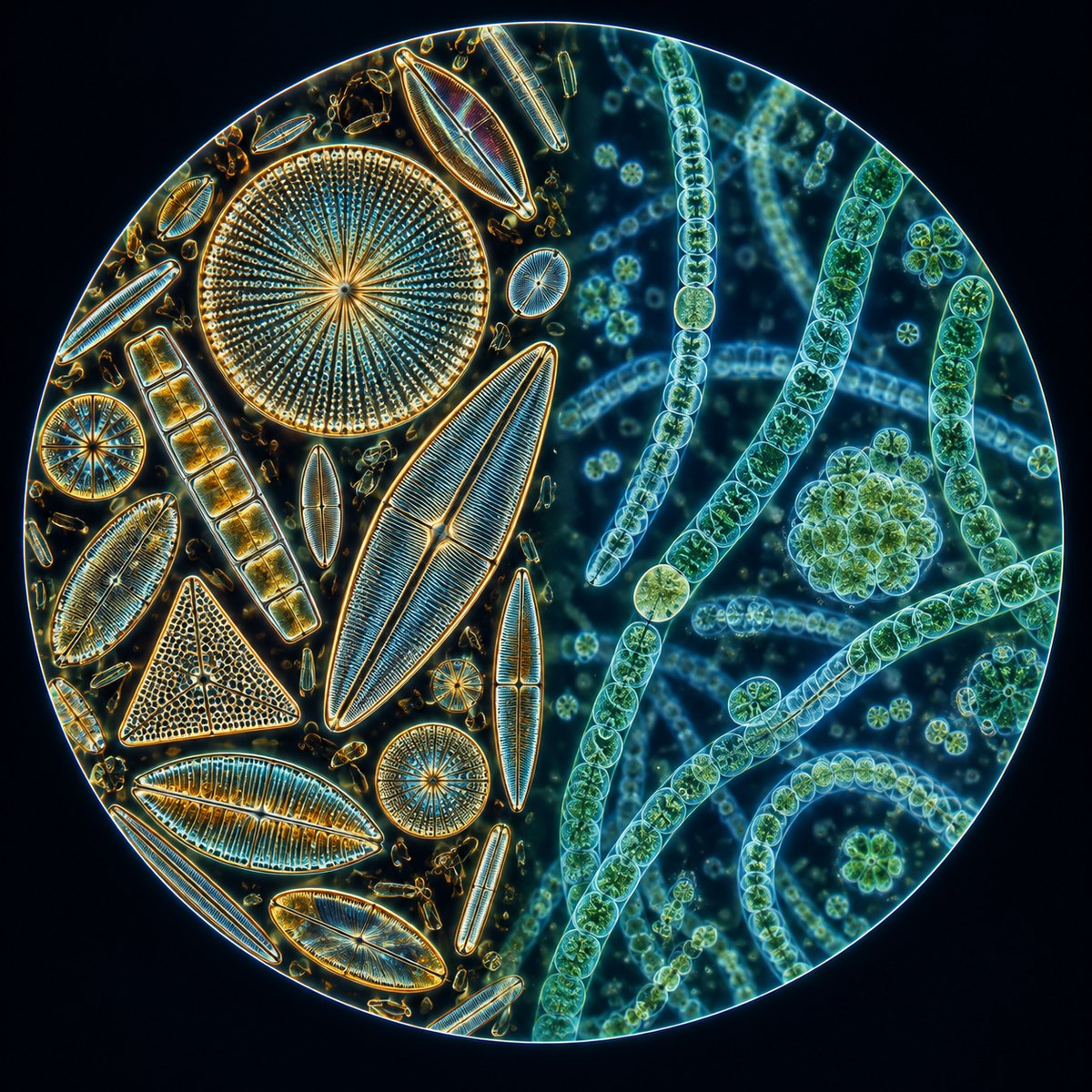
Observing Pond Water Under a Microscope
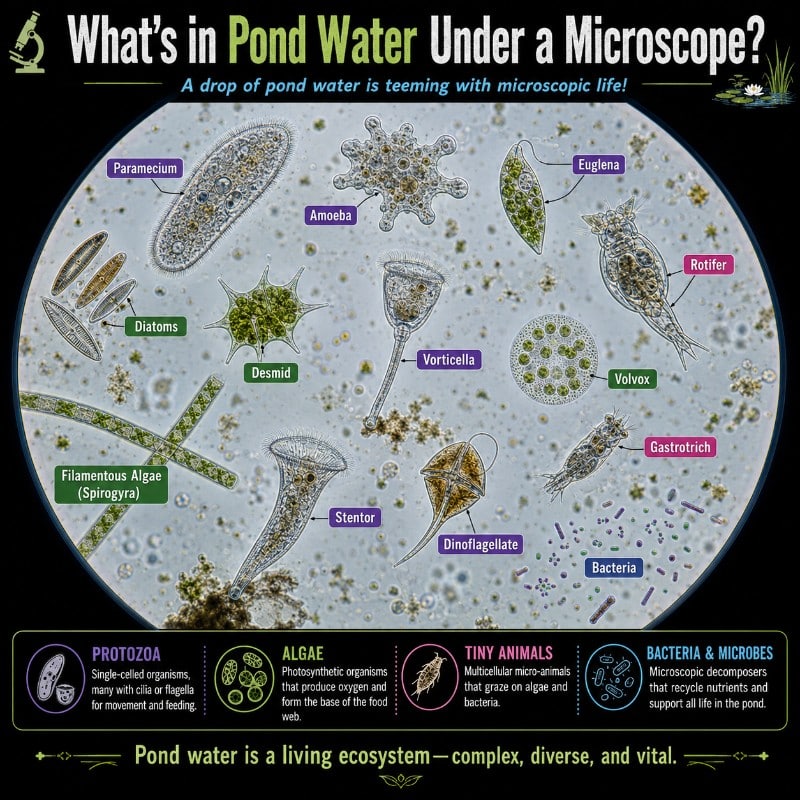
What’s in Pond Water Under a Microscope? A single drop of pond water contains a thriving ecosystem — protozoa like paramecium and vorticella gliding and pulsing, algae such as Spirogyra’s spiral chloroplast coiling through the frame, micro-animals like rotifers and water fleas jerking across your field of view, and non-living debris including pollen grains, detritus, …
Read more

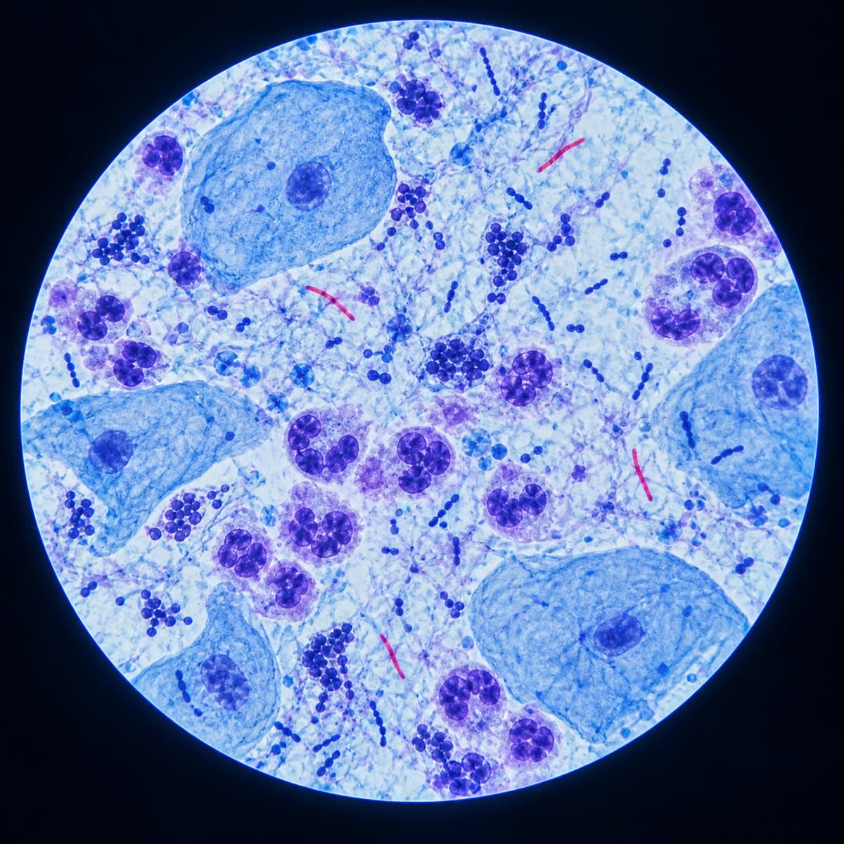
White Blood Cells Under a Microscope: How to ID All 5

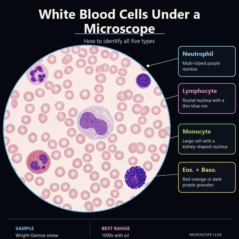
Five white blood cell types in a stained smear: neutrophil, lymphocyte, monocyte, eosinophil and basophil

White blood cells under a microscope appear as scattered, purple-nucleated cells standing out sharply against a pink carpet of red blood cells. Once you know what to look for, you can name every one of the five types in seconds. Nucleus shape, granule color, and size relative to a red blood cell are the only …

Read more

Aspergillus Under a Microscope: ID It vs Penicillium

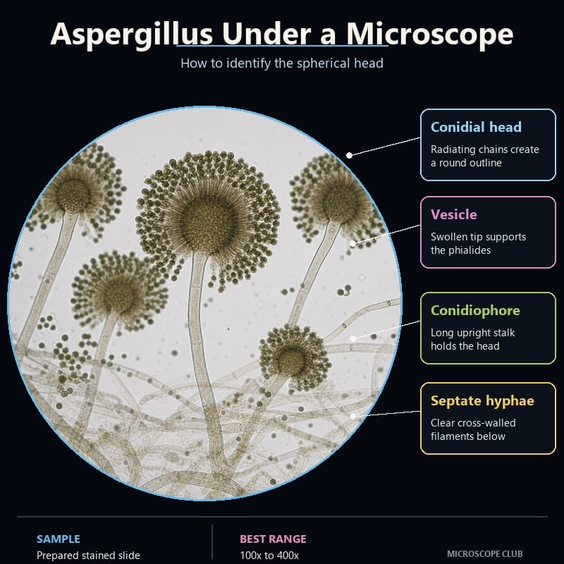
Aspergillus under a microscope showing spherical conidial heads, vesicles, conidiophores and septate hyphae

Aspergillus under a microscope reveals a long, unbranched stalk that swells into a round vesicle covered in radiating chains of spores. This distinctive “aspergillus head” is what separates the mold from every look-alike on a lab bench. Found a fuzzy black, yellow-green, or gray-green colony on bread, fruit, soil, or a damp windowsill? A compound …

Read more

Rotifers Under a Microscope: The Wheel That Never Spins

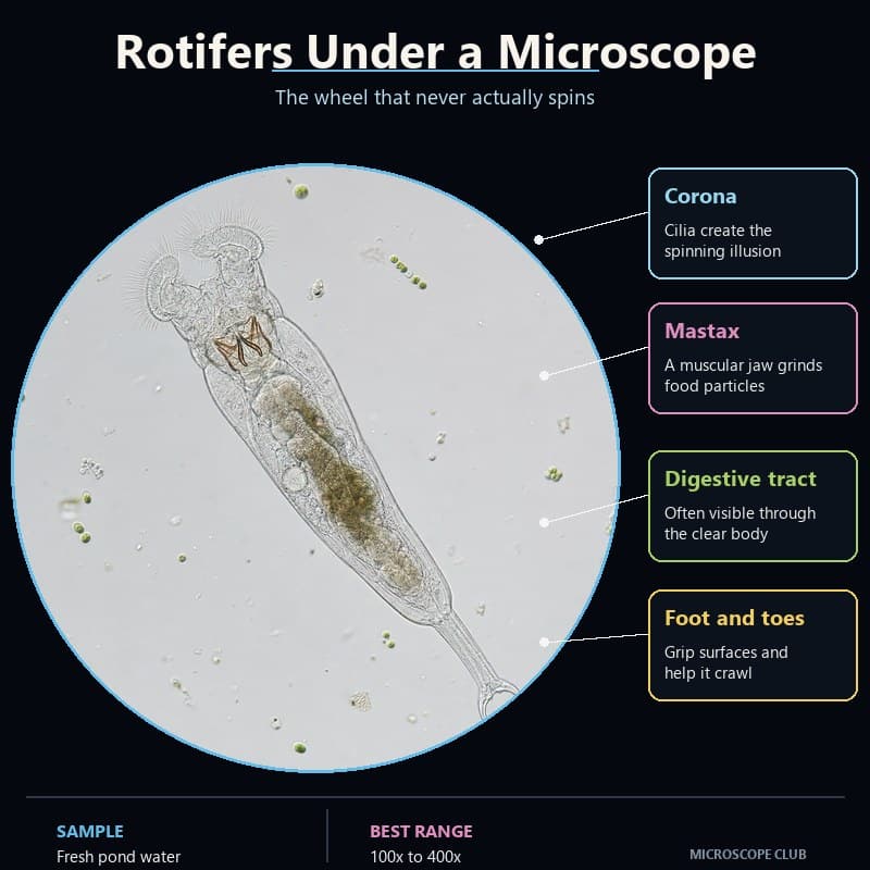
Live rotifer under a microscope showing the ciliated corona, mastax, transparent gut and two-toed foot

Rotifers under a microscope look like they’re wearing a spinning wheel on their head. That single detail is the reason they’re one of the most satisfying finds in a drop of pond water. They’re not protozoa and not tardigrades; they’re true microscopic animals, usually 100–500 micrometers long, named for a crown of beating cilia that …

Read more

Hydra Under a Microscope: Tentacles and Stinging Cells

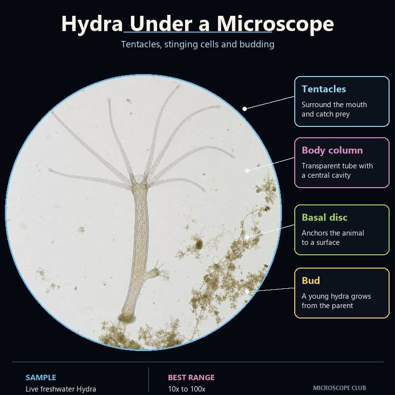
Live freshwater Hydra under a microscope showing tentacles, body column, basal disc and a lateral bud

A hydra under a microscope looks like a translucent thread glued to the glass, crowned with a starburst of tentacles armed with stinging cells. This freshwater cnidarian, usually just 1–20 mm long, is one of the best live specimens a hobbyist can view: relax it under the right conditions and you can watch it hunt, …

Read more

Bone Tissue Under a Microscope: Structure Explained

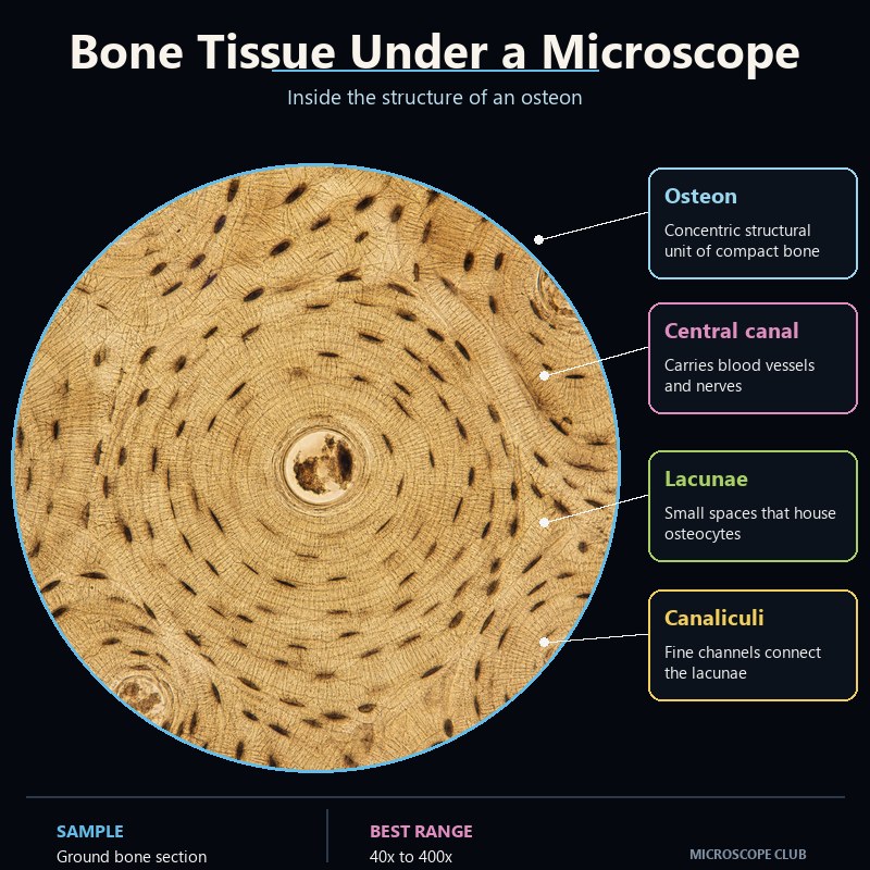
Compact bone tissue under a microscope showing an osteon, central canal, lacunae and canaliculi

Bone tissue under a microscope shows a striking bullseye pattern: repeating cylindrical units called osteons, each built from concentric rings of mineralized matrix wrapped around a central canal, dotted with tiny cell chambers and hair-thin connecting channels. What you’re actually viewing is never raw bone — it’s a prepared, decalcified or ground thin section, because …

Read more

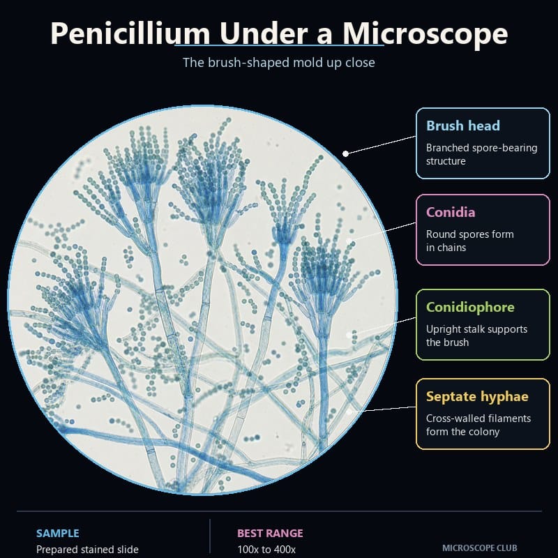
Penicillium Under a Microscope: The Brush vs Aspergillus

Penicillium conidiophores under a microscope showing characteristic blue brush-shaped heads and chains of conidia

Penicillium under a microscope reveals a blue-green mold whose stalk branches into a tiny brush — the penicillus — that holds long chains of round spores. That brush shape is also the fastest way to separate Penicillium from Aspergillus, the mold it gets confused with most often. This guide walks you through preparing a slide, …

Read more