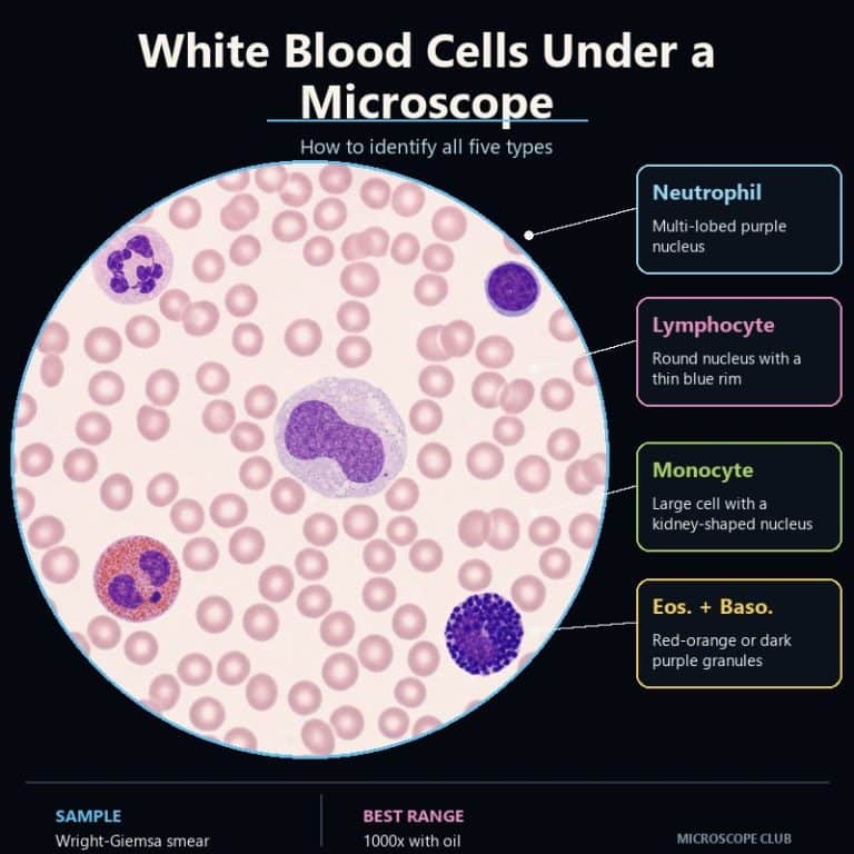
White blood cells under a microscope appear as scattered, purple-nucleated cells standing out sharply against a pink carpet of red blood cells. Once you know what to look for, you can name every one of the five types in seconds. Nucleus shape, granule color, and size relative to a red blood cell are the only three clues you need. This guide walks through each type the way a lab tech reads a differential count, then gives you a side-by-side chart to lock it all in.
The 3 Clues That Tell Every White Blood Cell Apart
Every white blood cell you’ll ever see under a scope gives up its identity through the same three clues, checked in the same order: size relative to a red blood cell, nucleus shape, and granule color. Lab techs run this exact sequence during a manual differential count, and it works just as well on a hobbyist’s stained smear as it does in a clinical lab.
Why the Red Blood Cell Is Your Built-In Ruler
A red blood cell is a reliable ruler because it never changes size: about 7-8 µm across, every time, on every slide. White blood cells range from roughly 7 µm (a small lymphocyte, barely bigger than the red cells around it) up to 20 µm (a monocyte, unmistakably the largest cell on the field), according to StatPearls’ histology reference on white blood cells. Holding every white cell up against its red-cell neighbors is the fastest first filter — it can rule a monocyte in or out before you’ve even looked at the nucleus.
Once you’ve placed the cell on the size scale, read the nucleus, then the granules, in this order:
- Compare size to a red blood cell. Roughly red-cell-sized or smaller usually means a lymphocyte. Distinctly the biggest cell on the slide usually means a monocyte. Mid-size points to one of the three granulocytes.
- Read the nucleus shape. Multi-lobed and segmented is a neutrophil. Large, round, and dark is a lymphocyte. Kidney-bean or horseshoe-shaped is a monocyte. Bi-lobed is an eosinophil or basophil.
- Check the granule color. Faint lilac points to a neutrophil. Red-orange points to an eosinophil. Dark blue-purple, often hiding the nucleus entirely, points to a basophil.
Neutrophils — The Multi-Lobed Nucleus You’ll See Most
Neutrophils are the cell you’ll identify most often on any human blood smear, making up roughly 40-60% of the white cells you count, per Cleveland Clinic’s overview of white blood cell types. The nucleus is the giveaway: two to five lobes connected by thin purple strands, like a string of beads or a linked sausage. An immature neutrophil, called a “band,” hasn’t segmented yet and shows a single C-shaped nucleus instead.
The cytoplasm is pale pink to lilac, with granules so fine and faint they read as a light dusting rather than distinct dots — subtle compared to the other granulocytes. At roughly 10-15 µm, a neutrophil sits solidly in the mid-size range, noticeably bigger than a lymphocyte but smaller than a monocyte. The memory aid “Never Let Monkeys Eat Bananas” — Neutrophil, Lymphocyte, Monocyte, Eosinophil, Basophil — lists all five types in descending order of abundance, and it’s worth memorizing before you sit down at the eyepiece.
Lymphocytes — Almost All Nucleus, Thin Blue Rim
Lymphocytes read as a dense purple marble with a thin blue shadow along one edge — almost no visible cytoplasm at all. The nucleus is large, round or slightly notched, and stains so darkly it can be hard to see where it ends and the cell membrane begins. What little cytoplasm shows up forms a narrow, pale sky-blue rim hugging one side of the nucleus.

Most small lymphocytes measure only 7-10 µm — close enough to a red blood cell’s 7-8 µm that beginners sometimes mistake them for oddly stained red cells at first glance, until the dark nucleus makes it obvious that’s wrong. Reactive or “large” lymphocytes can stretch closer to 14 µm, which is where the classic mix-up with monocytes happens. The one-line discriminator: a lymphocyte’s nucleus is round with a thin blue cytoplasm sliver, while a monocyte’s nucleus is kidney-shaped with abundant gray cytoplasm. Lymphocytes make up about 20-40% of the white cell population, second only to neutrophils.
Monocytes — The Big Cell With the Kidney-Bean Nucleus
Monocytes are the cell that makes a first-timer stop scanning and say “that one’s huge.” At 15-20 µm, a monocyte is the largest white blood cell by a clear margin, and size alone usually flags it before you’ve even read the nucleus. The nucleus itself is folded — kidney-bean, horseshoe, or crumpled-comma shaped — never lobed and never a simple round ball.
Cytoplasm is abundant and blue-gray, often described as having a frosted, ground-glass texture, sometimes dotted with small clear vacuoles that look like tiny bubbles. Monocytes are relatively uncommon on a normal smear, at roughly 2-8% of the total count, so finding one is a genuine “spot the odd one out” moment rather than a routine occurrence. See our guide to observing blood cells under the microscope for how monocytes and the other formed elements sit together on a full smear.

Eosinophils — Bright Red-Orange Granules You Can’t Miss
Eosinophils are the crowd-pleaser of the differential count: cytoplasm packed edge-to-edge with large, coarse, evenly sized granules that glow red-orange, often compared to fish roe or salmon eggs. Nothing else on a stained smear looks quite like it, which makes eosinophils one of the easiest cells to call correctly even for a first-time observer.
The nucleus is usually bi-lobed, giving it a “headphones” or spectacles silhouette, though the bright granules tend to steal all the attention before you even register the nucleus shape. At 10-15 µm, eosinophils sit in the same size range as neutrophils, so granule color — not size — is what separates the two. Eosinophils make up only about 1-4% of white blood cells on a normal differential. Elevated counts are commonly associated with allergic reactions or parasitic infection, though interpreting an abnormal count is a job for a qualified lab professional, not a hobby slide.
Basophils — Dark Granules That Hide the Nucleus
Basophils are the cell you hunt for and often don’t find. Cytoplasm is crammed with large, dark blue-purple to almost black granules, frequently so dense they obscure the bi- or multi-lobed nucleus underneath entirely. At roughly 10-14 µm, basophils overlap in size with neutrophils and eosinophils, so granule color — inky and dark rather than lilac or red-orange — is the clearest tell.
Basophils are the rarest white blood cell, at only about 0.5-1% of the total count. It’s entirely normal to scan a whole monolayer and count zero — that’s not a technique failure, it’s just how uncommon these cells are. If you do spot one, it’s usually unmistakable once you know what you’re looking for.
White Blood Cell Identification Chart
Once you’ve read each type on its own, this chart lines up all five side by side using the same three clues — nucleus shape, granules, and size — plus how often you’ll actually see each one.
| Type | Nucleus Shape | Granules / Cytoplasm | Size vs. RBC | % of WBCs | Quickest Tell |
|---|---|---|---|---|---|
| Neutrophil | 2-5 lobes, segmented | Faint, pale lilac | Larger (10-15 µm) | 40-60% | Most common cell + lobed nucleus |
| Lymphocyte | Large, round, dense | Thin sky-blue rim | ~RBC-sized (7-10 µm) | 20-40% | Almost all nucleus, tiny cytoplasm |
| Monocyte | Kidney-bean, folded | Gray, frosted, vacuolated | Largest (15-20 µm) | 2-8% | Biggest cell on the slide |
| Eosinophil | Bi-lobed | Coarse red-orange | Larger (10-15 µm) | 1-4% | Bright red-orange granules |
| Basophil | Lobed but hidden | Dense blue-purple/black | Larger (10-14 µm) | 0.5-1% | Dark granules hiding the nucleus |
How to Prepare and View a Blood Smear
A blood smear only reveals white blood cell types clearly after it has been fixed and stained — a fresh, unstained wet mount lets you see white cells only as faint, pale, grainy blobs, without enough detail to name a single type. Making a wet mount slide is a fine way to observe cells moving, but typing white blood cells requires a proper stain.
Most hobbyists get a cleaner, safer result from a prepared or commercial blood smear slide rather than drawing their own blood. If you do prepare your own smear, treat all blood — even your own — as a biohazard: wear gloves, use a single-use sterile lancet, never share a lancet between people, and dispose of sharps in an approved container, following CDC bloodborne pathogen guidelines.
- Fix and stain the smear. A Wright or Wright-Giemsa (Romanowsky) stain is the standard — it’s what gives nuclei their purple color and granules their characteristic hues.
- Scan at 40x to find the feathered edge — the thin monolayer near the far end of the smear where cells lie flat and separated rather than piled on top of each other.
- Identify at 400x using nucleus shape and cell size to place each cell into one of the five types.
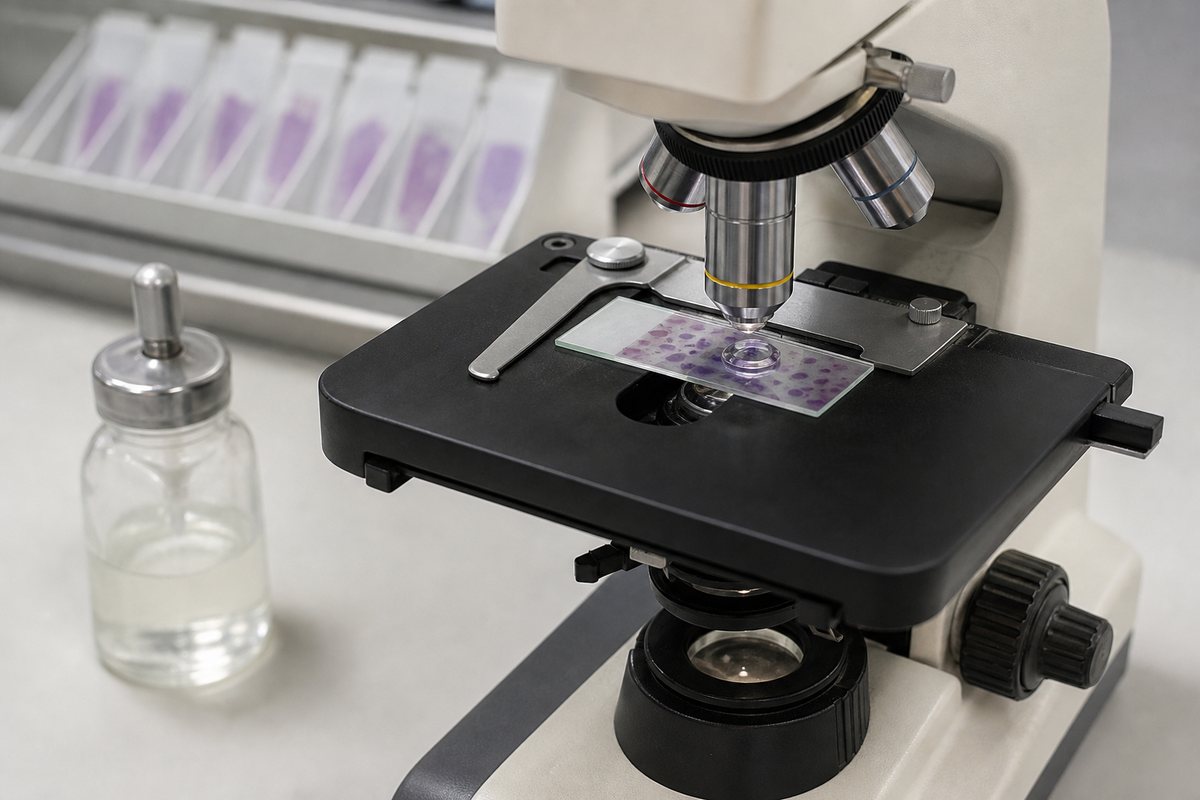
- Confirm granule detail at 1000x with an oil immersion objective — this is the only magnification fine enough to reliably distinguish neutrophil, eosinophil, and basophil granules by color and texture.
Work your compound light microscope‘s stage in a systematic zig-zag (“battlement”) pattern across the monolayer so you don’t recount the same field twice — that’s how a real differential count is done. Get your condenser and light level right, too; too much brightness washes out a neutrophil’s already-faint granules. If you need a refresher on getting a slide onto the stage cleanly, see how to prepare a microscope slide first.
This guide is for educational and microscopy-hobby purposes only and is not medical advice. Actual clinical differential counts, and any concerns about your own blood work, should be handled by a qualified clinical laboratory or healthcare professional — not a self-read hobby slide. For a look at what abnormal or malignant white cells can look like, see our piece on what cancer cells look like under a microscope.
Frequently Asked Questions
Which white blood cell is most common on a smear?
Neutrophils are the most common, typically making up 40-60% of the white blood cells on a normal differential count.
How do you tell a lymphocyte from a monocyte?
Check the nucleus and the cytoplasm together. A lymphocyte has a round, dense nucleus with only a thin sliver of blue cytoplasm. A monocyte, by contrast, has a folded, kidney-bean-shaped nucleus with abundant gray, frosted-looking cytoplasm and is noticeably the larger of the two.
What stain is used to see white blood cells?
A Wright stain, or the closely related Wright-Giemsa stain, is the standard Romanowsky-type stain used on blood smears. It’s what turns nuclei purple and gives each cell’s granules their characteristic colors.
Can you see white blood cells without staining?
Yes, faintly. In an unstained wet mount, white blood cells show up as pale, grainy blobs among the red cells. You can’t reliably tell the five types apart this way — the nuclear and granule detail that separates them only becomes visible after staining.
What magnification do you need to see white blood cells?
Scan at 40x to locate the thin monolayer area of the smear, identify cell types at 400x, and switch to a 1000x oil immersion objective to confirm fine granule and nuclear detail.
How big is a white blood cell compared to a red blood cell?
White blood cells are larger. A red blood cell is about 7-8 µm across. White blood cells range from about 7 µm for a small lymphocyte up to 20 µm for a monocyte — which is why the red blood cell works so well as a built-in size ruler.
Conclusion
Every white blood cell on a stained smear is identifiable with the same three checks: size against a red blood cell, nucleus shape, and granule color. Neutrophils give themselves away with a lobed nucleus and faint granules, lymphocytes with almost nothing but a dense round nucleus, monocytes with sheer size and a kidney-bean nucleus, eosinophils with unmistakable red-orange granules, and basophils with dark granules that bury the nucleus entirely. Basophils are rare enough that not finding one on a slide is normal.
Have you spotted all five types on a single slide, or is there one — basophils, usually — that’s given you the slip? Tell us what you found on your own smear in the comments below.
Related Specimen
To see white blood cells in their natural context alongside red cells and platelets, see our full guide to reading a blood smear.
Originally posted 2026-08-07 12:00:00.