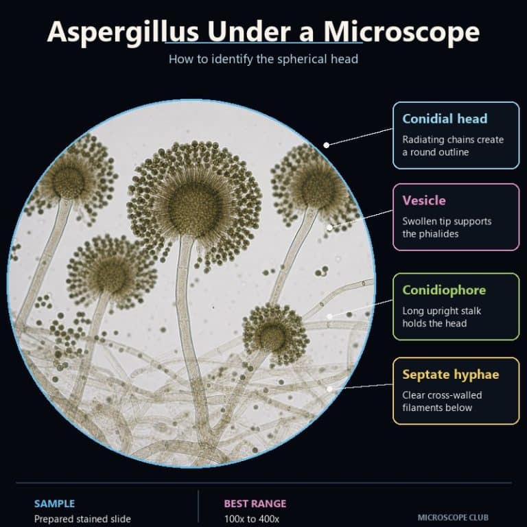
Aspergillus under a microscope reveals a long, unbranched stalk that swells into a round vesicle covered in radiating chains of spores. This distinctive “aspergillus head” is what separates the mold from every look-alike on a lab bench. Found a fuzzy black, yellow-green, or gray-green colony on bread, fruit, soil, or a damp windowsill? A compound microscope at 400x is the fastest way to confirm it’s Aspergillus rather than Penicillium or Rhizopus. This guide walks through that structure, how to prepare a slide that shows it clearly, and the exact features that tell the three most-confused molds apart.
The Aspergillus Head: Conidiophore, Vesicle, and Spore Chains
The aspergillus head is built from the ground up out of a handful of specialized cells. Once you can name each one, the whole structure stops looking like a random tangle and starts looking like a diagram. This is also the single feature that confirms an identification — everything else on the slide is supporting detail. For general context on what a mold colony looks like before you get to species level, see our guide to what mold looks like under a microscope.
Conidiophore, Vesicle, Phialides, and Spore Chains
The structure starts at a foot cell, a specialized cell within the hypha that anchors everything above it. From the foot cell rises the conidiophore (or stipe) — a long, erect stalk that, unlike Penicillium’s, never branches. At its tip the conidiophore swells into a spherical-to-flask-shaped vesicle, the single defining feature of the genus. Covering the vesicle are flask-shaped phialides. In uniseriate species they sit directly on the vesicle surface; in biseriate species a supporting layer of cells called metulae sits between the vesicle and the phialides. Each phialide then produces a long, dry chain of round conidia (spores) that radiates outward. It’s the mass of these spores — not the stalk or hyphae — that gives the colony its color.
Why It Looks Like a Sparkler
The genus name comes from the aspergillum, a liturgical holy-water sprinkler with a round, perforated head — a Catholic priest’s tool for scattering water over a congregation. Under the scope, the resemblance is exact: a handle-like stalk topped by a ball that showers outward in every direction. Students describe the same shape as a dandelion clock gone to seed, a firework frozen mid-burst, or a shower head. Whichever image sticks, that radiating symmetry from one round point is what you’re looking for.
Septate, Clear Hyphae — Why the Color Is in the Spores
Aspergillus hyphae are septate (divided by regular cross-walls) and hyaline, meaning clear and colorless. That’s a detail worth remembering precisely because it’s invisible at first glance. The black, yellow-green, or gray-green color you associate with the mold belongs entirely to the spore mass sitting on top of the heads. The threads carrying nutrients through the colony are transparent. This septation is also the fastest way to rule out Rhizopus, whose hyphae have no cross-walls at all.
How to See Aspergillus Under Your Microscope
Finding an intact aspergillus head takes the right magnification, a stain that makes clear structures visible, and a mounting technique gentle enough not to destroy the very thing you’re trying to look at.
What Magnification You Need
Start at 100x total magnification (a 10x objective) to scan the slide. At this power the conidial heads show up as tiny lollipops or pinheads standing off the hyphae, easy to spot even though you can’t yet make out their structure. Once you’ve located one, switch to the 40x objective for 400x total magnification, which is where the vesicle, phialides, and radiating spore chains all resolve clearly. A 40x or 60x stereo microscope will show you the fuzzy colony and its color, but it cannot resolve the head itself — that requires a compound scope. Oil immersion at 1000x is unnecessary overkill for this specimen; it’s built for structures far smaller than a conidial head.

Preparing the Slide
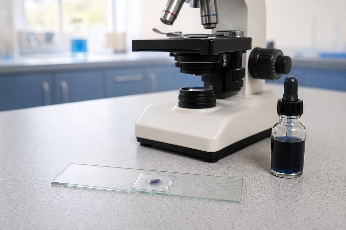
The standard fungal stain is lactophenol cotton blue (LPCB), which works in three ways. Cotton blue dyes the chitin in the fungal cell walls. Phenol inactivates the organism. Lactic acid preserves the structure so it doesn’t collapse on the slide. Against the pale blue background it produces, a clear, colorless head stands out sharply.
Mounting method matters as much as the stain. A standard wet mount slide made by teasing the sample apart with a needle usually shears the fragile spore heads clean off their stalks. Instead, use the gentler cellotape touch: press a short strip of clear tape lightly against the colony surface. Then lay the sticky side onto a drop of LPCB on a slide. A slide culture, where the fungus grows directly on a small block of agar under a coverslip, works even better if you have the time to set one up in advance.
- Gently touch a strip of clear tape to the edge of the colony, where heads are freshest and least clumped together.
- Lay the tape sticky-side down onto a drop of lactophenol cotton blue on a clean slide.
- Lower a coverslip at a slight angle to avoid trapping air bubbles.
- Scan at 100x total magnification until you spot an intact, lollipop-shaped head.
- Switch to 400x and re-center the head to confirm the vesicle, phialides, and radiating spore chains.
A Safety Note Before You Handle Mold
This article is educational, not medical advice. Aspergillus spores are the infectious and allergenic part of the organism. Work in a ventilated space, keep cultures sealed when not in use, and never deliberately inhale or sniff a colony to check its smell. Don’t grow mold samples at scale for a home lab — a small existing patch is plenty for slide prep. Readers who are immunocompromised or have asthma should avoid handling mold cultures directly. Anyone with respiratory symptoms after mold exposure should consult a doctor rather than self-diagnose from a microscope slide.

Aspergillus vs Penicillium vs Rhizopus — Telling Them Apart
These three molds get confused constantly because they all show up as fuzzy, colored patches to the naked eye, but under the scope their reproductive structures are unmistakably different. Aspergillus carries one swollen vesicle at the tip of an unbranched stalk. Bread mold (Rhizopus) looks nothing like either of them, with a completely different structure built around a spore sac instead of an open head.
| Feature | Aspergillus | Penicillium | Rhizopus |
|---|---|---|---|
| Head shape | Round vesicle with radiating spore chains | Branched, brush-like tip (no vesicle) | Closed sporangium (spore sac), not an open head |
| Hyphae | Septate (cross-walled) | Septate (cross-walled) | Aseptate (no cross-walls), broad |
| Vesicle present? | Yes — the defining feature | No — has a penicillus instead | No — has a sporangium instead |
| Spore arrangement | Long dry chains radiating from phialides | Chains from brush-like clusters of phialides | Spores packed inside a sac, released when it ruptures |
Penicillium takes its name from the Latin penicillus, “little brush.” That’s exactly what its conidiophore looks like — a branching structure ending in brush-like clusters, with no swollen vesicle anywhere. Rhizopus sits in an entirely different fungal phylum (Zygomycota/Mucoromycota). It shows root-like rhizoids anchoring broad, aseptate hyphae, topped by a sporangium: a closed sac full of spores rather than an open, radiating head. If your hyphae have visible cross-walls and the tip carries any kind of vesicle, you’re looking at Aspergillus, not Rhizopus. If there’s no vesicle and the tip branches into brush clusters, it’s Penicillium.
Identifying Aspergillus Species by Color
Aspergillus species are commonly told apart by the color of their spore mass, though color alone should never be the only evidence. A. niger and the true “black mold” Stachybotrys can both look black to the naked eye, and only the head structure confirms the genus.
| Species | Spore/Colony Color | Series | Note |
|---|---|---|---|
| A. niger | Black / dark brown | Biseriate | Large globose vesicle; spores cover the whole sphere; very common on food. |
| A. flavus | Yellow-green | Uni- and biseriate | Produces aflatoxins; roughened conidiophore. |
| A. fumigatus | Blue-green / smoky gray-green | Uniseriate | Flask-shaped vesicle with phialides on the upper half only, giving a columnar head; thermotolerant; the main human pathogen in the genus. |
| A. terreus | Cinnamon-buff / tan | Biseriate | Compact, columnar head. |
A. fumigatus is responsible for most cases of aspergillosis, a lung infection that is most serious in people with weakened immune systems, according to the CDC. A. flavus produces aflatoxins, compounds the National Cancer Institute classifies as carcinogenic — one more reason not to handle unknown mold colonies casually, even for a school project.
Common Beginner Mistakes When Viewing Aspergillus
Most failed Aspergillus slides come down to three habits, all easy to fix once you know what’s happening.
Teasing the sample too hard. Digging in with a needle to spread the colony shears every spore head off its stalk. That leaves you with a field of loose spores and bald vesicles instead of an intact head. Use the gentle sticky-tape touch instead — don’t mash the sample.
Staying at low power only. A 40x stereo scope shows the fuzzy colony and its color beautifully, but it will never resolve the vesicle, and without the vesicle you can’t confirm the genus. You need a compound scope at 400x to see the actual head.
Skipping the stain. Aspergillus hyphae are clear, and against a bright, unstained field they nearly vanish, along with the pale head sitting on top of them. A drop of lactophenol cotton blue is what makes the structure visible in the first place. A wide-open iris diaphragm compounds the problem by washing out what little contrast the unstained hyphae have — stop it down before you start scanning.
Frequently Asked Questions
Is Aspergillus dangerous, or can you get sick from it?
It can be, mainly for specific groups. Healthy people are rarely affected, but A. fumigatus can cause aspergillosis in people with weakened immune systems, and A. flavus produces aflatoxins linked to cancer risk. This is general education, not medical advice — anyone with concerning symptoms after mold exposure should see a doctor.
Does bleach kill Aspergillus spores?
Diluted bleach can kill spores on hard, non-porous surfaces, but it doesn’t reliably penetrate porous materials like drywall or wood, where the fungus can survive underneath. For lab samples specifically, a proper disinfectant or autoclaving is more reliable than a quick bleach wipe.
Where is Aspergillus found?
Almost everywhere. It’s a common saprophyte in soil, decaying plant matter, stored grain, house dust, damp building materials, and spoiling food — which is why it’s one of the first molds most people ever encounter.
Can I identify Aspergillus without a microscope?
Not reliably. Colony color and texture can narrow things down, but several molds share similar colors to the naked eye, and only the conidial head — visible at 400x — actually confirms the genus.
Can I use a smartphone microscope adapter to see the head?
A basic clip-on phone lens usually tops out well below the 400x needed to resolve a vesicle and phialides clearly. A proper compound microscope with a 40x objective is the reliable option for this specific structure.
How long does it take to prepare and view a slide?
A sticky-tape mount in lactophenol cotton blue takes about five minutes to prepare, and finding an intact head under the scope usually takes another five to ten minutes of scanning, depending on how established the colony is.
Does Aspergillus smell different from other molds?
Many molds share a similar musty, earthy odor, and it’s not a reliable way to tell species apart. More importantly, don’t deliberately sniff a colony to test this — inhaling spores directly is exactly what the safety guidance above warns against.
Conclusion
Once you know the sequence — foot cell, unbranched conidiophore, swollen vesicle, phialides, radiating spore chains — the aspergillus head stops being an intimidating blur. It becomes one of the more satisfying structures to find under a compound microscope. Pair that structural knowledge with a gentle sticky-tape mount in lactophenol cotton blue and the 400x view most beginners skip. You’ll be able to separate Aspergillus from Penicillium and Rhizopus with confidence rather than a guess.
Have you managed to catch an intact aspergillus head on a slide, or does your sample keep shattering before you get there? Tell us what colony you’re working from and what magnification you’re using in the comments below.
Related Specimen
For a comparison, see our full Penicillium mold review — another common mold specimen with a very different visual structure.
Originally posted 2026-08-06 12:00:00.