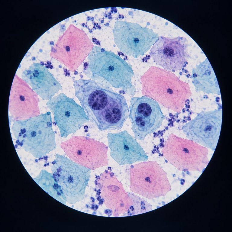
A Pap smear under a microscope looks like a scattered field of flat, Pap-stained cervical cells. Most are pink-orange or blue-green, each with a small, dark nucleus tucked into one corner. A cytotechnologist or pathologist scans that field for cells whose nucleus has grown too large, too dark, or is ringed by a telltale halo. Those changes are what separate a normal result from an abnormal one. This guide walks through exactly what normal and abnormal cervical cells look like, and what the resulting label — NILM, ASC-US, LSIL, or HSIL — actually means.
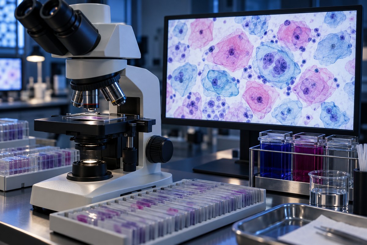
How a Pap Smear Slide Is Made and Stained
A Pap smear starts with a small brush or spatula sampling cells from the cervix and the transformation zone, the border where two types of tissue meet. Those cells are transferred onto a glass slide (the classic “smear”) or rinsed into a vial of liquid fixative for liquid-based cytology. The ThinPrep or SurePath methods are now used for most screening in the United States. Liquid-based prep spins the cells into a clean, even monolayer, which clears away the blood, mucus, and clumping that used to obscure classic smears.
Either way, the slide is stained with the Papanicolaou (Pap) stain, a multichromatic stain that does more than make cells visible — it labels their maturity. Hematoxylin stains nuclei a dark blue-purple. The counterstains then split the cytoplasm by cell type: mature, estrogen-exposed superficial cells pick up a warm pink-to-orange tone, while less mature intermediate and parabasal cells stay blue-green. That color split is a real diagnostic cue, not decoration — it tells the reader how mature the tissue is before they even look at a single nucleus. For a closer look at how any specimen goes from tissue to viewable slide, see this guide to preparing a microscope slide and staining it.
What a Normal Pap Smear Looks Like Under the Microscope
A normal field looks calm and roomy. Large, pale, polygonal cells overlap like floor tiles or fried eggs, each with a small, dense dot of a nucleus and plenty of clear space around it. Nothing crowds together, and no single nucleus dominates its cell. The CDC explains how the Pap test screens cervical cells for precancerous changes.
The Squamous Cells (Superficial vs. Intermediate)
Superficial squamous cells are the largest and most mature cells on the slide: flat, angular, pink-to-orange, with a tiny, condensed (pyknotic) nucleus that shows almost no internal detail. Intermediate squamous cells are similar in size and shape but stain blue-green, and their nucleus is slightly larger and more open, with fine, even chromatin. Both have a low nuclear-to-cytoplasmic (N:C) ratio — the nucleus takes up only a small share of the cell, which is exactly what a reader expects to see. Cervical squamous cells share this flattened, plate-like structure with skin cells, another type of squamous epithelium, since both tissues are built to withstand friction.
Smaller, rounder parabasal cells, with denser blue cytoplasm and a proportionally larger nucleus, show up more often in low-estrogen states — after menopause or shortly after childbirth. This is one of the most common beginner mistakes in reading a Pap smear: a higher N:C ratio in parabasal cells is normal for that hormonal context, not a sign of disease. Judging N:C ratio without checking the patient’s estrogen status is how a normal slide gets over-called.
Endocervical Cells and Why They Matter
A good sample also usually contains endocervical (glandular) cells, arranged in a honeycomb sheet or a picket-fence strip. Their presence tells the reader the transformation zone — the area most likely to develop precancerous change — was actually sampled. A slide without them may be flagged as “limited” or “unsatisfactory,” since it may have missed the exact tissue the test is meant to check. Neutrophils, mucus, and a scattering of normal bacteria are common background; you’ll frequently see the same white blood cells (neutrophils) that show up in almost any inflammatory or infected sample.

What Abnormal Cervical Cells Look Like
Abnormal cells jump out because the nucleus is too big and too dark for the cell it’s sitting in. The nucleus fills the room instead of staying in the corner. The visual red flags are consistent across every grade of abnormality; they just intensify as the grade gets more severe:
- Nuclear enlargement and a rising N:C ratio
- Hyperchromasia — a darker-staining nucleus, reflecting more DNA packed inside it
- Irregular nuclear membrane and coarse, clumped chromatin instead of a smooth, even texture
A second common beginner mistake is confusing genuine atypia with air-drying or degenerative artifact — swollen, washed-out nuclei that happen when a classic smear dries before it’s fixed. An experienced reader always judges nuclear detail on well-preserved cells, never on damaged ones. A third: getting absorbed in the neutrophils and mucus and missing the handful of truly abnormal cells hiding in a crowded corner of the slide. Abnormal cells are often sparse rather than widespread. Long-lasting infection with certain high-risk types of HPV is the underlying driver of most of these cellular changes.
Koilocytes — the Signature of HPV
A koilocyte is the clearest visual fingerprint of HPV infection on a Pap smear. It looks like the nucleus is sitting inside a clear, punched-out hole. A sharp-edged perinuclear halo surrounds an enlarged, dark, wrinkled (“raisinoid”) nucleus that’s often pushed off to one side, sometimes with two nuclei in the same cell. Koilocytes are the defining feature of the LSIL category described below.

Normal vs. Abnormal Cervical Cells — A Visual Comparison
| Feature | Normal squamous cell | Abnormal squamous cell |
|---|---|---|
| Overall cell size | Large, flat, polygonal | Often smaller and more rounded in higher-grade change |
| Nucleus size | Small relative to the cell | Enlarged, sometimes filling most of the cell |
| N:C ratio | Low | Elevated to markedly high |
| Chromatin | Fine, even, or condensed (pyknotic) | Coarse, clumped, unevenly distributed |
| Cytoplasm color | Pink-orange (mature) or blue-green (intermediate) | Variable; may appear otherwise normal around an abnormal nucleus |
| Telltale sign | Small, uniform, centrally placed nucleus | Perinuclear halo (koilocyte), hyperchromasia, irregular nuclear membrane |
Reading the Result — What NILM, ASC-US, LSIL, and HSIL Actually Mean
Cytology reports use the Bethesda System to translate what the reader saw down the microscope into a standard category. This is an educational summary of what each category generally means — not a diagnosis, and not a substitute for a conversation with a clinician about an individual result. The NCI provides a patient-facing guide to normal, unsatisfactory and abnormal Pap test results.
| Bethesda category | What the cells look like | Plain-English meaning (educational) |
|---|---|---|
| NILM | Normal squamous and endocervical cells, low N:C ratio | Negative for intraepithelial lesion or malignancy — a normal result |
| ASC-US | Slight nuclear enlargement or atypia, not enough for a clear lesion | Atypical squamous cells of undetermined significance — the least reproducible, “not sure” category; often followed by an HPV test |
| LSIL | Koilocytes plus mild nuclear atypia | Low-grade squamous intraepithelial lesion — corresponds to HPV effect; commonly resolves on its own |
| HSIL | Smaller, more immature-looking cells with a markedly high N:C ratio and irregular, dark nuclei | High-grade squamous intraepithelial lesion — carries a higher risk of progression and usually needs follow-up |
Educational summary — not a diagnosis. Two rarer categories worth knowing by name are ASC-H (atypical squamous cells, cannot exclude HSIL) and AGC (atypical glandular cells). At the more serious end of the spectrum, cytology can also detect features consistent with how cancer cells look under the microscope, though that finding always requires confirmation.
It’s worth repeating: a Pap smear is a screening test, not a diagnosis. According to the National Cancer Institute, an abnormal result is typically followed up with colposcopy and biopsy before any diagnosis is made. Most abnormal results — especially ASC-US and LSIL — are HPV-related changes that regress on their own rather than progressing to cancer. Current screening guidelines from the American College of Obstetricians and Gynecologists lay out general age- and interval-based recommendations, but they are exactly that — general guidance, not a personal schedule. Anyone with questions about their own results or screening interval should raise them with their own clinician.
The Magnification and Microscope Used
Pap smears are read with standard light (brightfield) microscopy on stained, fixed cells — no special optics required. A reader starts with a systematic low-power scan across the whole slide, typically around a 10x objective, looking for any cell groups that look out of place. Once something catches the eye, they drop to high power to study nuclear detail up close. That means a 40x objective for roughly 400x total magnification, or oil-immersion 100x for 1000x total. That same rhythm of “scan wide at low power, then zoom in at high power for detail” is the same discipline used when reading a blood smear step by step.
Experienced readers judge nuclei on the single best-preserved cells they can find, not on damaged or overlapping clumps. They also know abnormal cells tend to cluster, so finding one usually means slowing down to search the area around it rather than moving on. There’s a genuine trade-off between prep methods, too. Liquid-based cytology gives a clean, even layer that’s faster to screen, but the classic smear sometimes preserves how cells sit together in their original architecture, a clue the dispersed liquid-based prep can lose.
The Famous Cells That Started With a Cervical Smear
Cervical cytology has one especially well-known chapter in cell biology. In 1951, cells taken from a cervical tumor biopsy at Johns Hopkins Hospital were cultured and, unlike almost every cell line before them, kept dividing indefinitely in the lab. Those became the immortal HeLa cells taken from a cervical tumor — named, imperfectly, for the patient Henrietta Lacks. HeLa cells have since been used in an enormous share of modern cell biology and vaccine research. Their origin traces directly back to the same tissue this article has been describing.
Frequently Asked Questions
Why might a Pap smear need to be repeated?
The most common reason is an unsatisfactory or limited sample. Examples include too few cells, obscuring blood or mucus, or no endocervical cells to confirm the transformation zone was reached. A repeat sample doesn’t necessarily mean anything was found; it usually just means the slide wasn’t adequate to read confidently.
Can you see cancer on a Pap smear?
Cytology can show cell features consistent with cancer, but a Pap smear alone cannot diagnose it. Any result suggesting malignant or highly abnormal cells is confirmed with colposcopy and biopsy before a diagnosis is made.
What’s the difference between a Pap smear and liquid-based cytology (ThinPrep)?
Both sample the same cervical cells. A classic Pap smear spreads them directly onto a glass slide. Liquid-based cytology (ThinPrep or SurePath) suspends them in fixative first and deposits them as a clean, even layer, which reduces obscuring blood and mucus.
How often should you get a Pap smear?
Current U.S. guidelines from groups like ACOG set general age- and interval-based recommendations for cervical cancer screening, but these are population-level guidelines, not personalized instructions. The right interval for any individual should come from their own clinician.
Is a Pap smear the same as an HPV test?
No. A Pap smear (cytology) looks at the cells themselves for abnormal changes, while an HPV test checks for the virus that causes most of those changes. The two are often run together, a practice known as co-testing.
Conclusion
Under the microscope, a Pap smear comes down to a simple visual question. Does this nucleus look the way a healthy nucleus should — small, evenly stained, and sitting quietly inside its cell — or has it grown too large, too dark, or gained a telltale halo? Normal cells are calm and uniform. Abnormal ones stand out because the nucleus takes over, and the specific pattern of that change is what maps onto categories like ASC-US, LSIL, and HSIL.
Have you ever looked at stained cervical or other epithelial cells under your own microscope? Or wondered what your own Pap result actually meant once you saw the terminology written out? Share what you noticed, or what still confuses you, in the comments below.