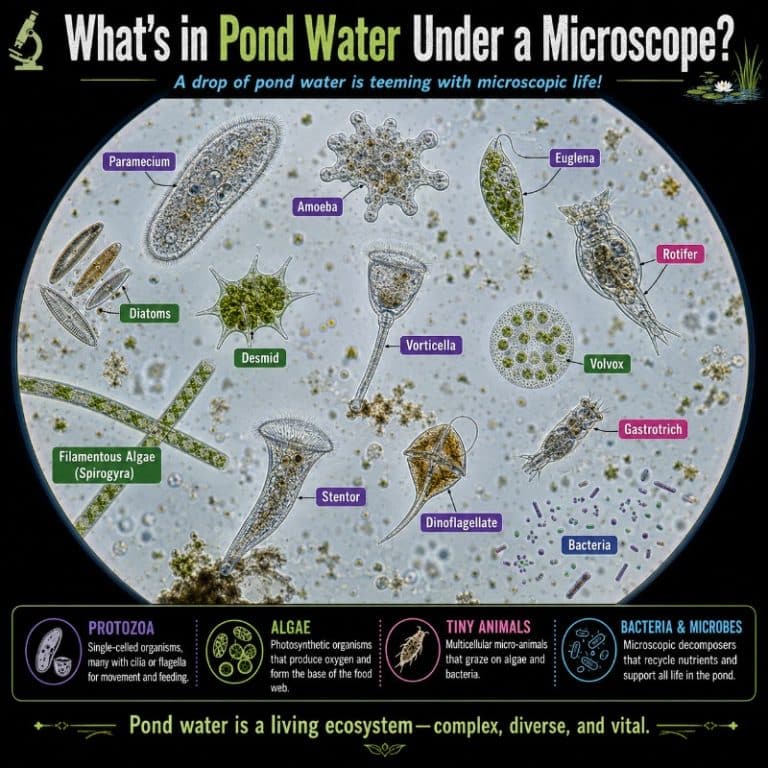
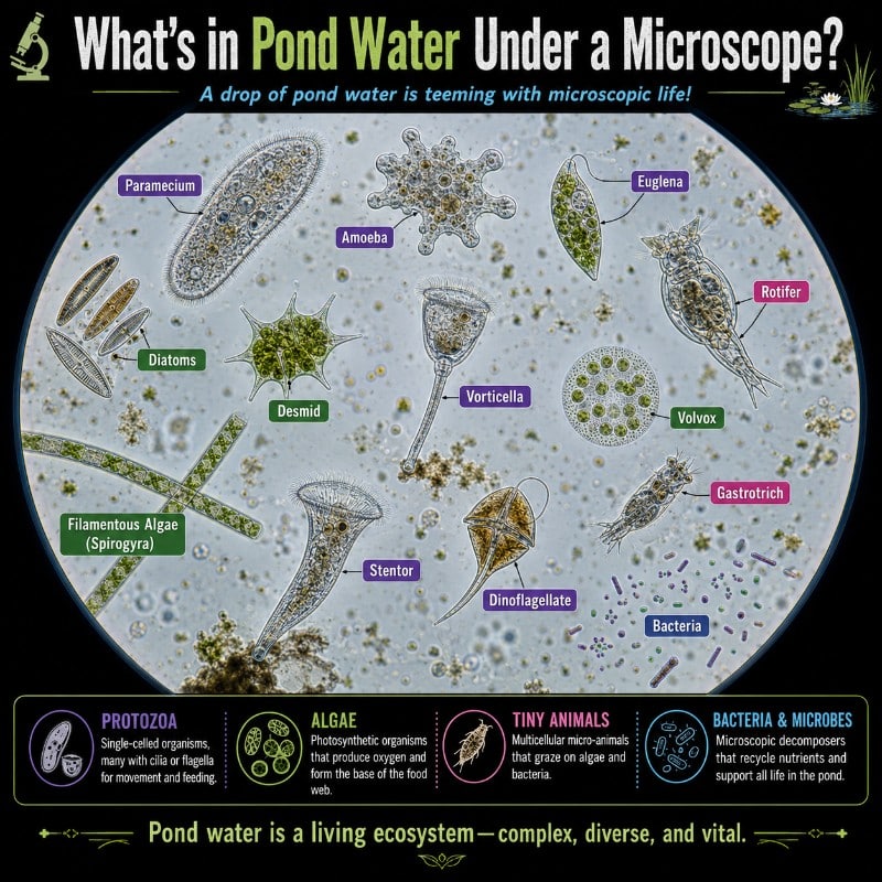
What’s in Pond Water Under a Microscope?
A single drop of pond water contains a thriving ecosystem — protozoa like paramecium and vorticella gliding and pulsing, algae such as Spirogyra’s spiral chloroplast coiling through the frame, micro-animals like rotifers and water fleas jerking across your field of view, and non-living debris including pollen grains, detritus, and air bubbles that can fool beginners. Whether you’re sampling a backyard pond or a farm dam, here is exactly what you are likely to see and how to tell it apart.
| Organism | What it looks like | Size | Magnification needed | Why it’s there |
|---|---|---|---|---|
| Paramecium | Slipper / footprint shape, smooth gliding motion, covered in fine cilia, visible oral groove | 50–330 µm (~120 µm common) | 40–100× to spot; 400× for detail | Bacterivore — feeds on bacteria and algae in decaying matter |
| Vorticella | Inverted bell on a coiled stalk that springs shut in a flash | Bell 35–50 µm; stalk up to ~1 mm | 40–100× | Filter-feeder attached to debris or plant surfaces |
| Euglena | Green, spindle-shaped, single whip-like flagellum, red eyespot, rubbery “metaboly” wriggling | 40–100 µm | 100–400× | Photosynthesises and feeds — abundant in sunlit, nutrient-rich water |
| Amoeba | Shapeless, flowing blob with no fixed form; moves by extending finger-like pseudopodia | 100–800 µm | 100–400× | Engulfs bacteria and other cells; found in bottom sediment |
| Rotifer | Spinning “wheel” of beating cilia (corona) at the head; foot with tiny toes often anchored to a particle | 100–500 µm | 40–100× | Filter-feeds on algae and bacteria; extraordinarily durable in variable conditions |
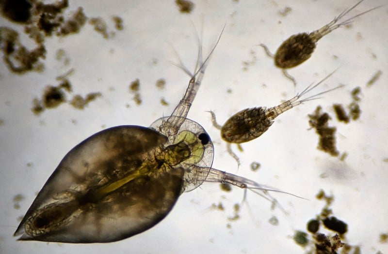
| Daphnia (water flea) | Transparent body, jerky hopping swim, beating heart visible inside, single compound eye | 0.2–5 mm | Naked eye to 40× | Grazes on algae; key link in the aquatic food chain |
| Cyclops (copepod) | Single median eye, teardrop body, two egg sacs dangling from females, darting escape bursts | 0.5–3 mm | Naked eye to 40× | Predatory crustacean; eats algae, rotifers, and small invertebrates |
| Hydra | Tubular body, ring of tentacles armed with stinging cells; contracts to a blob when disturbed | 1–20 mm extended | Naked eye to 40× | Anchors to submerged plants and debris; ambush predator of small crustaceans |
| Spirogyra | Unbranched green filament; distinctive helical spiral chloroplast winding through each cell | Filaments; cells 10–100 µm wide | 100–400× | Photosynthetic; forms tangled surface mats in still, sunlit water |
| Volvox | Hollow rolling green sphere built of hundreds of cells; daughter colonies visible inside | Colony ~0.5 mm | 40–100× | Colonial alga; rolls through the water column using coordinated flagella |
| Diatoms | Glassy, geometric silica shells (frustules); brown-gold colour; radial or bilateral symmetry | 2–200 µm | 100–400× | Photosynthetic; major oxygen producers; shells persist in sediment |
| Desmids / Closterium | Crescent or symmetrical paired semi-cells; bright green; crisp clean edges | 20–500 µm | 100–400× | Found in soft, slightly acidic water; sensitive indicator of water quality |
| Cyanobacteria (blue-green algae) | Filaments or clumps; blue-green to olive; Oscillatoria slowly glides and oscillates | Filaments / colonies | 100–400× | Photosynthetic bacteria; can bloom toxically in warm, nutrient-rich water |
| Nematodes | Unsegmented, thread-like worms thrashing in a whipping S-motion | 0.1–2.5 mm | 40–100× | Decomposers and bacterivores; found in sediment and plant debris |
| Bacteria | Tiny rods or dots; only faint motion detectable on most school scopes | 0.5–5 µm | ~1000× oil immersion only | Decomposers; present everywhere but not resolvable on a basic 400× microscope |
The Main Organisms You’ll See
Protozoa — Paramecium, Vorticella, Euglena, and Amoeba

Protozoa are single-celled, animal-like protists and they are usually the first things to catch your eye. Paramecium is the classic pond-water introduction organism: shaped like a worn slipper and covered in thousands of hair-like cilia, it glides purposefully in one direction and reverses when it hits an obstacle. Under 400× magnification you can make out the oral groove — a channel along its side that sweeps food into the cell. Britannica’s overview of Paramecium is a solid reference if you want to read further about its biology. Vorticella is harder to spot at first because it sits still while it filter-feeds, then suddenly recoils its spring-like stalk in a fraction of a second — one of the fastest movements in the microscopic world. Look for it attached to bits of debris or plant material at the edge of your slide.
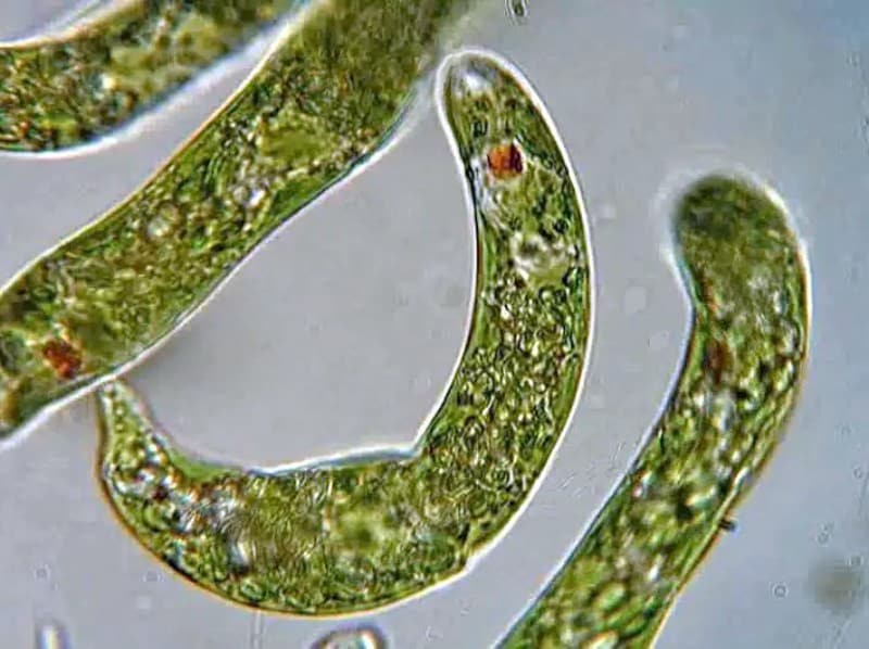
Euglena is fascinating because it bridges the animal-plant divide: it photosynthesises in light but can also absorb nutrients in darkness. Under the microscope it appears as a bright green spindle that writhes and contorts — a behaviour called metaboly — and if you look carefully at 400× you can spot the red eyespot (stigma) near the front end. Amoeba, by contrast, has no fixed shape at all. It oozes slowly across the slide, extending and retracting pseudopodia as it flows around food. Because amoeba moves so slowly and can blend in with debris, many beginners miss it entirely — try scanning the edges of plant fragments in your sample.
Algae — Spirogyra, Volvox, Diatoms, and Desmids
The green things in pond water are not all the same. Spirogyra forms long unbranched filaments that tangle into soft green mats on the water surface. Each cell is defined by a distinctive helical chloroplast that spirals inside like a green corkscrew — there is nothing else in pond water that looks quite like it. You can read much more in our detailed guide to Spirogyra’s spiral chloroplast.
Volvox is a colonial alga that builds a hollow sphere of hundreds of cells connected by cytoplasmic strands. Under low power you can watch it rotate slowly as its cells beat their flagella in coordinated waves. Smaller daughter colonies are often visible inside the parent sphere. We have a full article on the rolling green colonies of Volvox if you want to go deeper. Diatoms are arguably the most beautiful microorganisms in fresh water — their intricate silica shells display perfect geometric symmetry and catch the light with a glassy sheen. Under polarised light they can be spectacular. Diatoms are also ecologically crucial: NOAA estimates phytoplankton like diatoms produce roughly half of Earth’s oxygen. Desmids like Closterium are crescent-shaped green algae typical of clean, slightly acidic water; their presence is often taken as a sign of good water quality.

Micro-animals — Rotifers, Daphnia, Cyclops, Hydra, and Nematodes
These are multicellular animals, not single cells, and many are visible without a microscope or with very low magnification. Rotifers look otherworldly: the corona of beating cilia at their head spins so fast it resembles a turning wheel, which is how they got their name. They are extraordinarily tough — some species can survive desiccation for years — and they are among the most abundant animals in fresh water globally. Daphnia (water fleas) are crustaceans whose transparent bodies let you see organs in action. A beating heart, developing eggs, and a branching alimentary canal are all visible at 40× through their glassy carapace. Their jerky, hopping swimming motion is unmistakable.
Cyclops copepods are named for their single central eye. Females carry two distinctive egg sacs that look like a pair of grapes trailing behind them. They are fast and will often dart out of your field of view when disturbed. Hydra is one of the few cnidarians (jellyfish relatives) that lives in fresh water. Extended, it looks like a tiny transparent tube with a crown of tentacles armed with stinging cells that can paralyse small crustaceans. When disturbed it contracts instantly to a featureless blob — if you see something like that on a submerged surface, give it a minute to re-extend. For microscopic worms like nematodes, look in the sediment; their thrashing S-shaped motion makes them easy to identify even at 40×.
Bacteria and Cyanobacteria — Why You Can Mostly Only Guess at Bacteria
Bacteria are everywhere in pond water but they are essentially invisible on a standard compound light microscope. At 0.5–5 µm, individual bacterial cells are at or below the resolution limit of a 400× scope with a 10× eyepiece. What you may see at high magnification is a cloud of tiny dots jiggling randomly — that is Brownian motion affecting the smallest particles, not organisms you can resolve. To actually see bacterial shapes you need approximately 1000× magnification with oil immersion. Seeing bacteria under the microscope is a whole separate discipline; for pond water beginners, it is more honest to say bacteria are confirmed by their effects — the decomposing matter they consume — rather than direct visual ID.

Cyanobacteria (commonly called blue-green algae in Australia) are an important exception. These are bacteria, not true algae, but they form filaments and colonies large enough to see at 100–400×. Species like Oscillatoria form blue-green or olive chains of cells that slowly glide and rock back and forth. The green surface scum you might mistake for algae could be a cyanobacterial bloom — which matters for safety reasons covered below.
What Magnification Do You Need?
One of the most common mistakes beginners make is turning straight to maximum magnification. On a standard compound light microscope, the objectives typically give you 40×, 100×, and 400× (using a 10× eyepiece), and sometimes 1000× with oil immersion. Here is how to match magnification to what you are looking for:
- Naked eye to 40× — Daphnia, cyclops, hydra, nematodes, Volvox, and rotifers. These organisms are big enough to see without optics or at very low power. Using 400× on a Daphnia is like trying to examine a football from 10 centimetres away — you get a confusing blur of tissue. Use a dissecting (stereo) microscope for larger pond animals if you have one; it shows them in full context at natural scale.
- 100–400× — Paramecium, Euglena, amoeba, diatoms, Spirogyra, desmids, cyanobacteria. This is the productive range for most protists and algae. Start at 100× to find organisms, then switch to 400× for detail.
- 1000× oil immersion — Bacteria. Not achievable on most entry-level scopes and requires a specific oil-immersion objective and immersion oil. Without it, individual bacteria are simply not resolvable. Dark-field microscopy for faint organisms can improve contrast for bacteria at lower magnifications, but you still cannot resolve their shapes clearly.
A key concept here is field of view at low magnification — at 40× you see a wide area and moving organisms stay visible long enough to track; at 400× your field shrinks dramatically and fast swimmers like Euglena will zip across and vanish in milliseconds. Start low, find your organism, then zoom in.
Living Organism or Just Debris?
This is the single most-asked question from people at their first pond-water session. The field of view looks chaotic — particles of every size jiggle, drift, and occasionally zip across the slide. Here is how to read the motion:
Self-directed movement is the giveaway for life. A living organism moves with purpose — it steers, accelerates, turns, responds to obstacles, or maintains a consistent direction against the current. Paramecium reverses when it bumps into something. Vorticella springs shut and reopens. Euglena turns towards the light.
Brownian motion is the most common source of confusion. Very small non-living particles — bits of cell wall, fine detritus, mineral grains — jiggle continuously because water molecules are constantly colliding with them at random. This motion has no direction: the particle shakes but goes nowhere. It is fastest for the very smallest particles. If something is vibrating in place but not travelling, it is Brownian motion, not a microbe.
Water current drift happens when everything on the slide moves in the same direction at the same speed. This is caused by water flowing under the coverslip (often due to evaporation pulling liquid from the edge). A swimming organism will deviate from this drift; debris just rides it.
Air bubbles are an immediate tell: perfectly circular with a thick, dark rim and no internal structure. They do not move on their own. If you see a large, very dark-edged circle, it is air, not a giant cell.
What Changes With the Seasons
The diversity and abundance of pond microorganisms changes dramatically across the year. Where you sample also matters as much as when.
Spring brings the first algal burst. As water temperatures rise and daylight increases, Volvox, Spirogyra, and diatom populations explode — this is why ponds go green in spring. The water may look murky and green, but under the microscope it is rich with photosynthetic cells.
Summer brings peak biodiversity. Protozoa are abundant, rotifers peak, and Daphnia and cyclops populations reach their highest numbers. Summer is also the season to be careful: warm, nutrient-rich water is ideal for cyanobacterial blooms. In Australia, state water authorities issue blue-green algae alerts regularly during summer, particularly for dams and irrigation reservoirs. If you see a thick scum with an unpleasant smell, treat that sample with extra caution — see the safety section below.
Autumn brings a different character. Decaying leaf litter boosts detritus in the water and with it comes a surge in detritus-feeding ciliates and bacteria. The water may turn brownish and look less appealing but is still full of microscopic activity.
Winter gives the clearest water — fewer algae, fewer crustaceans, cooler temperatures suppressing microbial metabolism. Hardy ciliates still persist, and diatoms often remain active in cooler water. Winter samples are great for practising identification because there is less visual noise.
Where you sample matters: scrapings from submerged aquatic plants will yield different organisms to a draw from the surface scum or a syringe pushed into the bottom sediment. For maximum diversity, collect three small samples from different microhabitats and combine them, or examine them separately to compare.
How to Collect and Prepare a Pond Water Sample
Good results start with good collection. A hasty sample from the middle of open water often yields much less than a targeted scraping from near vegetation.
- Collect from microhabitats. Use a dropper or a small syringe to sample from three spots: the surface film (especially any green scum), the water just around submerged plant stems, and near the bottom sediment. Label each container.
- Use a wet mount. Place one small drop on a clean glass slide. Lower a coverslip at an angle to push air bubbles out as you lay it down. See our guide on preparing a wet-mount slide for step-by-step instructions — the quality of your mount determines whether you can see fine features like cilia and flagella.
- Slow fast swimmers. Paramecium and Euglena move too quickly at 400× to observe easily. Two options: add a few fibres of cotton wool or a thread of lens tissue to the drop before adding the coverslip — organisms get briefly tangled and slow down. Alternatively, add a tiny amount of methyl cellulose solution (available from scientific suppliers), which thickens the water just enough to reduce speed without killing the organisms.
- Observe promptly. Evaporation sets in quickly, especially at the edges of the coverslip. If you want to spend time with a slide, seal the edges with clear nail varnish, but be aware organisms will gradually run out of oxygen.
- Dispose responsibly. Rinse the slide in a small container of water before washing, and pour that rinse water down the household drain rather than directly into a garden bed or waterway, to avoid introducing organisms to environments where they don’t belong.
Is Pond Water Safe to Look At?
For most samples from a healthy pond, the risk during a normal microscopy session is very low — you are looking at, not interacting with, tiny amounts of water on a glass slide. That said, a few sensible precautions apply, especially in Australia where cyanobacterial blooms are a genuine summer hazard.
- Wash hands thoroughly after handling the sample, slides, and coverslips. This is non-negotiable, especially before eating or touching your face.
- Cover any cuts on your hands before collecting. Open wounds should be covered with waterproof dressings.
- Do not ingest the sample or pipette water by mouth — use a bulb pipette or syringe only.
- Do not aerosolise the sample (e.g., shaking the container vigorously with the lid off).
- Cyanobacterial blooms require extra caution. If the water has a paint-like scum or an unpleasant earthy or musty smell, treat it as potentially toxic. The WHO notes that cyanotoxins can affect the liver, nervous system, and skin even in small amounts. Avoid direct skin contact, do not sample from bloom water with young children present, and keep pets away from the shoreline. In Australia you can check current blue-green algae alerts through your state’s environment or water authority website.
- Supervising children: pond water microscopy is a fantastic classroom and home activity. Adult supervision for collection is recommended, and the handwashing rule should be enforced consistently.
If you’re setting up for a classroom or working with a beginner microscope for kids, take water from a well-maintained ornamental pond or a school water tank rather than a potentially contaminated agricultural dam or runoff channel. The organisms are just as interesting and the safety risk is lower.
Frequently Asked Questions
What organisms can you see in pond water under a microscope?
The most common organisms visible in pond water are paramecium, vorticella, euglena, amoeba, rotifers, Daphnia (water fleas), cyclops copepods, hydra, Spirogyra, Volvox, diatoms, desmids, cyanobacteria, and nematode worms. Bacteria are also present but require ~1000× magnification with oil immersion to resolve individually — they are not visible as distinct cells on most school or hobbyist microscopes.
What magnification do you need to see pond water organisms?
It depends on the organism. Larger animals like Daphnia, cyclops, and hydra are best observed at 10–40× or under a dissecting microscope — they are too big for high-power objectives. Protozoa like paramecium and Euglena are best at 100–400×. Diatoms and desmids need 100–400× for their fine surface detail. Bacteria require ~1000× oil immersion and are effectively invisible on a standard 400× compound microscope.
Is it safe to look at pond water under a microscope?
Yes, with basic hygiene precautions. Wash your hands before and after handling samples, avoid ingesting or aerosolising the water, and cover any cuts. In summer, if you suspect a cyanobacterial (blue-green algae) bloom — paint-like surface scum, strong smell — treat the sample with extra caution and check your state water authority’s current algae alerts before collecting.
What are the green things in pond water under a microscope?
Green material can be several different organisms. Spirogyra forms unbranched filaments with a distinctive spiral chloroplast. Volvox appears as hollow rolling spheres of green cells. Euglena is a green spindle-shaped single cell with a flagellum and red eyespot. Desmids are crescent or symmetrical paired bright-green cells. Cyanobacteria form blue-green filaments or colonies. Each has a very different appearance at 100–400× so they are worth learning to distinguish.
What is the most common organism in pond water?
Paramecium is often cited as the most commonly encountered organism in pond water microscopy because it is large enough to see easily (~120 µm), fast-moving enough to attract attention immediately, and universally present in nutrient-rich fresh water. In practice, rotifers and cyanobacteria are probably more abundant by numbers in many ponds, but paramecium is the most reliably spotted by beginners.
How do you prepare a pond water slide?
Place a single small drop of pond water on a clean glass slide. Lower a coverslip at an angle to minimise air bubbles. Observe within a few minutes while organisms are still active. To slow fast-moving organisms, add a few cotton wool fibres to the drop before covering. For a more detailed walkthrough, see our guide on preparing a wet-mount slide.
Why is my pond water moving under the microscope?
Several things cause movement. Self-directed motion (swimming, gliding, contracting) means you are looking at a living organism. Random jiggling of tiny particles that stays in place is Brownian motion — not life, just physics. If everything drifts in one direction it is water current under the coverslip caused by evaporation. Perfectly round dark-rimmed circles that do not move on their own are air bubbles.
Can you see bacteria in pond water with a light microscope?
Not with a standard 400× compound microscope. Individual bacteria are 0.5–5 µm — at or below the resolution limit of light optics at that magnification. You may see a faint cloud of jiggling dots (Brownian motion of the smallest particles) but not resolvable bacterial shapes. To see bacteria clearly you need approximately 1000× magnification with an oil-immersion objective and oil applied directly to the coverslip.
Conclusion
Pond water is one of the richest, most accessible specimens you can put under a microscope. A single drop can contain dozens of species across every kingdom — protozoa gliding and springing, algae photosynthesising in geometric splendour, crustaceans with visible heartbeats, and worms thrashing through the frame. Understanding what magnification each organism needs, how to distinguish life from physics, and how the community changes across seasons turns a chaotic blur into a comprehensible world. The table and descriptions above are designed to sit beside your microscope as a reference guide while you work through your slide.
Have you tried a pond water session yourself? We’d love to hear what you found — whether it was a textbook paramecium or something that completely stumped you. Drop a description or a photo in the comments below and let’s see if we can help you identify it.