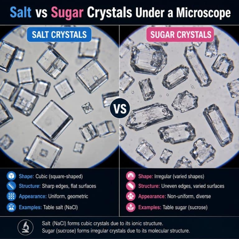
Under a microscope, salt and sugar crystals look completely different. Table salt (sodium chloride) forms small, transparent cubes with sharp 90° corners, while table sugar (sucrose) grows into larger, glassy, elongated prisms with slanted, non-right-angle ends. That difference in shape is a direct readout of each substance’s internal crystal lattice — and it means you can reliably tell them apart without ever tasting them.
Salt vs Sugar Crystals — the Quick Answer
The table below captures the key differences at a glance. Everything that follows explains why.
| Property | Table Salt (NaCl) | Table Sugar (Sucrose) |
|---|---|---|
| Crystal system | Cubic (isometric) | Monoclinic |
| Shape under microscope | Cubes / square blocks | Elongated slanted prisms |
| Corner angles | ~90° (right angles) | Oblique (not 90°) |
| Typical grain size | 0.1–0.5 mm (100–500 µm) | 0.3–0.5 mm (300–500 µm) |
| Relative size | Smaller, more uniform | Generally larger, more irregular |
| Transparency | Transparent / translucent | Transparent, glassy, gem-like |
| Magnification needed | 40×–100× | 40×–100× |
What Salt Crystals Look Like Under a Microscope
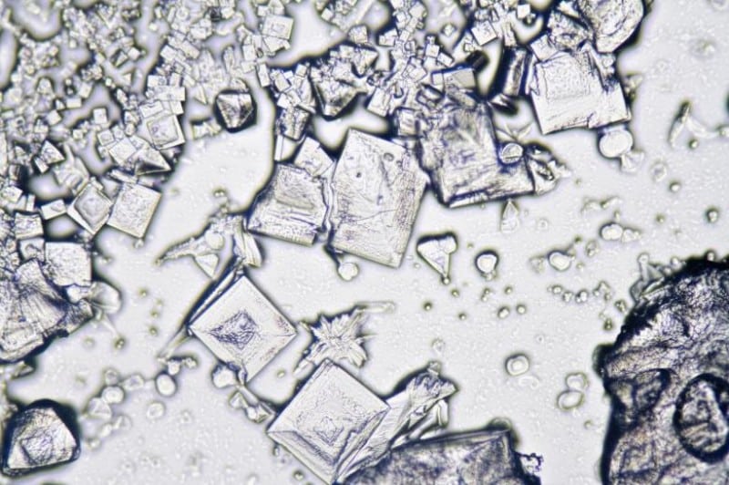
At 40× to 100× magnification, table salt crystals appear as transparent to slightly milky cubes or rectangular blocks. The faces are flat and the edges are clean and straight. Corners meet at crisp right angles — they look almost machine-cut. Under good lighting you can often see that several grains are stacked or stepped on top of one another, because the cubic faces lock together neatly.
One feature worth looking for is the hopper pit — a stepped, pyramid-shaped depression in the face of the cube. This forms because the edges and corners of a growing salt crystal attract ions faster than the flat face centres do, leaving a shallow hollow. It is not an impurity or defect; it is a normal growth feature and is actually a useful identification clue unique to salt.
In terms of size, a grain of standard table salt is roughly 100–500 µm across. That is just about visible to the naked eye as a fine white grain, but at 40× the cubic geometry becomes unmistakeable. You do not need a high-powered research microscope — a basic compound light microscope or even a 10× hand loupe will reveal the shape clearly.
Why Salt Forms Cubes (the Cubic Lattice)
Sodium chloride has a face-centred cubic (FCC) lattice. Inside the crystal, sodium ions (Na⁺) and chloride ions (Cl⁻) alternate in a precise three-dimensional grid, each ion surrounded by six neighbours of the opposite charge in an octahedral arrangement. Every unit cell — the repeating building block — is a perfect cube with equal sides and all 90° angles.

Because the internal unit cell is cubic, the crystal grows outward by adding identical cubic layers in all three equivalent directions. The external shape that results is also cubic. This is the fundamental principle: a crystal’s outer form reflects its inner geometry. The flatness, the right angles, the stepped hopper pits — all of these features trace directly to the symmetry of the NaCl lattice. For a closer look at what different salt varieties reveal, see our dedicated article on salt under the microscope, including close-up images of the hopper structure.
You can read more about the ionic bonding that holds this lattice together at Encyclopaedia Britannica’s entry on sodium chloride.
What Sugar Crystals Look Like Under a Microscope
Table sugar crystals look quite different. Under the microscope they appear as large, glassy, elongated blocks — longer in one direction than the others, with ends that are cut at an angle rather than squared off. The corners are not right angles; they are oblique, giving the crystal a slightly lopsided, slanted appearance. Because sucrose refracts light strongly, the crystals can catch the light beautifully — almost gem-like — with a clarity that surprises many first-time observers.

Granulated sugar grains are typically 300–500 µm, which makes them generally larger and more irregular in outline than table salt grains. Under 40× the size difference is noticeable: sugar crystals tend to be chunkier and more visually prominent on the slide, even though the two substances look nearly identical to the naked eye.
One misconception is worth clearing up immediately: a sugar cube (the kind you drop into tea) is not a single sucrose crystal — it is a compressed block of thousands of granulated crystals bound together. Individual sucrose crystals are the irregular prisms described above, not cubes of any kind.
Why Sugar Crystals Are Slanted, Not Cubic (the Monoclinic Lattice)
Sucrose (C₁₂H₂₂O₁₁) is a disaccharide — a glucose molecule bonded to a fructose molecule. Its unit cell belongs to the monoclinic crystal system, which means two of the three lattice angles are 90° and the third is oblique (not 90°). Because the repeating building block is not a cube, the crystal cannot grow as a cube — it grows as an elongated prism with slanted faces instead.
This is the same principle as with salt, applied in reverse: the low symmetry of the monoclinic lattice produces a crystal with lower external symmetry — elongated shape, bevelled ends, non-right-angle corners. There is no way to make a cubic external form out of a monoclinic internal geometry. The shape you see under the microscope is the lattice geometry made visible. Khan Academy’s introduction to crystalline solids covers the geometry of unit cells clearly if you want to go deeper on the underlying chemistry.
Side-by-Side — How to Tell Salt and Sugar Apart
If you have two unlabelled white powders and access to a microscope or even a good hand loupe, here is a reliable identification procedure. Do not taste unknown white powders — this rule applies in any lab or kitchen-science setting.
- Look at the corners. Salt corners meet at right angles (~90°). Sugar corners are oblique. This is the single most reliable visual cue.
- Check the overall shape. Salt grains are compact cubes or near-cubes. Sugar grains are elongated — noticeably longer in one direction, like a stubby rectangular prism with angled ends.
- Compare size. Sugar crystals in a batch of standard granulated sugar tend to run larger and less uniform than table salt. If one group of crystals looks noticeably bigger and chunkier, it is likely sugar.
- Look for hopper pits. A stepped, hollow depression in the flat face of a crystal is characteristic of salt. Sugar crystals do not form this feature.
- Observe light refraction. Sugar refracts light more strongly and looks more gem-like or glassy. Salt crystals transmit light well but are somewhat less dazzling.
- Edge regularity. Salt edges are very straight and regular. Sugar edges have the same transparency but the oblique angles make the overall outline look less geometrically perfect.
With 40× magnification and good lighting, an experienced observer can tell these two substances apart in seconds. Even a 10× hand loupe in bright natural light is enough to distinguish the cube vs prism shapes reliably. The same analytical approach — identifying a solid from its crystal habit — applies to surprising everyday materials like sand under a microscope, where the variety of grain shapes is equally remarkable.

How to Observe Salt and Sugar Crystals Yourself
This is one of the easiest and most rewarding microscopy activities for students, hobbyists, and kitchen scientists. You need very little equipment and the prep time is under five minutes.
What You Need
- A compound light microscope (40×–100× objective) or a strong hand loupe (10×).
- Plain glass microscope slides and cover slips.
- Table salt and granulated sugar — standard supermarket varieties work perfectly.
- A toothpick or small spatula for transferring a few grains.
Method — Dry Mount
- Place a clean slide on a flat surface. Use a toothpick to transfer a small pinch of salt crystals — about 10–20 grains — to the centre of the slide. Do the same on a second, labelled slide with sugar.
- Do not add water. Both salt and sugar are highly soluble; water will dissolve the crystals immediately and destroy the shapes you want to observe. A dry mount is essential for shape observation.
- Gently lower a cover slip over the grains to hold them in place.
- Place the slide on the stage and start at your lowest power objective (typically 4× or 10×) to locate the grains, then switch to 40× for detail. For most observations 40×–100× total magnification is ideal.
- For lighting: transparent crystals can wash out in full bright-field illumination. Slightly close the iris diaphragm or lower the condenser to increase contrast. Dark field lighting makes the edges of transparent crystals pop dramatically — it is worth trying if your scope supports it.
- Compare both slides directly. Place them side by side and move between them. The shape difference is immediately apparent once you know what to look for.
For a detailed walkthrough of slide preparation in general, see our guide on how to prepare a microscope slide. If you are running a kitchen-science activity with younger students, our guide to the best binocular microscopes covers which entry-level scopes handle this kind of observation well.
Crystal observation is also a gateway to other structured solids. Once you have compared salt and sugar, try watching snowflakes and their crystal symmetry — the hexagonal branching is another stunning example of lattice geometry made visible. For another natural crystal formation that surprises first-time observers, see our article on kidney stones under the microscope. For a broader scientific perspective on crystal structure and physical properties, the American Chemical Society’s crystallography overview is a solid reference, and the National Geographic Education entry on crystals offers accessible context for high school students.

Frequently Asked Questions
What is the difference between salt and sugar crystals under a microscope?
Salt crystals form small, transparent cubes with sharp 90° corners — a result of their cubic (face-centred cubic) lattice. Sugar crystals form larger, glassy, elongated prisms with oblique (non-right-angle) corners — a result of their monoclinic lattice. The shape difference is clearly visible at 40×–100× magnification.
What shape are salt crystals under a microscope?
Salt (sodium chloride) crystals are cubic. Under the microscope they appear as compact square or rectangular blocks with flat faces and right-angle corners. Some crystals also show a hopper pit — a stepped, hollow depression in the face caused by faster growth along the edges than across the flat face centre.
What shape are sugar crystals under a microscope?
Sugar (sucrose) crystals are monoclinic prisms. Under the microscope they look like elongated, glassy rectangular blocks with slanted, bevelled ends. The corners are oblique rather than square, and the crystals are generally larger and less uniform in outline than salt grains.
How can you tell salt and sugar apart under a microscope?
The most reliable cues are shape and corner angle. Salt forms compact cubes with right-angle (~90°) corners; sugar forms elongated prisms with oblique corners. Salt may also show hopper pits on its flat faces. At 40× magnification the difference is obvious — no tasting required.
Why are salt crystals cube-shaped?
Because sodium chloride has a face-centred cubic (FCC) internal lattice, where Na⁺ and Cl⁻ ions alternate in a perfectly symmetric three-dimensional grid. The external shape of a crystal reflects the geometry of its repeating unit cell — and when that unit cell is cubic, the crystal grows as a cube.
What magnification do you need to see salt and sugar crystals?
40×–100× total magnification is more than sufficient. Even a 10× hand loupe in good natural light reveals the shape difference. You do not need a high-powered or oil-immersion objective — this is one of the most accessible microscopy observations you can do at home or in a school lab.
Are sugar crystals bigger than salt crystals?
Generally yes. Standard granulated sugar crystals typically range from 300–500 µm, while common table salt grains range from 100–500 µm. In practice, sugar grains tend to look larger and more irregular under the microscope. The exact size depends on the grade used — caster sugar runs finer than standard granulated, while rock salt or sea salt flakes run much coarser than table salt.
Can you see salt and sugar crystals without a microscope?
You can see that they are small white grains, but the crystal shapes are not resolvable with the naked eye. A 10× hand loupe is the minimum needed to distinguish cubic from prism shapes. A compound light microscope at 40× makes the comparison straightforward and is the recommended tool for a proper side-by-side observation.
Conclusion
Salt and sugar look almost identical as white powders, but under a microscope they are as different as a cube from a slanted prism. Table salt’s face-centred cubic lattice produces neat, right-angled cubes — sometimes with striking hopper pits in their faces. Table sugar’s monoclinic lattice produces larger, glassy, oblique-angled prisms. Those shapes are not accidental; they are the direct, visible consequence of each substance’s internal atomic geometry. Understanding that connection — external crystal form as a readout of internal structure — is one of the most elegant ideas in materials science, and you can explore it with a basic microscope and a few grains from your kitchen bench.
Have you compared salt and sugar under your own scope, or tried this as a classroom activity? We would love to hear what you observed — did you catch the hopper pits in the salt, or spot the gem-like sparkle of the sugar prisms under oblique lighting? Drop your observations or questions in the comments below.