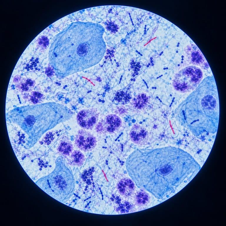
Sputum under a microscope reveals a crowded, cell-packed world: inflammatory cells, invading bacteria, and — in the classic diagnostic case — the slender red rods of tuberculosis. A stained sputum smear is how laboratories screen for TB, confirm bacterial pneumonia, and flag fungal infection, often before any other test result comes back.
This article is for educational purposes only and is not medical advice or a diagnostic guide. If you have a persistent cough, blood in your sputum, or suspect a respiratory infection, see a licensed healthcare provider. Sputum testing is performed and interpreted by clinical laboratories, not at home.
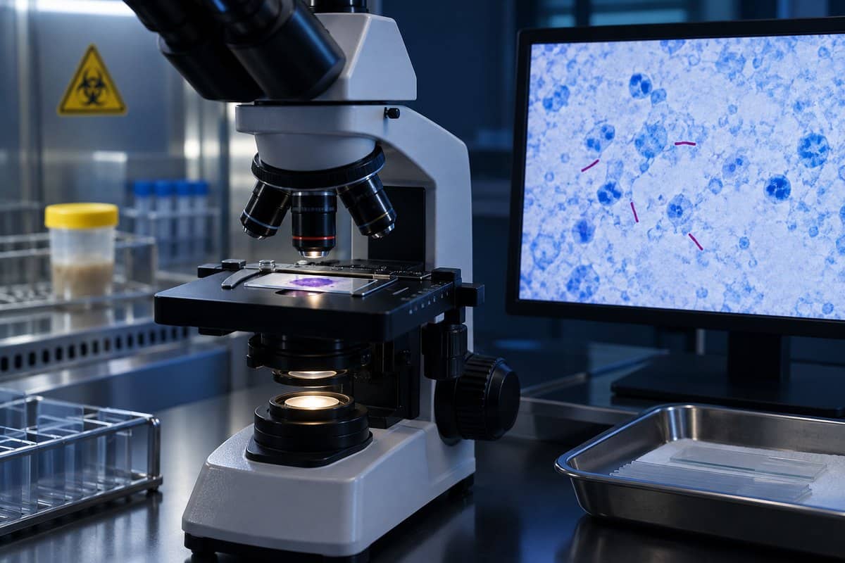
Sputum vs Saliva — Why the Sample Decides Everything
Sputum is mucus and cellular debris coughed up from the lower respiratory tract — the bronchi and lungs — by a deep, productive cough. Saliva is simply spit from the mouth, and the two are not interchangeable, even though patients often use “phlegm” loosely for either one. A lab can tell the difference under the microscope before it ever reads for bacteria. A genuine lower-airway sample carries neutrophils (infection-fighting white blood cells) and alveolar macrophages, the resident scavenger cells of the lung. A sample that’s mostly saliva instead shows sheets of squamous epithelial cells — large, flat cells shed from the mouth and throat — with almost nothing else. The CDC outlines safe sputum collection and specimen requirements for tuberculosis evaluation.
This distinction matters clinically. According to screening criteria used by hospital microbiology labs, a specimen with more than 25 squamous epithelial cells per low-power field is typically rejected outright. That’s because it’s really oropharyngeal contamination, not lower-airway material. A good specimen instead shows a favorable ratio of white blood cells to squamous cells. The microscope, not the naked eye, is what decides whether a sample is worth testing at all.
How a Clean Sputum Sample Is Collected
Labs ask patients to produce the sample from a deep chest cough, ideally the first cough of the morning, before eating or drinking, when overnight secretions have pooled in the airways. Clearing the throat and coughing gently first helps avoid mixing in saliva. This is a collection instruction given by clinical staff, not a self-diagnosis step — what the sample shows is read and interpreted entirely by trained laboratory personnel.
How a Sputum Smear Is Made and Stained
Turning a raw sputum sample into something readable under the microscope follows a short, standardized sequence:
- Collect the deep-cough specimen in a sterile container.
- Smear a thin layer of the sample onto a glass slide.
- Heat-fix the slide, which bonds the cells and any bacteria to the glass so they survive the staining steps.
- Stain the smear — with a Gram stain for suspected bacterial infection, or a Ziehl-Neelsen (acid-fast) stain for suspected TB.
- Read the slide under the microscope, scanning field by field.
The Gram stain separates bacteria into two broad groups by cell-wall chemistry: Gram-positive organisms trap crystal violet dye and stay purple, while Gram-negative organisms lose it during decolorization and pick up a pink counterstain instead. It’s the first-line stain for suspected bacterial pneumonia.

The Ziehl-Neelsen stain works on a completely different principle, and it’s why Mycobacterium tuberculosis gets its own dedicated test. Mycobacteria have a thick, waxy cell wall built from mycolic acid, which resists the Gram stain almost entirely. Instead, technicians apply carbolfuchsin dye under heat, which forces it past the waxy wall. Once inside, the mycolic acid holds the red dye so tightly that even a harsh acid-alcohol rinse can’t strip it back out. Everything else in the smear decolorizes and takes up a blue methylene-blue counterstain, but the mycobacteria stay red. That resistance to acid decolorization is literally where the term “acid-fast” comes from, a mechanism laid out in detail in open microbiology lab teaching materials on the Ziehl-Neelsen method. A newer fluorescent stain, auramine-rhodamine, works on the same waxy-wall principle but makes bacilli glow yellow-green, which is faster to scan for high-volume labs.
What You Actually See Under the Microscope
At low power, a stained sputum smear looks like a patchy blue-purple landscape — strings and clumps of mucus with darker specks caught in the mesh, similar in spirit to how bacteria appear under a microscope in other clinical specimens. Zooming in on those specks is where the diagnosis actually happens. Its TB laboratory course also explains how acid-fast bacilli are assessed in stained sputum smears.
Neutrophils show up as small, dark cells with a distinctive multilobed nucleus — they look like little clusters of connected dots rather than one smooth blob. Their sheer number signals an active, purulent infection. Alveolar macrophages are noticeably larger, with a “dusty,” granular cytoplasm that sometimes contains visible black carbon flecks. That’s a genuine sign the sample came from deep in the lung rather than the mouth. Squamous epithelial cells, by contrast, are big, flat “fried-egg” cells with a small central nucleus; a field full of these is the tell-tale sign of a saliva-contaminated sample. Eosinophils, when present in numbers, point toward asthma or an allergic airway process rather than a bacterial one — the same white blood cell types covered in a full identification guide to all five white blood cell types.

Bacteria are visible directly on a Gram-stained smear. Gram-positive cocci that cluster in pairs — diplococci — are a classic clue for Streptococcus pneumoniae, one of the most common causes of bacterial pneumonia. Gram-negative rods take up the pink counterstain instead, the same visual pattern seen when E. coli shows up as pink rods at 1000x in other specimens. Fungal elements — yeast cells or branching hyphae — appear on a KOH prep or special fungal stains, most often in patients with weakened immune systems.
The Classic TB Picture — Red Rods on a Blue Field
Against a flat blue background, acid-fast bacilli appear as thin, slightly curved red or pink rods, often beaded — a dashed-line look rather than a solid stroke — and sometimes lying together in loose parallel bundles called cords. Technicians read these slides under oil immersion at roughly 1000x total magnification, because a dry lens simply can’t resolve rods this fine. The bacilli can be sparse — as few as one to nine organisms across 100 fields in a “scanty” result. That’s why a careful reader scans slowly and systematically before ever calling a smear negative. The video below shows a real Ziehl-Neelsen-stained sputum smear with visible acid-fast bacilli, the same red-rods-on-blue appearance described above.
The more bacilli visible on the slide, the higher the reported grade — from scanty up through 1+, 2+, and 3+ — and the more infectious the patient is considered to be. Reading these slides is a close cousin of reading a stained blood smear step by step, in that both depend on methodical, field-by-field scanning rather than a single glance.
What a Sputum Test Checks For
A single sputum smear can point toward several different diagnoses, depending on which stain is used and what turns up. The table below summarizes the main targets:
| Target | Stain or Prep | What It Looks Like | What It Suggests |
|---|---|---|---|
| Tuberculosis (M. tuberculosis) | Ziehl-Neelsen (acid-fast) | Thin red/pink rods on a blue background | Possible TB; needs culture or molecular confirmation |
| Bacterial pneumonia | Gram stain | Purple cocci/rods (Gram-positive) or pink rods (Gram-negative) | Likely bacterial cause, e.g. S. pneumoniae |
| Fungal infection | KOH prep / fungal stain | Yeast cells or branching hyphae | Possible fungal airway infection |
| Airway inflammation type | Gram stain / cell count | Dominant neutrophils vs dominant eosinophils | Bacterial process vs asthma/allergic process |
What Sputum Microscopy Can’t Tell You
A stained smear is fast and cheap, but it has real limits, and treating it as a final answer is a mistake. A positive acid-fast smear means acid-fast bacilli are present — it does not by itself confirm M. tuberculosis, because other, non-tuberculous mycobacteria are also acid-fast. Under CDC guidance on TB laboratory diagnosis, culture remains the gold standard for confirming TB disease, and rapid molecular tests such as NAAT (nucleic acid amplification testing) can confirm the species faster than culture alone. Smear microscopy is also less sensitive than culture, so a negative smear does not rule out TB. It simply means the bacterial load, if any, was below what the technician could see.
Two more limits are worth separating clearly. First, sputum color observed by the naked eye — green, yellow, blood-streaked — is not a microscope finding at all; it’s a different kind of observation and shouldn’t be confused with what a stained smear actually shows. Second, viruses are far too small for a light microscope to resolve, sitting well below its roughly 200-nanometer limit. Conditions like influenza or COVID-19 are never diagnosed by looking at a sputum smear — they require PCR or antigen testing instead.
Frequently Asked Questions
Why do labs ask for three sputum samples instead of one for TB testing?
Because TB bacteria can be shed inconsistently, a single sample can miss them even when the infection is present. Collecting specimens on separate occasions — often spaced eight to twenty-four hours apart — improves the odds of detecting acid-fast bacilli if they’re there, which is why multiple-specimen collection is standard guidance for suspected pulmonary TB.
Can you see viruses like the flu or COVID-19 in a sputum smear?
No. Viruses are roughly one hundred times smaller than bacteria and sit below the resolution limit of a standard light microscope. Viral respiratory infections are confirmed with PCR or antigen tests, not by examining a stained sputum smear.
Is a sputum sample the same thing as a phlegm sample?
Functionally, yes — “phlegm” is the everyday word for the same lower-airway mucus that clinicians call sputum. The lab still checks it under the microscope for quality before testing, regardless of which word the patient uses to describe it.
How long does it take to get sputum smear results back?
A smear can typically be stained and read within a day, much faster than a culture, which can take one to several weeks to grow mycobacteria. That speed is exactly why smear microscopy is still used as a same-day screening step even though culture is the more definitive test.
What happens if a patient can’t cough up sputum on their own?
Clinical staff can induce a sample using a saline mist that encourages a deeper cough, or in some cases collect a sample by suctioning the airway directly. Either way, obtaining and interpreting the sample stays a task for trained clinical personnel, not something done at home.
Does the color of sputum predict what the smear will show?
Not reliably. Color is a naked-eye clue that can suggest inflammation or blood, but it doesn’t tell you what’s actually happening at the cellular level. Only the stained microscope slide reveals whether neutrophils, macrophages, bacteria, or acid-fast rods are actually present.
Conclusion
Under a stained microscope smear, sputum tells a layered story: whether the sample itself is genuine lower-airway material or mostly saliva, which cells are responding to the problem, and — in the highest-stakes case — whether acid-fast rods point toward tuberculosis. The stains do the heavy lifting, turning an otherwise invisible mix of cells and bacteria into a readable, color-coded picture that a trained technician can interpret in minutes, even though confirming a diagnosis like TB still takes culture or molecular testing to finish the job.
Have you ever wondered what your own lab results were actually looking at under the lens? Tell us what questions you still have about sputum, staining, or TB screening in the comments below.