Rotifers under a microscope look like they’re wearing a spinning wheel on their head. That single detail is the reason they’re one of the most satisfying finds in a drop of pond water. They’re not protozoa and not tardigrades; they’re true microscopic animals, usually 100–500 micrometers long, named for a crown of beating cilia that only looks like it’s rotating. Here’s how to find one, mount it, and see exactly what’s really happening at the “wheel.”
What Is a Rotifer?
A rotifer is a microscopic, multicellular freshwater animal in the phylum Rotifera — not a single-celled protozoan, and not a plant. Most species run 100 to 500 µm long, with a fair number sitting around 200 µm (roughly three times the width of a human hair, which is about 70 µm). The body is usually transparent or lightly amber-tinted, which is a gift for the observer. You can often see the gut, the pulsing mastax, and even eggs through the body wall without staining anything.
The old common name, “wheel animalcule,” comes straight from Latin — rota (wheel) and fera (bearing). It was coined by early microscopists who watched the same illusion you’re about to see. Rotifers aren’t rare. They’re one of the most common animals you’ll pull out of pond water, moss, or aquarium sediment, and most species are completely harmless to handle.
How to Find Rotifers (Where to Collect a Sample)
Rotifers show up wherever there’s standing freshwater with some organic life in it. Clear tap water will not have any — you need the green or brown “gunk” zone.
- Pond water: Scoop from the edge of a pond, ditch, or puddle, favoring areas with algae, decaying leaves, or plant debris rather than open clear water.
- Moss and lichen: Squeeze a handful of wet moss into a jar, or soak dry moss in water for 20–30 minutes. Many rotifers (especially the bdelloid group) survive complete drying and revive within minutes of rewetting.
- Aquarium detritus: The sludge at the bottom of an established aquarium is a reliable source, especially if it hasn’t been cleaned recently.
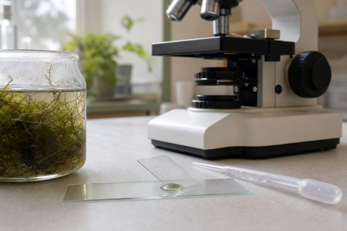
One trick worth using every time: let a jar of pond water with a pinch of dry moss sit for one to three days on a windowsill. Rotifer numbers bloom noticeably over that window, and you’ll find far more under the lens than in a sample taken straight from the source.
How to View Rotifers Under Your Microscope
You don’t need a stain or a fixed slide — rotifers are far more interesting alive and moving. Follow these steps:
- Collect a sample from the green/brown zone of pond water, or squeeze out moss that’s been soaking.
- If using dry moss, rewet it and wait 20–30 minutes before sampling, giving dormant animals time to revive.
- Pipette a small drop onto a glass slide and lower a cover slip at an angle to avoid trapping air bubbles.
- Start at 40x (using your lowest-power objective) to scan the field and locate a moving speck.
- Center the animal in the field of view before increasing magnification — don’t jump straight to high power.
- Climb to 100x, then 400x, adjusting focus in small steps as the corona and mastax come into resolution.
- Slow the animal down by adding a strand of cotton fiber to the drop, or a trace of methyl cellulose, so it can’t swim out of frame.
- Watch the corona first, then shift focus slightly back to catch the mastax pulsing behind it.
Two adjustments make a bigger difference than any fancy technique: reduce the light and close the iris diaphragm. Rotifers are transparent, and too much light washes out the body entirely. Lower contrast settings reveal the outline and internal structures the way a stain would, without killing the animal. Work reasonably fast, too; a small drop dries out quickly under the lamp’s heat and crumples the animal before you get a good look. A slightly bigger drop under the cover slip buys you more observation time.

The Corona — and Why the “Wheel” Doesn’t Actually Spin
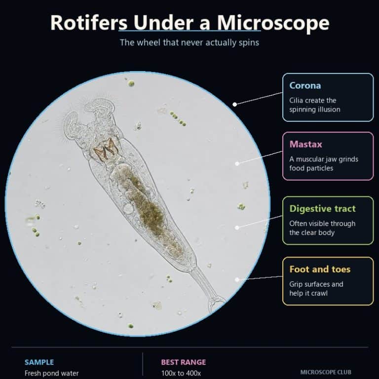
The corona is the ciliated crown at a rotifer’s head end. At 100x it resolves into a shimmering, flickering halo that genuinely looks like two tiny gears turning in opposite directions. That’s the whole reason for the name “wheel animal” — but nothing at the corona is rotating. What you’re seeing is a metachronal wave. Each cilium beats a fraction of a second after its neighbor, in a fixed sequence around the ring, and that phase-shifted timing creates the optical illusion of continuous rotation. It’s the same principle as a stadium wave — no seat actually travels around the stadium, but the wave looks like it moves.
Push to 400x and watch a single cilium instead of the whole ring, and the illusion collapses immediately: it beats back and forth, it doesn’t spin. The corona’s real job is functional, not decorative. The coordinated beating generates a water current that pulls in bacteria, algae, and organic detritus and sweeps it toward the mouth. In many species, that same current also drives swimming.
This detail is well documented outside hobbyist circles too — the Britannica entry on Rotifera describes the same ciliary mechanism behind the “false wheel” effect.

The Mastax — The Grinding Jaw You Can Watch Working
Just behind the corona sits the mastax, a muscular pharynx that houses a set of hardened jaws called the trophi. Under the microscope it shows up as a small, dark, rapidly pulsing knot — it looks almost like a tiny beating heart, except it’s grinding rather than pumping. That constant chewing motion is one of the most reliable ways to confirm you’re looking at a rotifer and not something else drifting through the same sample. Few other pond-water animals have a visibly working jaw right behind the head.
The trophi grind the bacteria, algae, and detritus that the corona’s current sweeps toward the mouth. Their exact shape varies by species and is actually how specialists tell rotifer species apart under higher magnification. You don’t need that level of detail to enjoy watching one work, but it’s worth knowing the pulsing you’re seeing has a name and a purpose.
The Foot, Toes, and How Rotifers Move
Many rotifers, especially bdelloid species like Philodina and Rotaria, have a tapering “foot” at the tail end that ends in one to four toes. Glands in the foot secrete an adhesive that lets the animal cement itself to a surface — a leaf, a strand of algae, the underside of a cover slip. That lets it feed while anchored rather than expend energy swimming constantly.
Rotifers move in two distinct ways, and it’s worth watching for both. Free-swimming individuals glide and spin smoothly through the water, propelled by the corona’s ciliary current. When crawling, the same animal switches to a leech-like or inchworm motion. It extends the head, grips with it, then loops the foot forward and anchors with the toes before releasing the head again. Seeing an animal switch from a graceful swim to this looping crawl in the same field of view is one of the more memorable things a beginner notices.
What You’ll See at Each Magnification
Knowing what to expect at each step saves you from losing the animal or mistaking it for debris.
- 40x: Transparent, elongated specks darting through the debris field, easy to overlook. Look for the ones that stop moving and appear to extend a “head” — that pause-and-extend behavior is a strong rotifer tell before you can see any real detail.
- 100x: The body resolves into a translucent, often slightly amber, vase- or worm-shaped animal. The front end blurs into a shimmering, flickering halo — the corona in motion, and at this power it genuinely looks like two small wheels spinning.
- 400x: The “wheel” breaks apart into individual cilia rippling in sequence rather than rotating. Behind the head, the mastax pulses like a small dark heartbeat. Gut contents are often visible, and sometimes an egg attached near the tail.
Rotifers vs Tardigrades vs Protozoa (Quick ID)
Rotifers, tardigrades, and protozoa all turn up in the same moss and pond-water samples, and beginners regularly mix them up. This quick comparison should stop that.
| Feature | Rotifer | Tardigrade | Protozoa |
|---|---|---|---|
| Cell type | Multicellular animal | Multicellular animal | Single-celled |
| Movement | Ciliated swim / leech-like crawl | Bear-like walk on 8 stubby legs | Cilia, flagella, or pseudopods |
| Key feature | Corona (ciliated “wheel”) | Claws, no corona | No organs, no corona |
If it has a wheel-like ciliated head and either swims smoothly or loops like a leech, it’s a rotifer. If it has eight stubby legs and lumbers along like a microscopic bear, that’s a tardigrade (water bear). If it’s a single cell with no visible internal organs, you’re looking at a protozoan such as an amoeba, paramecium, or euglena.
Frequently Asked Questions
Are rotifers plankton, animals, or protozoa?
Rotifers are true multicellular animals, and they’re often classified as part of the zooplankton community because they drift and swim in open water alongside other tiny organisms. They are not protozoa — protozoa are single-celled.
What do rotifers eat?
Bacteria, single-celled algae, protozoa, and organic detritus, all filtered from the water current their corona generates and ground up by the mastax.
Are rotifers harmful to humans or parasites?
No. The vast majority of rotifer species are harmless free-living microfauna with no interaction with humans at all. As with any pond-water sample, wash your hands after handling it as general good practice.
Can rotifers survive being completely dried out?
Yes — many bdelloid rotifers enter a dormant state called anhydrobiosis when their habitat dries up, and can revive within minutes of being rewetted, sometimes after months or years dry. That’s the reason a pinch of dry moss soaked in water is such a reliable way to find them.
How do you keep rotifers alive to observe them?
Keep the sample cool, avoid direct lamp heat for extended periods, and use a large enough drop that it won’t dry out mid-observation. There’s no need to feed or culture them for a single viewing session — a fresh sample from pond water or rewetted moss is enough.
Conclusion
Rotifers reward a closer look precisely because the first thing you notice about them is an illusion. The “wheel” that gives them their name is really a metachronal wave of beating cilia. The mastax behind it is a genuine grinding jaw you can watch working in real time. The foot and toes let the same animal switch between swimming and a leech-like crawl. Once you know what the corona, mastax, and foot actually are, a jar of week-old pond water or a pinch of rewetted moss stops looking like debris. It starts looking like a working animal instead.
Have you found rotifers in your own samples, or been fooled by the spinning-wheel illusion before you knew what was really happening? Tell us what you saw — and at what magnification — in the comments below.
Related Specimen
Another freshwater specimen worth finding in the same sample: see our full Hydra anatomy and observation guide.