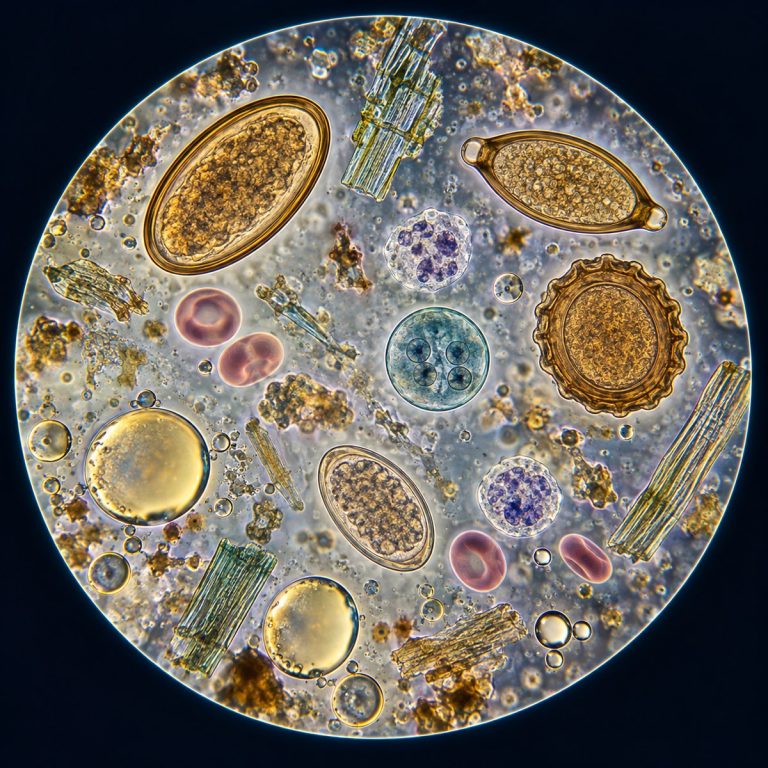
A stool sample under a microscope reveals six things a laboratory scientist is trained to look for. These are parasite eggs and larvae, protozoa, red and white blood cells, fat globules, undigested muscle fibers, and a handful of tell-tale crystals. Each finding has its own shape, size, and color at the eyepiece, and each one points toward a different explanation for a patient’s symptoms. Here is what a lab technician actually sees on the slide, and what each finding can mean.
This article is educational, not diagnostic. It describes how a trained laboratory professional examines a stool specimen — it is not a guide for testing your own sample at home. Fecal material is a biohazard, and only a lab with proper containment, stains, and equipment should prepare or interpret a slide like this. If you have been asked to provide a stool sample, talk to your doctor about what the results mean; never self-diagnose from a description like this one.
Before any of the individual findings make sense, it helps to know the six-item checklist a lab technician works through on every sample:
- Parasite eggs and larvae (ova and parasites) — helminth worms like Ascaris, hookworm, and whipworm.
- Protozoa — single-celled organisms such as Giardia and Entamoeba, seen as either active trophozoites or dormant cysts.
- Red blood cells — a sign of bleeding or inflammation somewhere in the lower gut.
- White blood cells (fecal leukocytes) — a marker of invasive, inflammatory infection.
- Fat globules — evidence the gut isn’t absorbing fat properly.
- Undigested muscle fibers and crystals — signs of poor digestion or an allergic/parasitic reaction.
How a Stool Sample Is Prepared for the Microscope
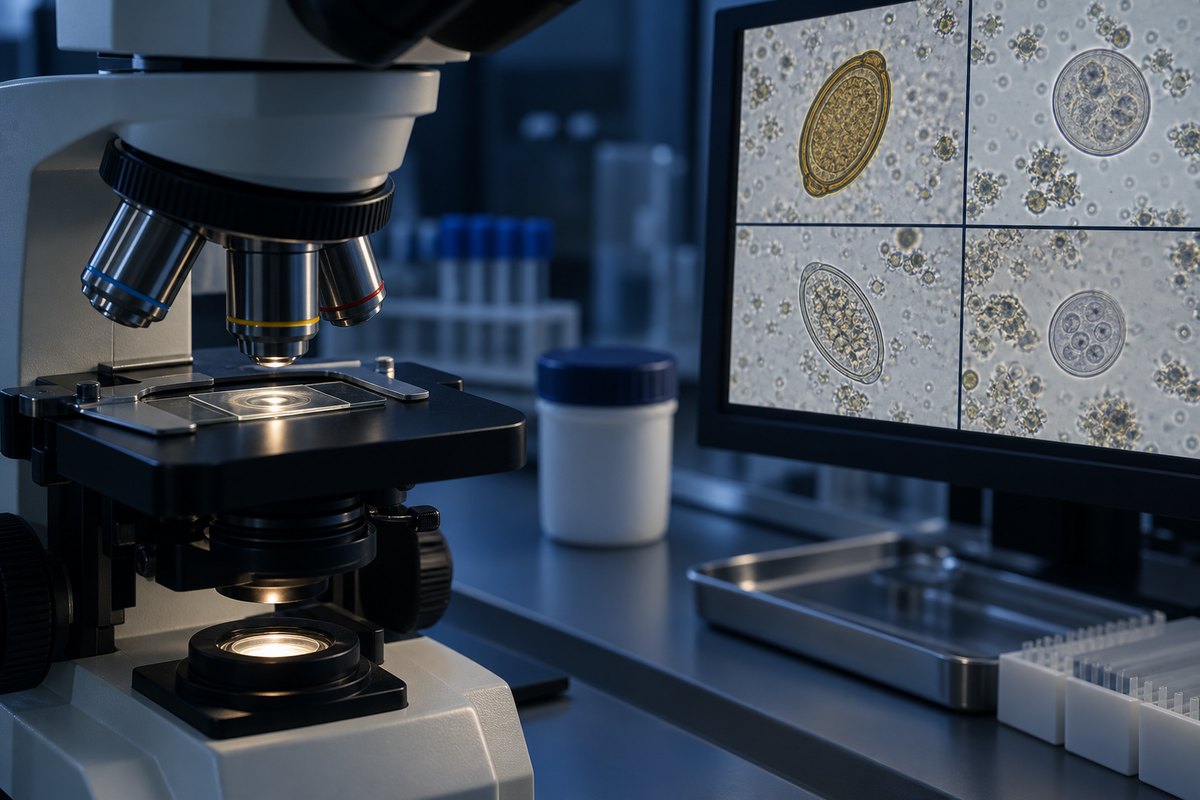
A stool sample is prepared for the microscope in four stages: a macroscopic check, a wet mount, a concentration step, and a permanently stained smear. That last stage is only needed when a definitive species identification is required. Skipping straight to the slide without this groundwork is how findings get missed or misread. The CDC’s DPDx resource details how fresh and preserved stool specimens are processed for microscopy.
Macroscopic Check First
Before any slide is made, the technician records the specimen’s color and consistency — formed, loose, or watery — and notes any visible mucus, streaks of blood, or worm segments. This macroscopic pass takes seconds, but it tells the lab where to look once the sample is on a slide. A mucoid or bloody streak, for instance, is exactly where amoebic trophozoites tend to concentrate.
The Saline and Iodine Wet Mount
A direct wet mount comes next: a small amount of the sample is mixed with normal saline on one slide and with iodine on a second. The saline mount shows live motility — protozoal trophozoites and flagellates swimming, plus eggs, larvae, and blood cells — so it’s examined first, while everything is still moving. Iodine stains the nuclei and glycogen inside protozoan cysts to make their internal structure easier to identify. It kills motility outright, though, which is why it’s always the second prep, never the first. Both mounts are scanned systematically at 10× magnification, then dropped to 40× to confirm anything that looks suspicious.
A common beginner mistake is jumping straight to 40× and getting lost in a sea of debris. The coverslip has to be scanned edge-to-edge at 10× first, with 40× reserved for confirming specific structures already spotted. A wet mount that’s smeared on too thick is just as much of a problem: a heavy prep goes opaque and hides everything underneath it. The prep should be thin enough that you could read newsprint through it, and it needs to be read promptly. The edges dry out and crystallize within minutes, throwing artifacts into the field that can be mistaken for something real.

Concentration and Permanent Stained Smears
Because ova, cysts, and larvae can be scarce, most labs run a concentration technique — typically formalin-ethyl acetate sedimentation — to pull the sparse material into a small pellet. That pellet is far easier to scan than raw stool. For a definitive protozoan species call, a permanent stained smear (trichrome or iron-hematoxylin) is examined under 100× oil immersion across at least 200 to 300 fields. Special stains fill in the gaps routine staining can’t reach: a modified acid-fast stain for Cryptosporidium, Cyclospora, and Cystoisospora, plus UV autofluorescence for Cyclospora oocysts. Sudan III or IV is used separately to make fat globules light up orange-red.
Parasite Eggs and Larvae (Ova and Parasites)
Parasite eggs are the classic “ova and parasites” (O&P) finding, per the CDC’s DPDx parasite identification reference. Each species has a shell shape distinctive enough to identify on sight once you know what to look for. Ascaris lumbricoides eggs are golden-brown ovals with a thick, bumpy, quilted-looking rind. Hookworm eggs (Ancylostoma or Necator) are thin-shelled, clear, and oval, with a segmented embryo visible inside. Trichuris trichiura, the whipworm, produces a barrel or lemon-shaped egg with a distinctive clear “plug” glowing at each pole. Taenia tapeworm eggs are round with a thick, radially striated shell, and Schistosoma eggs carry a characteristic spine off to one side or the tip. Strongyloides stercoralis is usually spotted as a motile larva rather than an egg at all. Its reference tables compare the microscopic morphology of intestinal parasites in different preparations.
One frequently confused point worth flagging: pinworm (Enterobius vermicularis) is not diagnosed from a routine stool sample. Its eggs are laid around the anus at night, so it’s picked up with a cellophane-tape swab of the perianal skin, not stool microscopy. For a closer look at how helminth worms and their eggs appear under magnification more broadly, see this guide to what parasitic worms and their eggs look like under a microscope.

Protozoa: Giardia, Amoebas, and Other Organisms
Protozoa show up in stool as either a motile trophozoite or a dormant, thicker-walled cyst, and the two most clinically important genera look nothing alike. Giardia duodenalis trophozoites are pear-shaped and translucent, with two nuclei that genuinely read as a tiny cartoon face — a look lab staff commonly describe as an “owl face” or “smiley.” That look comes from four pairs of flagella powering the cell. Its cysts are smooth, oval capsules roughly 11 to 14 micrometers across that drift rather than swim. Entamoeba histolytica trophozoites are only clinically significant when they contain ingested red blood cells. That single detail is what separates the invasive, disease-causing form from a harmless commensal amoeba living in the same gut. Its cysts can carry up to four nuclei.
Two organisms need special stains to see at all. Cryptosporidium oocysts are tiny, only 4 to 6 micrometers, and appear acid-fast pink; Cyclospora cayetanensis oocysts are slightly larger at 8 to 10 micrometers and autofluoresce blue-green under UV light. A classic beginner mistake at this stage is mistaking an air bubble for a cyst. Bubbles have a thick, uniform black rim and no internal detail, while a real cyst shows visible internal structure once you focus through it.
Red and White Blood Cells in Stool
Red blood cells in a stool sample appear as flat, pale-gold discs, and their presence points to bleeding or inflammation somewhere in the lower gastrointestinal tract. Possible causes include dysentery, colitis, hemorrhoids, or, less commonly, a tumor. Labs distinguish frank blood, visible to the naked eye, from occult blood detected only by testing or microscopy. For a broader look at how these cells appear under magnification generally, see how red blood cells appear under a microscope.
White blood cells — specifically fecal leukocytes — look grainier and slightly larger than red cells, with a lumpy interior. Their presence, especially neutrophils, is a genuinely useful diagnostic discriminator: it signals inflammatory, invasive diarrhea caused by organisms like Shigella, Salmonella, Campylobacter, invasive E. coli, or C. difficile, as well as noninfectious causes like ulcerative colitis. Fecal leukocytes are typically absent in toxin-mediated bacterial diarrhea and in viral diarrhea, which is exactly why the finding matters clinically. See more on how white blood cells seen under the microscope appear in general.
Fat Globules, Muscle Fibers, and Crystals
Fat globules are large, perfectly round droplets that stain a vivid orange-red under Sudan III or IV, floating separately from everything else on the slide. Excess fat in stool — a condition called steatorrhea — suggests the gut isn’t absorbing fat properly, which shows up in conditions like celiac disease or chronic pancreatitis.
Undigested muscle fibers look like pale rectangular blocks with faint horizontal stripes, almost like tiny wooden planks — the cross-striations are the giveaway. More than about 10 fibers per field suggests creatorrhea, a sign of poor digestion often tied to the same pancreatic insufficiency that causes steatorrhea. Starch granules and plant fibers are noted the same way when present in excess.
Charcot-Leyden crystals are clear, glassy, needle- or spindle-shaped structures, sharp at both ends, formed from the breakdown of eosinophils. Finding them points toward a parasitic infection or an eosinophilic, allergic process in the colon. Budding yeast cells, usually Candida, are another structure noted on the same slide, though they’re rarely the primary finding.
What the Findings Can — and Can’t — Tell You
| Finding | What it looks like under the microscope | What it can indicate |
|---|---|---|
| Helminth eggs (Ascaris, hookworm, whipworm, etc.) | Species-specific oval or barrel shells, 25–75 µm | Intestinal worm infection |
| Giardia trophozoite/cyst | Pear-shaped “owl face” cell or smooth 11–14 µm oval cyst | Giardiasis |
| Entamoeba histolytica trophozoite | Ameboid cell, disease form contains ingested RBCs | Invasive amebiasis (only if RBCs present) |
| Red blood cells | Flat, pale-gold discs | Bleeding/inflammation: dysentery, colitis, hemorrhoids |
| White blood cells (fecal leukocytes) | Grainy, lumpy-interior cells | Invasive/inflammatory diarrhea |
| Fat globules | Round droplets, orange-red with Sudan stain | Steatorrhea, malabsorption |
| Muscle fibers | Rectangular blocks with cross-striations | Creatorrhea, poor digestion |
| Charcot-Leyden crystals | Clear needle/spindle shapes | Parasitic or eosinophilic reaction |
A stool microscopic exam has real limits worth knowing. It cannot identify bacterial species — routine microscopy only shows bacteria as a general mass, never a named pathogen. That’s why a suspected bacterial infection needs a stool culture or PCR test rather than microscopy alone. A negative result doesn’t rule parasites out, either, because ova shedding is intermittent: the NIH’s guide to the ova and parasite (O&P) test notes that labs commonly ask for three samples collected on different days before calling a result truly negative. And finding Entamoeba trophozoites on a slide isn’t automatically a diagnosis of disease — as covered above, it’s specifically the ingested-red-blood-cell form that marks the invasive, symptomatic infection.
Frequently Asked Questions
Can you see bacteria in a stool sample under a microscope?
Only as a general mass, not as a named species. Identifying which bacteria are present — Salmonella versus Campylobacter, for example — requires a stool culture or PCR test, not routine microscopy. For a look at how individual bacterial cells actually appear under magnification, see gut bacteria under a microscope.
What magnification is used to examine stool?
A lab typically scans the whole slide at 10× first, then confirms suspicious structures at 40×. For a permanent stained smear used to confirm a protozoan species, the read happens at 100× under oil immersion.
Is an O&P test the same as a stool culture?
No. An ova and parasite (O&P) test is a microscopic exam looking for parasite eggs, larvae, and protozoa. A stool culture grows bacteria from the sample to identify a specific species. They answer different questions and are often ordered together.
How is an O&P exam different from a fecal occult blood test?
An O&P exam is read under a microscope to find parasite eggs, larvae, and protozoa by sight. A fecal occult blood test is a chemical or immunoassay screen for blood that isn’t visible to the naked eye, and it never involves looking at the sample under magnification. A lab can run both on the same specimen, but they’re answering completely different questions — one is looking for organisms, the other for a chemical signal of bleeding.
How should a stool sample be collected before testing?
A doctor’s office typically provides a clean, dry collection container and asks that the sample avoid contact with urine, water, or toilet paper. Because parasites and cells degrade quickly at room temperature, the sample should reach the lab promptly — usually within an hour for motility studies, or per whatever preservative and timing instructions the lab provides.
Is a rapid antigen test the same as looking at stool under a microscope?
No. A rapid antigen or immunoassay test — commonly used for Giardia or Cryptosporidium — chemically detects one organism’s proteins and skips the microscope entirely. It’s faster for that single target, but unlike a full microscopic exam, it won’t reveal blood cells, fat, muscle fibers, or any other parasite that wasn’t specifically tested for.
Conclusion
A stool sample under a microscope is a genuinely small, low-cost window into six categories of disease: parasites, protozoa, blood cells, fat, muscle fibers, and crystals. Each has a shape and stain that a trained eye can read in minutes. The preparation matters as much as the finding itself — the right wet mount, stain, and magnification are what separate a real parasite from an air bubble, or a harmless amoeba from a dangerous one.
Have you seen a prepared stool slide yourself, in a classroom or a lab rotation? Tell us what stood out to you — or what you found hardest to identify at first — in the comments below.