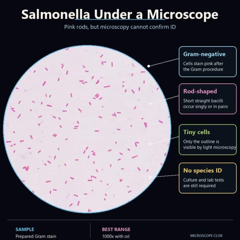
Salmonella under a microscope appears as tiny, straight, pink rods after a Gram stain — small enough that you need 1000x oil immersion just to resolve their outline. A light microscope can show you that shape, size, and color, but it cannot tell you the rods are Salmonella specifically. That confirmation only comes from a lab culture and a battery of biochemical tests.
What Salmonella Looks Like Under the Microscope
Shape and Arrangement
Salmonella is a bacillus — a straight rod with rounded ends, occurring mostly as single cells rather than chains or clusters. Picture a grain of rice shrunk down to a speck you can barely separate from the background stain. On a well-made slide, the rods scatter singly across the field, each one a short, solid-colored dash with no branching and no obvious grouping pattern.
Size and Why There’s No Internal Detail
Each cell measures roughly 0.7–1.5 µm wide by 2–5 µm long, which sits right at the resolving edge of a standard light microscope. That’s why the rods look flat and featureless — no visible nucleus, no internal texture, just a uniform pink capsule. Beginners often assume something is wrong with the focus when they see this. It isn’t. A flat, solid-colored rod at this scale is the correct result, not a broken scope or a bad lens.
Color and the Gram-Negative Reaction
Salmonella stains pink to red because it is Gram-negative. The pink comes entirely from the safranin counterstain, not from any natural pigment in the bacterium. Gram-negative cells have a thin peptidoglycan layer sandwiched between an inner and outer membrane. That thin layer can’t hold onto the crystal-violet-iodine complex once alcohol decolorizer hits it, so the cell loses its purple color and picks up the pink counterstain instead. Gram-positive bacteria, like the ones covered in the Gram-positive bacteria in yogurt, keep the thick peptidoglycan wall that traps the violet dye and stay deep purple. The mechanism behind that color split is well documented in general microbiology cell-wall material from Oregon State’s open microbiology textbook.
Want a broader sense of what other bacterial cells look like under magnification, before or after this one? What bacteria look like under a microscope covers the range of shapes you’ll run into on a typical slide.
How a Salmonella Slide Is Prepared and Viewed
The image above comes from a standard lab Gram-stain workflow — not something to replicate at home on a real food or clinical sample. In a microbiology lab working under proper biosafety controls, technicians follow these steps:
- Spread a thin smear of the bacterial culture onto a slide — thin enough that you could almost read text through the wet film.
- Heat-fix the smear with two or three quick passes over a flame to adhere the cells without cooking or distorting them.
- Flood with crystal violet, then Gram’s iodine, then decolorize briefly with alcohol.
- Counterstain with safranin, rinse, and blot dry.
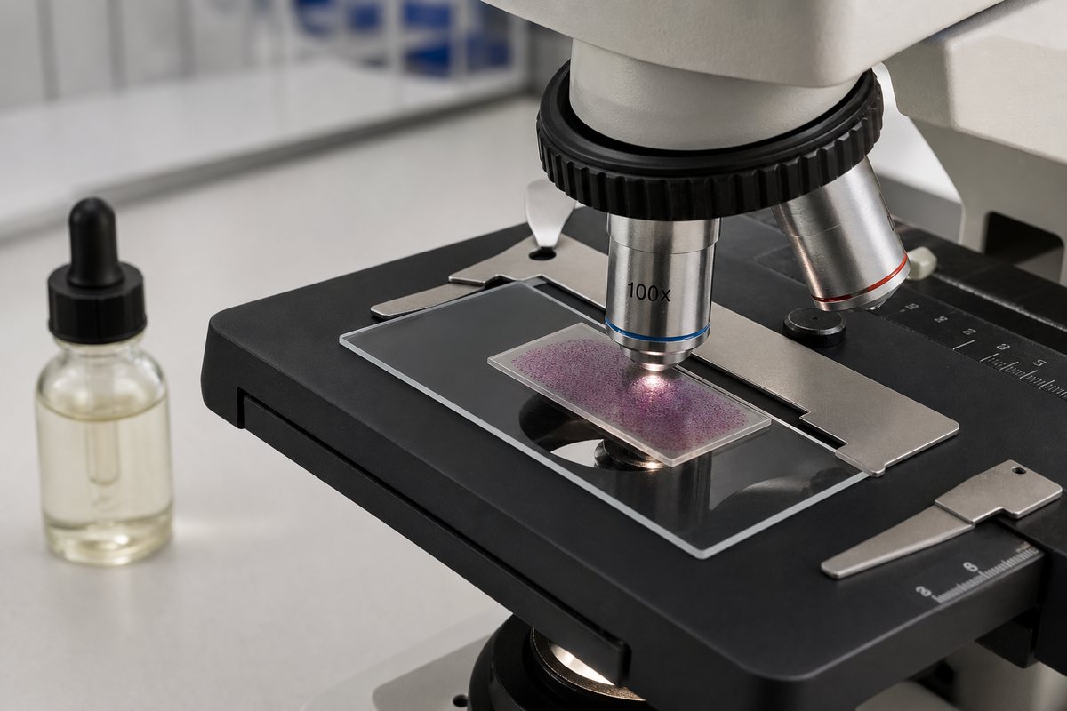
- View at 1000x total magnification under oil immersion.
That 1000x figure isn’t arbitrary — it’s the eyepiece power multiplied by the objective power. A 10x eyepiece paired with a 100x oil-immersion objective gives 10 × 100 = 1000x, which is the minimum practical magnification for resolving rods this small. Below that, the cells blur into indistinct specks. If you’re new to slide prep in general, prepare microscope slides walks through the basics before you tackle a stained bacterial smear.

Two mistakes ruin most student slides. Spreading the smear too thick dries into an opaque purple mass that never resolves into individual rods — thin is always better than thick. And over- or under-decolorizing throws off the whole reading: too little alcohol and the slide stays purple, masking the true Gram-negative reaction; too much strips the color entirely. Watching the alcohol runoff turn from purple to clear, then stopping immediately, is the entire skill.
Motility and Flagella — What You Can and Can’t See
Most Salmonella serovars are motile, moving by peritrichous flagella — thin, whip-like appendages distributed all around the cell. Two notable exceptions, S. Gallinarum and S. Pullorum, are non-motile. But here’s the catch: flagella are not visible on a standard Gram-stained slide under a light microscope. They’re too thin to resolve at this magnification and staining method, and by the time the cells are heat-fixed for Gram staining, they’re dead and stuck to the glass anyway.
Seeing motility itself requires a completely different preparation — a fresh wet mount or hanging-drop slide viewed while the cells are still alive. That’s the same technique described in how to make a wet mount slide. Trying to catch flagellar movement on a heat-fixed Gram slide is a common beginner mistake; the cells simply can’t move once they’ve been fixed and stained.

Salmonella vs E. coli Under the Microscope
This is the question most people actually want answered, and the honest answer is: you can’t tell them apart by looking. Salmonella and E. coli are both Gram-negative rods of overlapping size, and under a light microscope they are effectively indistinguishable. The same is true for most of the family Enterobacteriaceae they both belong to. If you’ve compared notes with E. coli under a microscope, the pink rods you saw there look essentially the same as Salmonella’s.
| Feature | Salmonella | E. coli | Staphylococcus (Gram-positive) |
|---|---|---|---|
| Shape | Straight rod (bacillus) | Straight rod (bacillus) | Round cluster (coccus) |
| Size | ~0.7–1.5 × 2–5 µm | ~0.5–1.0 × 2–3 µm (overlaps) | ~0.5–1.5 µm diameter |
| Gram reaction / color | Negative — pink | Negative — pink | Positive — purple |
| Motility | Usually motile, peritrichous flagella | Usually motile, peritrichous flagella | Non-motile |
| How you actually tell them apart | Not by shape or color — by lactose fermentation, H₂S production, citrate use, and serotyping | ||
Salmonella and E. coli split apart biochemically, not visually. Most Salmonella don’t ferment lactose, so they show up colorless on MacConkey agar where E. coli turns pink. Most Salmonella also produce hydrogen sulfide, giving black colonies on XLD or Hektoen agar — a reaction E. coli doesn’t show. That biochemical divergence, not anything you can see under the lens, is what a lab actually relies on.
Why a Microscope Alone Can’t Confirm Salmonella
This article is educational, not medical or food-safety advice. A microscope — home, school, or lab-grade — can only ever show you morphology: rod shape, approximate size, and a Gram-negative reaction. That combination narrows a sample down to “Gram-negative rod, probably Enterobacteriaceae.” It does not, and cannot, confirm Salmonella specifically, because E. coli, Shigella, and several other genera in the same family look identical under the lens.
Confirming Salmonella requires three things. First, a lab culture step — typically on MacConkey, XLD, or Hektoen Enteric agar. Second, a panel of biochemical tests: lactose fermentation, H₂S production, citrate utilization, and more. Third, serotyping of the O, H, and Vi antigens using the Kauffmann–White scheme. That’s the reference method the FDA’s Bacteriological Analytical Manual and the NCBI StatPearls entry on Salmonella both describe. If you suspect Salmonella contamination in food or a possible infection, the appropriate step is a lab test or a healthcare provider — never a visual check under a hobby microscope. Salmonella causes salmonellosis, a foodborne illness that the CDC estimates affects over a million people in the US each year. It should be treated with the same seriousness whether or not you can put it under a lens.
Frequently Asked Questions
Can you ever see Salmonella flagella directly, with any technique?
Yes, but not with a routine Gram stain. Specialized flagellar stains that build up a thick coating on the thin filament, dark-field microscopy, or transmission electron microscopy can all make flagella visible. None of those are part of a standard diagnostic workflow — they’re used in research or teaching settings specifically to demonstrate flagellar structure.
What magnification do you need to see Salmonella?
You need 1000x total magnification — a 10x eyepiece combined with a 100x oil-immersion objective (10 × 100 = 1000x). Below that, the rods are too small to resolve into a clear shape.
How is Salmonella actually identified in a lab?
Through a combination of selective culture media (MacConkey, XLD, or Hektoen Enteric agar), biochemical tests (lactose fermentation, H₂S production, citrate utilization), and serotyping of O, H, and Vi antigens under the Kauffmann–White scheme. Microscopy plays a supporting role at best — it can’t finish the identification on its own.
Can I use a home microscope to check if my food has Salmonella?
No. A home or school microscope can show you a Gram-negative rod, but it cannot distinguish that rod from E. coli or several other harmless bacteria that look identical at this scale. Suspected food contamination should be reported to the appropriate food-safety authority or handled through lab testing, not a visual check.
Why do all Gram-negative rods look the same under a microscope?
Because Gram staining only reveals cell wall structure — thick peptidoglycan (Gram-positive, purple) versus thin peptidoglycan with an outer membrane (Gram-negative, pink). It says nothing about the genus-level biochemistry that actually separates one Enterobacteriaceae species from another.
Is it safe to look at Salmonella slides in a school lab?
Prepared, fixed slides used for demonstration are standard teaching material and are handled under normal lab safety practices. Live culturing or handling of actual pathogenic Salmonella is a BSL-2 procedure that should only happen under trained supervision — it is not a home activity.
Can size alone tell Salmonella apart from E. coli?
No. Salmonella runs slightly larger on average (2–5 µm long versus E. coli’s roughly 2–3 µm), but the ranges overlap enough that size is not a reliable way to distinguish the two under a microscope.
Conclusion
Under a microscope, Salmonella is a small, unremarkable-looking Gram-negative rod — pink, featureless, and visually identical to several bacteria that pose no risk at all. That’s not a limitation of your equipment; it’s a real limitation of light microscopy itself. The honest takeaway is that shape and color can only get you to “Gram-negative rod,” never to a confirmed species. Actual identification belongs to culture, biochemistry, and serotyping in a lab.
Have you looked at a Gram-stained bacterial slide yourself, in a classroom or otherwise? Tell us what you saw — and whether the flat, featureless look caught you off guard the first time — in the comments below.