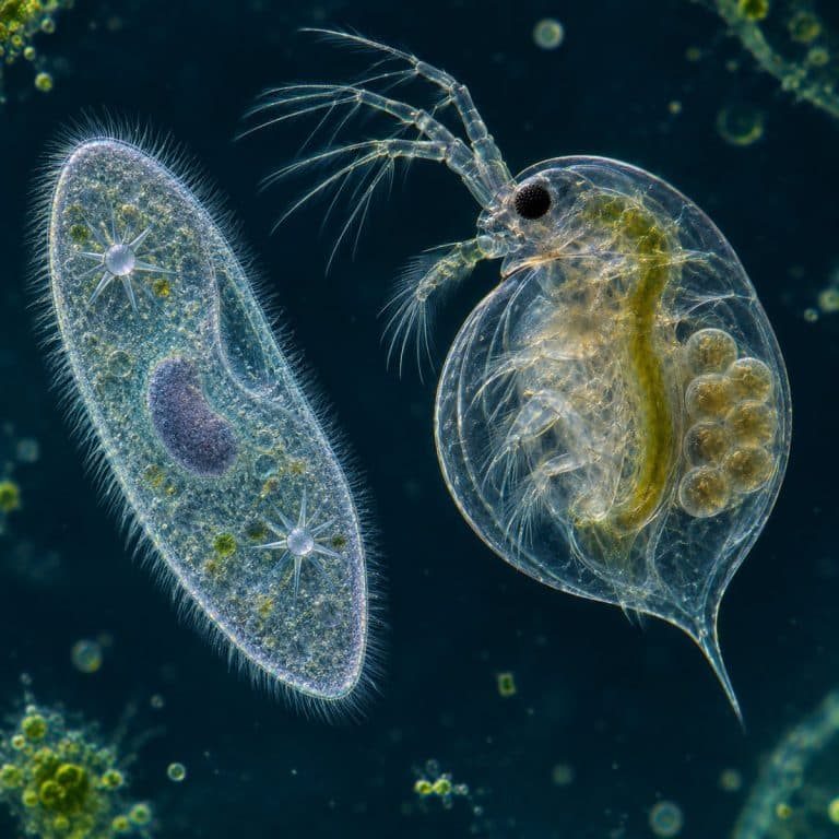
Unicellular vs multicellular organisms comes down to one question: how many cells share the work? A unicellular organism is a single cell that handles every life process on its own. A multicellular organism is built from many specialized cells that split those jobs and cannot survive apart. Yeast, amoeba, and paramecium are unicellular, Volvox sits on the border, and hydra, plants, and you are multicellular.
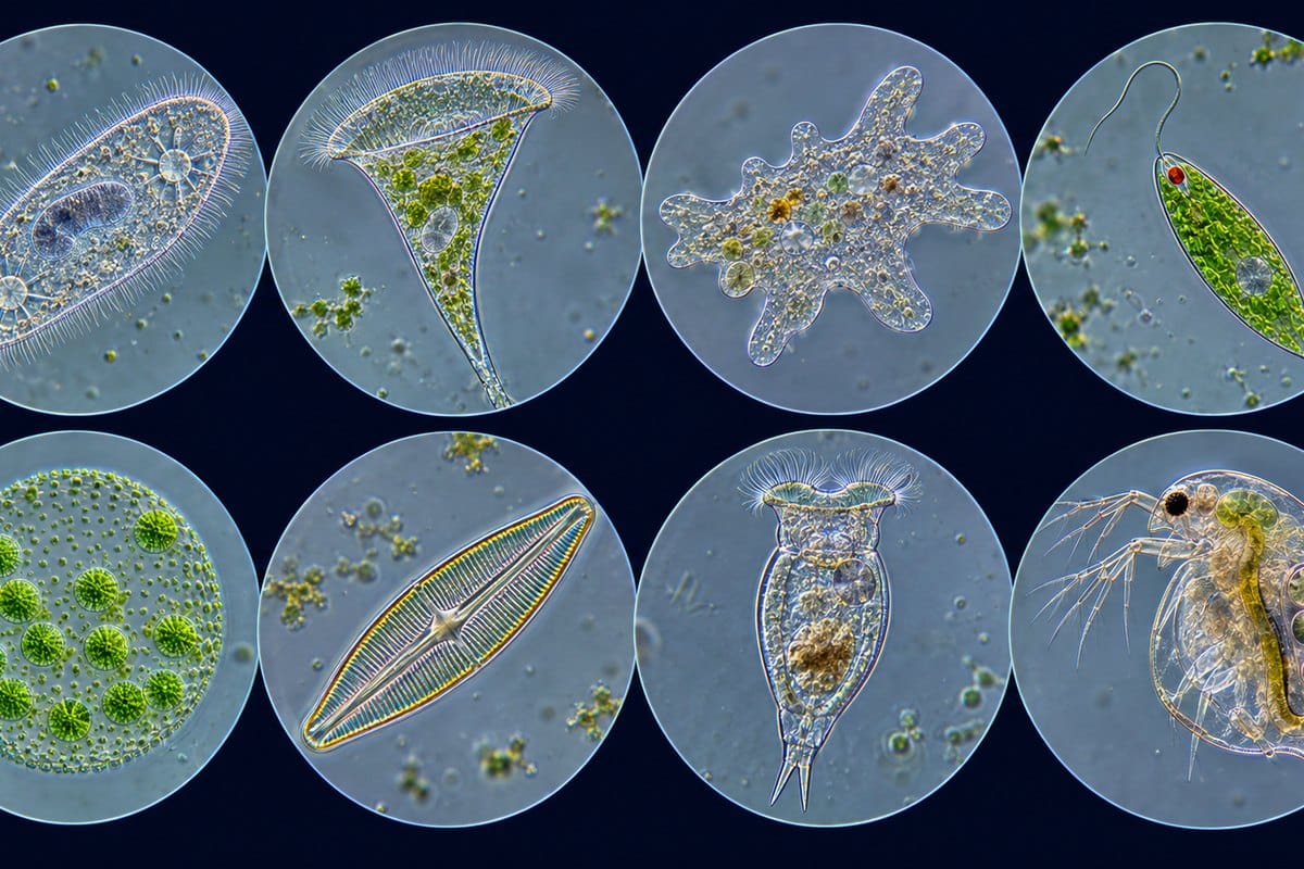
Key Differences: One Cell vs Many Specialized Cells
| Feature | Unicellular | Multicellular |
|---|---|---|
| Number of cells | One | Many: about 1,000 in a rotifer, roughly 30–37 trillion in a human |
| Specialization | None; one cell does everything | Cells differentiate into tissues and organs |
| Life processes | All inside one cell, using organelles | Shared among cells, tissues, and organ systems |
| Size | Mostly 1–700 µm; rare giants | Microscopic rotifers to a ~94 m giant sequoia |
| Surface area to volume | High; diffusion alone supplies the cell | Low for the whole body; needs blood or xylem and phloem |
| Reproduction | Mostly asexual (binary fission, budding); some conjugation | Mostly sexual via gametes; many also asexual (hydra budding) |
| Growth | The cell enlarges, then divides | More cells by mitosis, plus cell enlargement |
| Lifespan | Short per cell; division resets it | Usually longer; cells die and are replaced |
| Damage tolerance | Damage to the one cell can kill the organism | Losing some cells is survivable; many regenerate |
| Origin | First life, at least 3.5 billion years ago | Evolved later, independently at least 25 times |
Specialization is the difference that matters most. A paramecium swims, eats, and expels water with one cell, while your body assigns those jobs to muscle, gut, and kidney cells that die without each other. Size and reproduction follow from that split. A big body can’t feed itself by diffusion, so it needs transport systems, and a body of specialized cells usually reproduces through single-celled gametes. See OpenStax’s cell-organization overview for additional context.
How One Cell Does Every Job
A single-celled eukaryote runs its whole life with organelles instead of organs. A paramecium beats thousands of cilia to swim, sweeps food down an oral groove, and pumps out excess water through two star-shaped contractile vacuoles. Its large macronucleus runs everyday functions, and a small micronucleus handles reproduction and conjugation. An amoeba moves by extending pseudopodia (temporary arm-like bulges of cytoplasm), engulfs food by phagocytosis (wrapping around it), and uses its own contractile vacuole to pump out excess water. Learn what each part does in the organelles inside a eukaryotic cell. See OpenStax’s protist examples for additional context.
That complexity is why “unicellular” never means “simple,” and never means “prokaryote.” Amoeba, paramecium, and yeast all have a nucleus, which makes them eukaryotes. Bacteria and archaea are the unicellular prokaryotes, as explained in the difference between prokaryotes and eukaryotes.
What Changes When Cells Work Together
Multicellular organisms divide labor among differentiated cells. Cells with the same job group into tissues (animals have four basic types: epithelial, connective, muscle, and nervous), tissues form organs, and organs form organ systems. Plants use three tissue systems: dermal, vascular, and ground. Cells stay connected through adhesion and chemical signals, so the body acts as one unit.
Every sexually reproducing multicellular organism starts as a single cell, the zygote. Repeated mitosis and differentiation turn that one cell into a body, and the growing body needs blood or vascular tissue to move what diffusion can’t. For a close look at one specialized cell type, see specialized muscle tissue.
Unicellular and Multicellular Examples Across the Major Groups
| Group | Unicellular | Multicellular |
|---|---|---|
| Bacteria | E. coli, Salmonella, Streptococcus, single-cell cyanobacteria | None truly; filamentous Anabaena (with specialized heterocysts) and Myxococcus fruiting bodies are simple edge cases |
| Archaea | Halobacterium, methanogens | None |
| Protists | Amoeba proteus, Paramecium, Euglena, diatoms, Chlamydomonas | Kelp and seaweeds, Spirogyra filaments; Volvox is the colonial edge case |
| Fungi | Yeast (Saccharomyces cerevisiae) | Molds (Penicillium, Rhizopus), mushrooms |
| Plants | None | Mosses, ferns, onions, oaks, grasses |
| Animals | None | Hydra, rotifers, tardigrades, insects, humans |
Plants and animals are always multicellular, and bacteria and archaea are essentially always single-celled. Fungi split both ways: yeast is unicellular, while molds and mushrooms are multicellular. Some “yeasts,” such as Candida, can even switch to multicellular-looking hyphal (threadlike) forms, so yeast is unicellular in its everyday form, not by definition.

See the Difference Under Your Own Microscope
A basic compound microscope plus a stereo microscope (or your 4× objective) covers the whole range. It runs from a 5 µm yeast cell to a hydra you can see with your eyes. Doing all five in one session is the quickest way to understand this topic.
| Specimen | Type | Size | Start at | What proves its type |
|---|---|---|---|---|
| Yeast | Unicellular (fungus) | 3–10 µm | 400× | Buds on oval cells |
| Paramecium | Unicellular (protist) | 180–300 µm | 100× | Cilia, oral groove, contractile vacuoles |
| Amoeba | Unicellular (protist) | 220–750 µm | 100× | Pseudopodia, flowing cytoplasm |
| Volvox | Colonial or simple multicellular | 0.35–1 mm | 40× | Thousands of linked cells, daughter colonies |
| Hydra | Multicellular (animal) | 1–20 mm | 40× or stereo | Tentacles, buds, contraction |
- Collect. Take one drop each from the surface film, mid-water near plants, and bottom sediment of a pond jar. Samples look best within 24–48 hours; keep the lid loose for air. More collecting tips are in what lives in a drop of pond water.
- Mount. Use a wet mount for yeast, paramecium, and amoeba (see making a simple wet mount slide). Use a depression slide for Volvox and hydra, because a pressed cover slip crushes them.
- Start low. Begin at the “Start at” power in the table. Starting Volvox or hydra at 400× leaves you with a green blur or an empty field.
- Cut the light. Close the iris diaphragm and lower the condenser. Near-transparent cells wash out under bright light; microscope contrast methods explain why.
- Keep it wet. Wet mounts dry within 10–20 minutes under a warm lamp, and amoebae ball up and stop moving. Ring the cover slip with petroleum jelly or add a drop of water at the edge.
Yeast: Budding Cells at 400×
Yeast reproduces by budding, and a pinch of baker’s yeast in warm sugar water for 10–15 minutes gives you the most buds. At 400× you’ll see a field of clear-to-pale-gray ovals like scattered grains of rice, some with a smaller bubble stuck to one end. Add methylene blue: dead cells turn solid blue while live cells stay pale. Each cell is separate and complete, which is what unicellular means; how yeast cells look under a microscope shows more detail.
Amoeba and Paramecium: Single Cells That Hunt
An amoeba looks like a gray, grainy blob until you watch it for 30 seconds. Then granular cytoplasm pours forward into a bulging pseudopod like slow lava, and the shape never holds still. Amoebae are rare in clear water, so pipette from the bottom of a jar after it has settled for an hour, or sample the underside of decaying leaves (our guide to finding an amoeba under a microscope).

A paramecium is a translucent slipper zipping across the field and spinning on its long axis. At 400× you can see the shimmering fringe of cilia, the oral groove, and a contractile vacuole that fills and collapses every few seconds like a slow heartbeat. See watching a paramecium under the microscope for staining and focusing tips.
Volvox: A Colony on the Edge of Multicellularity
Volvox appears as bright green spheres rolling and tumbling through the water like tiny planets. At 40–100×, darker daughter spheres are visible inside the parent, and the surface resolves into a fine dot pattern of thousands of cells. Hold a jar up to the light first: pinhead green specks moving through the water are your cue. Volvox blooms in warm months, so collect from the sunlit surface layer (Volvox colonies under the microscope).
Hydra: Tissues You Can See With a Stereo Microscope
Hydra is a pale tan or green stalk (green when symbiotic algae live inside it) with 5–8 thread-like tentacles. Touch the jar and it contracts into a tiny ball, then slowly stretches back out. Buds appear as small side branches with their own mini-tentacles. Hydra cling to plant stems and jar walls instead of floating. Let the jar sit undisturbed for a day, then look for tiny threads hanging from the glass on the side facing the light. The two tissue layers and stinging cells make this the clearest example of tissue-level specialization a hobbyist can see (observing hydra under a microscope).
The Gray Zone: Colonies, Slime Molds, and Giant Single Cells
Volvox is the classic gray-zone case. A Volvox carteri colony has about 2,000 small somatic (body) cells that swim and sense light but cannot reproduce, plus about 16 large gonidia (reproductive cells) that do the reproducing. The somatic cells die when the parent releases its daughters. Because that is division of labor, many biologists count Volvox as the simplest true multicellular organism, while others call it colonial. Compare Gonium, a colony of 4–32 identical cells that each could live alone: that is colonial, not multicellular.
Other organisms blur the line from different directions:
- Slime mold (Dictyostelium discoideum): lives as separate amoebae until food runs out, then about 100,000 cells aggregate into a multicellular slug and fruiting body.
- Choanoflagellates: single-celled (sometimes colonial) protists and the closest living relatives of animals, hinting at how animal multicellularity began.
- Giant single cells: the bacterium Thiomargarita magnifica is one cell about 1 cm long, visible to the naked eye.
- The size paradox: a rotifer has about 1,000 cells and measures 100–500 µm, so it can be smaller than Amoeba proteus, a single cell reaching about 700 µm (rotifers under a microscope).
Cell count and size can’t settle the question. The real test is specialization and interdependence.
Common Misconceptions About Single-Celled and Multicellular Life
- “Unicellular means prokaryote.” False. Amoeba, paramecium, Euglena, and yeast are single-celled eukaryotes.
- “Single-celled means microscopic.” Not always. Caulerpa, an alga, is one multinucleate (many-nuclei) cell that can stretch for meters.
- “Multicellular means big.” The nematode C. elegans has 959 somatic cells as an adult and is about 1 mm long.
- “Unicellular means simple.” A paramecium has a feeding groove, an excretory system, two kinds of nucleus, and a form of sexual exchange.
Why Multicellularity Evolved More Than 25 Times
Multicellularity has evolved independently at least 25 times, according to Grosberg and Strathmann’s review. Biologists propose that larger size, escape from small predators, and division of labor each gave groups of cells an edge. Complex multicellularity, with many cell types and tissues, arose in only about six lineages: animals, land plants, red algae, brown algae, and fungi (twice).
Animals trace back to unicellular ancestors, which is why choanoflagellates matter so much to this story. Plant biologist Karl Niklas reviews the developmental side of this transition in his paper on the origins of multicellularity.
Frequently Asked Questions
Are viruses unicellular?
No. Viruses aren’t cells at all. They have no organelles and can’t carry out life processes or reproduce without a host cell, so they are neither unicellular nor multicellular.
How many cells are in the human body?
About 30–37 trillion. One 2016 PLoS Biology estimate put human cells at roughly 30 trillion, while a 2013 estimate reached about 37.2 trillion. Those cells fall into roughly 200 classic types, and single-cell atlases now list several hundred.
Which came first, unicellular or multicellular life?
Unicellular life. The earliest life dates to at least 3.5 billion years ago, while the oldest widely accepted multicellular eukaryote fossil, the red alga Bangiomorpha pubescens, is about 1.05 billion years old. Animals appear roughly 600 million years ago or later.
Can you see a single-celled organism without a microscope?
A few, yes. Amoeba proteus shows up as a whitish speck, and the “sailor’s eyeball” alga Valonia ventricosa reaches 1–4 cm. Most single cells, including yeast and paramecium, are too small to resolve by eye.
Why does my paramecium swim out of view before I can focus?
Paramecium swims about 1 mm per second, which is fast at 100×. Add a wisp of cotton fibers to the drop, or a drop of methyl cellulose such as Protoslo, to slow it down.
Should I wash my hands after handling pond water?
Yes. It’s good practice after any pond sample. Rinse your pipettes and slides as well, and keep samples away from food.
Conclusion
The test for unicellular vs multicellular is not size or cell count but whether cells specialize and depend on each other. Yeast, amoeba, and paramecium run a whole life in one cell, hydra and humans share the work across tissues, and Volvox shows where that line blurs.
Have you tried this yourself? Tell us which specimen surprised you most, or where your pond samples came from, in the comments below.