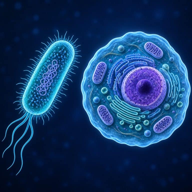
Prokaryotic and eukaryotic cells split every living thing on Earth into one of two camps, and the split comes down to a single structure. Eukaryotic cells keep their DNA sealed inside a membrane-bound nucleus; prokaryotic cells don’t, leaving their genetic material loose in the cytoplasm. Everything else that separates the two — size, organelles, DNA shape, how the cell divides — follows from that one distinction.
The Core Difference: A Nucleus or Not
A prokaryotic cell lacks a membrane-bound nucleus. Its DNA sits free in a region of the cytoplasm called the nucleoid — not enclosed by any membrane, just concentrated in one area. A eukaryotic cell contains its DNA inside a true nucleus, wrapped in a double-membrane nuclear envelope that separates it from the rest of the cell. The names tell the story: “eukaryote” comes from Greek eu (“true”) and karyon (“kernel,” referring to the nucleus), while “prokaryote” uses pro (“before”). Every other difference between these two cell types is downstream of this one architectural choice. For a concise side-by-side reference, see OpenStax’s comparison of prokaryotic and eukaryotic cells.
Prokaryotes vs Eukaryotes: Comparison Table
The table below lines up the seven differences that matter most, side by side.
| Feature | Prokaryotic Cells | Eukaryotic Cells |
|---|---|---|
| Nucleus | None — DNA sits in the nucleoid | True, membrane-bound nucleus |
| Membrane-bound organelles | Absent | Present (mitochondria, ER, Golgi, etc.) |
| Cell size | 0.1–5 µm | 10–100 µm |
| DNA structure | Single circular chromosome, plus plasmids | Multiple linear chromosomes wrapped around histones |
| Ribosomes | 70S | 80S |
| Reproduction | Binary fission | Mitosis (meiosis for gametes) |
| Examples | Bacteria, archaea | Plants, animals, fungi, protists |
Cell Size — Why Eukaryotes Are So Much Bigger
Most prokaryotic cells measure just 0.1 to 5 micrometers (µm) across, with typical bacteria landing around 1 to 2 µm. Eukaryotic cells run 10 to 100 µm — roughly ten times the diameter of a typical bacterium. Once you account for three dimensions, that works out to somewhere between 1,000 and 10,000 times the volume. That size gap isn’t incidental — a cell without internal compartments can only grow so large before diffusion alone can’t move nutrients and waste fast enough to keep it alive. Membrane-bound organelles let eukaryotic cells get away with being bigger by subdividing the workload.
Genetic Material — Circular vs Linear DNA
Prokaryotes absolutely have DNA — the idea that they don’t is one of the most common mix-ups in intro biology. It’s just organized differently. A prokaryotic cell’s main genome is typically one circular chromosome sitting in the nucleoid, often with smaller circular DNA rings called plasmids floating alongside it; that DNA generally isn’t wrapped around histone proteins. A eukaryotic cell’s DNA is split into multiple linear chromosomes, each one spooled around histone proteins and packaged as chromatin inside the nucleus. Bacteria and archaea (the two prokaryotic domains) sit alongside domain Eukarya — protists, fungi, plants, and animals. For a straightforward classroom contrast, “bacteria vs. anything with a nucleus” covers most of what you’ll encounter, as Britannica’s overview of the domain system lays out in more depth.
Organelles and Internal Structure
Prokaryotic cells have no membrane-bound organelles — no mitochondria, no endoplasmic reticulum, no Golgi apparatus, no chloroplasts, no lysosomes. Eukaryotic cells have all of these, each one doing a specialized job behind its own membrane. Both cell types do have ribosomes, though — ribosomes aren’t membrane-bound, so they don’t count as organelles in this comparison, and every living cell needs them to build proteins. The ribosomes themselves differ: prokaryotic ribosomes are the smaller 70S type, while eukaryotic cytoplasmic ribosomes are the larger 80S type. That structural gap is exactly why certain antibiotics can shut down a bacterium’s protein production without touching your own cells. The mechanism is explained in more detail in NCBI’s molecular biology reference on ribosome structure. It’s also the clue behind one of biology’s best origin stories. Mitochondria and chloroplasts carry their own circular DNA and their own 70S ribosomes because they descend from free-living prokaryotes that an ancestral eukaryotic cell engulfed and never let go. That relationship is known as endosymbiotic theory, summarized well in Khan Academy’s lesson on endosymbiosis. OpenStax also outlines the organelles that distinguish eukaryotic cells.
How They Reproduce
Prokaryotic cells reproduce by binary fission: the single circular chromosome copies itself, the cell elongates, and it splits into two identical daughter cells. There’s no spindle apparatus involved, and the whole process can take as little as 20 minutes under good conditions — one reason bacterial cultures grow so fast. Eukaryotic cells divide by mitosis, a multi-stage process that uses a spindle apparatus to separate duplicated chromosomes evenly between two daughter cells. Eukaryotic cells that produce sex cells (sperm and eggs, or their equivalents) use a related process called meiosis, which halves the chromosome count so the two parents’ genetic material can combine.

Examples of Each
Prokaryotes are almost always single-celled organisms. E. coli, a rod-shaped prokaryote you can stain and view at 1000x, is the textbook bacterial example, alongside Salmonella, another Gram-negative bacterium. Both are classification examples here, not a health warning — though the CDC’s E. coli overview is worth a look if you want the full picture on the genus. Cyanobacteria (photosynthetic bacteria) and the various archaea round out the prokaryotic side.
Eukaryotes cover a much wider range, from single cells to entire multicellular organisms — and that’s a common trap: eukaryote does not mean “big organism.” A single amoeba is a eukaryote, and so is a single yeast cell. On the plant side, onion epidermal cells, a classic plant (eukaryotic) specimen, show off a cell wall and a visible nucleus at low power. On the animal side, your own cheek cells, a textbook eukaryotic animal example, work just as well with a cotton swab and a stain. Among single-celled eukaryotes, four organisms make easy pond-water finds that put a face on the category: yeast, a single-celled eukaryote (a fungus), the amoeba, a shape-shifting eukaryotic protist, Paramecium, a ciliated eukaryotic cell, and Euglena, a eukaryote that blurs the plant/animal line.
Can You See the Difference Under a Microscope?
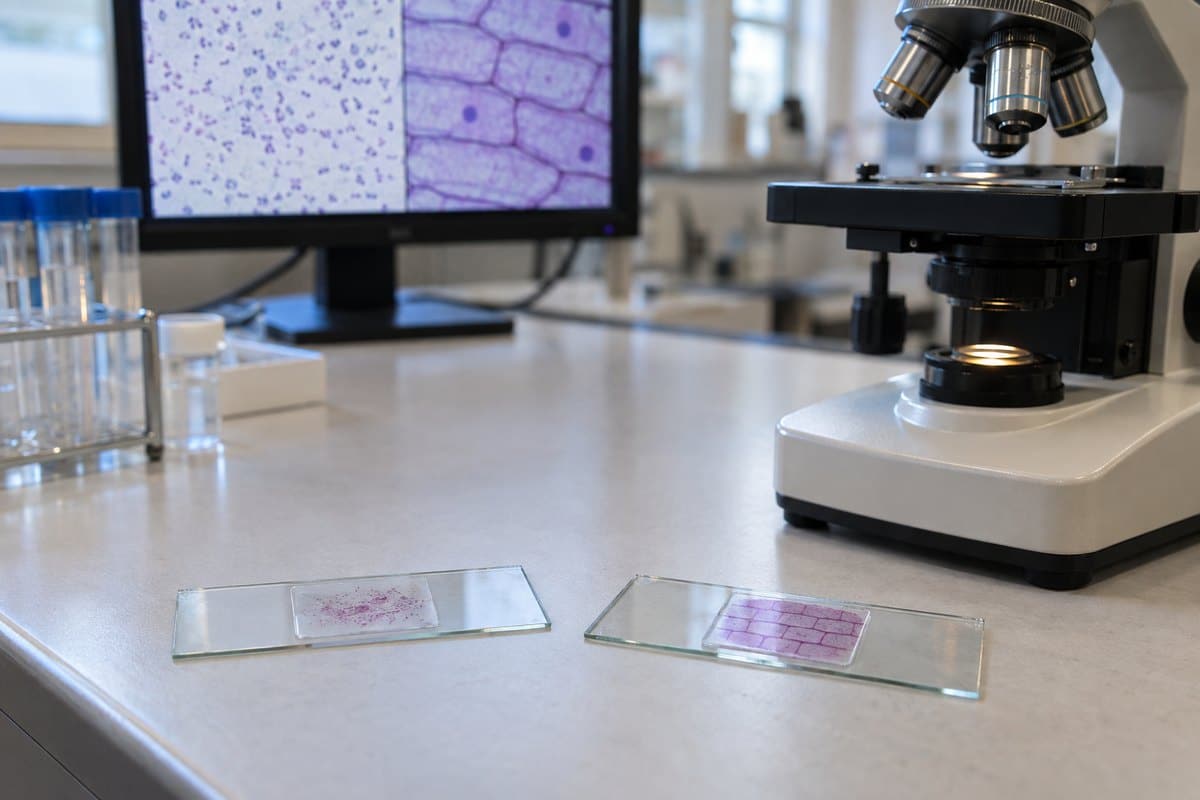
At 400x on a standard school light microscope, bacteria look like tiny colorless or faintly stained dots and short rods, drifting or vibrating in place. They resemble grains of pepper scattered across the field, with no visible internal parts at all. Put a stained eukaryotic cheek cell in the same field at the same power and the contrast is immediate: a big, flat, fried-egg shape with a clearly darker dot — the nucleus — sitting near its center. Onion cells read like a brick wall of neat rectangular boxes, each one often showing its own visible nucleus. That size gap is the moment it clicks: a bacterium is a speck next to a plant cell that fills half the field of view.

The most common beginner mistake is hunting for a nucleus or organelles inside a bacterium — you won’t find one, because prokaryotic internal structure sits at or below the resolution limit of a light microscope. The fix is simple: don’t look for what isn’t there. A second mistake is cranking the magnification to chase bacteria and losing them entirely. They need staining plus oil immersion at 1000x to show up reliably. At low power, they’re nearly invisible against the background — easy to mistake for debris or an air bubble. A stain like methylene blue or a Gram stain is really what makes the prokaryote/eukaryote difference visible on a slide — the presence or absence of a stained, dark nucleus is the fastest visual tell between the two. If you’re prepping your own slide, prepare a slide to view these cells yourself. One honest limitation worth knowing up front: a light microscope is enough to confirm a eukaryotic nucleus, but it can’t resolve a prokaryote’s internal structure. For that level of detail, you need electron microscopy, which resolves prokaryotic detail a light scope can’t.
Frequently Asked Questions
Are humans prokaryotes or eukaryotes?
Humans are eukaryotes. Every cell in your body — skin, muscle, blood, nerve — has a membrane-bound nucleus, which is the defining trait of a eukaryotic cell.
Which came first, prokaryotes or eukaryotes?
Prokaryotes came first, by a wide margin. The fossil record puts the earliest prokaryotic life at roughly 3.5 billion years ago, while eukaryotic cells didn’t appear until around 1.5 to 2 billion years ago — likely arising when one prokaryote engulfed another, the event behind endosymbiotic theory.
Do prokaryotes have mitochondria?
No. Mitochondria are membrane-bound organelles, and prokaryotic cells have none. Prokaryotes still generate energy — just using their cell membrane and cytoplasm directly, without a dedicated organelle to do it.
Do all eukaryotic cells have a cell wall?
No. Plant cells (cellulose) and fungal cells (chitin) have cell walls, but animal cells do not. Most prokaryotes, by contrast, do have a cell wall, typically built from peptidoglycan.
Can a prokaryotic organism be multicellular?
Essentially no — prokaryotes are unicellular. Some bacteria form colonies or filaments that look multicellular, but each cell in the group still functions independently, unlike the coordinated, specialized cells of a true multicellular eukaryote.
Are viruses prokaryotes or eukaryotes?
Neither. Viruses aren’t classified as cells at all — they have no nucleus, no cytoplasm, and no ribosomes of their own, and they can only reproduce by hijacking a host cell’s machinery.
Why does having a nucleus matter so much?
Enclosing DNA in a nucleus lets a eukaryotic cell separate the jobs of copying genetic instructions from reading them, and it protects the genome while making room for the more complex, compartmentalized machinery that supports a much larger, more specialized cell.
Conclusion
Every difference in this comparison — size, organelles, DNA shape, how the cell divides — traces back to one structure: whether the cell keeps its DNA inside a membrane-bound nucleus. Once that single fact is locked in, the rest of the comparison table follows almost automatically. It’s the concept that sits underneath every specimen you’ll ever put on a slide, from a rod of E. coli to a sheet of onion cells.
Have you looked at both cell types side by side under your own microscope? Tell us what you noticed — the size difference, the visible nucleus, anything that surprised you — in the comments below.