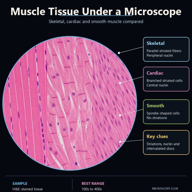
Muscle tissue under a microscope reveals its identity through four telltale features: striations, nuclei count and position, cell shape, and intercalated discs. Skeletal muscle is long, cylindrical, striated, and packed with nuclei pushed to the cell’s edge. Cardiac muscle is branched and striated too, but its cells carry dark connecting lines called intercalated discs. Smooth muscle has no striations at all, just spindle-shaped cells with a single nucleus in the middle. Once you know what to check, and in what order, telling the three apart on a stained slide takes seconds.
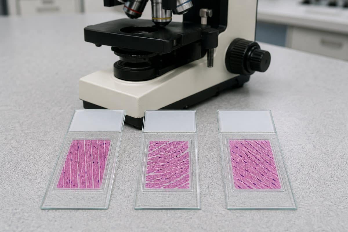
The 4 Features That Separate the Three Muscle Types
Four visible features do all the work of separating skeletal, cardiac, and smooth muscle on a prepared slide. Check whether the cell is striated, how many nuclei it has and where they sit, what shape the cell is, and whether intercalated discs are present. Every one of these shows up clearly on a standard hematoxylin and eosin (H&E) stained slide viewed at 100x–400x. That’s how muscle tissue is studied in almost every classroom and lab. Under H&E, nuclei stain purple-blue and the surrounding cytoplasm stains pink. So the four features stand out as color and shape contrasts rather than subtle details you have to hunt for.
The table below lines up all three types under that same stain and magnification context. Most references describe skeletal, cardiac, and smooth muscle in separate sections and leave the reader to build the comparison themselves.
| Feature | Skeletal Muscle | Cardiac Muscle | Smooth Muscle |
|---|---|---|---|
| Striations | Yes, clear and regular | Yes, same banding as skeletal | No |
| Nuclei (number & position) | Many, at the periphery | Usually one, central | One, central |
| Cell shape | Long, cylindrical, unbranched | Short, branched | Spindle-shaped (fusiform) |
| Intercalated discs | Absent | Present (the giveaway) | Absent |
| Voluntary / involuntary | Voluntary | Involuntary | Involuntary |
| Where found | Skeleton, tongue, diaphragm, extraocular muscles | Myocardium (heart wall) only | Walls of hollow organs: gut, vessels, uterus, bladder, airways |
Skeletal Muscle Under the Microscope
Skeletal muscle fibers run as long, unbranched cylinders, some stretching several centimeters despite being only 10–100 micrometers across. In a longitudinal section — cut along the length of the fiber — the fibers look like long pink ribbons lying in parallel. Each is crossed by faint, regular cross-lines that resemble a barcode. Those lines are striations, produced by the ordered stacking of actin and myosin filaments into repeating units called sarcomeres, alternating dark A-bands with lighter I-bands. The nuclei are small, dark purple, and sit flattened against the outer edge of the fiber rather than floating in the middle. A fiber can carry dozens of these peripheral nuclei because it forms from the fusion of many individual muscle precursor cells during development.
Cut the same tissue in cross-section and the picture changes completely. Instead of ribbons, you see a tile floor of tightly packed pink polygons, each rimmed with one or two purple dots pressed against the outer wall. No striations are visible here, because you’re looking down the length of the fiber instead of along it — a common point of confusion for anyone new to the slide. Skeletal muscle is the only one of the three types under voluntary, somatic nervous control. It’s what you’re looking at in tissue from the limbs, trunk, tongue, diaphragm, or the muscles that move the eyes.
Cardiac Muscle Under the Microscope
Cardiac muscle cells, or cardiomyocytes, are shorter and stockier than skeletal fibers. Instead of running as straight parallel lines, they fork and rejoin like a branching river seen from above. Each cell carries a single nucleus — occasionally two — sitting in the center rather than pushed to the edge. That’s the first clue that separates cardiac from skeletal muscle, even before you look for anything else. Cardiac muscle is striated, built from the same actin-myosin sarcomere arrangement as skeletal muscle, so striations by themselves cannot tell the two apart. A beginner’s most common mistake is calling any striated tissue “skeletal” without checking further.
What separates cardiac muscle from every other tissue in the body is the intercalated disc. It’s a dark, often step-like or staircase-shaped line crossing the fiber wherever two cardiomyocytes meet end to end. At low power these can look like just another striation, so switch to the 40x objective (about 400x total magnification) and ease back the condenser light slightly. The thin dark lines pop into contrast once you do. Structurally, intercalated discs pack gap junctions for electrical coupling between cells and desmosomes for mechanical anchoring. That’s exactly how the heart contracts as one coordinated unit rather than as loose individual cells. Cardiac muscle is involuntary and, unlike skeletal or smooth muscle, is found in exactly one place in the body: the wall of the heart, the myocardium.

Smooth Muscle Under the Microscope
Smooth muscle cells are spindle-shaped, or fusiform — wide in the middle and tapering to a point at each end — and they show no striations whatsoever. Actin and myosin are present in the cytoplasm, but they aren’t organized into the regular sarcomere repeats that create banding. That’s why the cytoplasm looks smooth and uniformly pink under H&E stain — the origin of the tissue’s name. Each cell has a single nucleus, elongated and oval, often described as cigar-shaped or football-shaped. If the cell happens to be contracted at the moment it was fixed, that nucleus can look wavy or corkscrewed rather than straight.
Smooth muscle sheets show a distinctive tell in cross-section that skeletal and cardiac muscle don’t. Because each spindle-shaped cell’s nucleus sits only in its widest middle section, a cut through the tapered end of one cell shows plain pink cytoplasm with no nucleus at all. Scan a field of smooth muscle in cross-section and you’ll see round or oval profiles of varying size, and only some of them contain a visible purple nucleus. That “only some circles have a dot” pattern is one of the fastest ways to confirm smooth muscle at a glance. It’s worth not confusing these cells with fibroblasts in nearby connective tissue: smooth muscle nuclei are plumper and more cigar-shaped, while fibroblast nuclei are thinner and flatter. Smooth muscle is involuntary and lines the walls of hollow organs — the gastrointestinal tract, blood vessels, uterus, bladder, and airways.
How to Identify Muscle Tissue Under a Microscope (Step by Step)
With an unknown slide in front of you, work through the four features in this order rather than trying to take in everything at once:

- Check for striations. No striations at all means smooth muscle — stop here. If you do see banding, move to step 2. (Make sure you’re looking at a longitudinal section first; striations only show along the length of a fiber, not in cross-section.)
- Look for branching and intercalated discs. If the striated cells are branched and you can find dark, step-like intercalated discs crossing between cells, it’s cardiac muscle.
- Confirm with nucleus position. Unbranched, striated fibers with many nuclei pressed against the outer edge confirm skeletal muscle; branched, striated cells with one central nucleus confirm cardiac muscle.
This decision order matters because the reverse approach — starting with nuclei instead of striations — is slower and easier to get wrong on a busy field of view.
How to View Muscle Tissue Yourself (Slides, Stain, Magnification)
Muscle tissue is essentially invisible without preparation. Fresh or unstained tissue gives almost no contrast between structures, so what you’re actually examining is a thin, prepared, sectioned slide stained with hematoxylin and eosin. Hematoxylin binds to the nuclei and stains them purple-blue; eosin stains the surrounding cytoplasm pink. Start scanning at 100x to find a region with clearly parallel, undamaged fibers, then switch to 400x (a 40x objective) to bring out striations and intercalated discs. Closing the iris diaphragm slightly as you go increases contrast and makes both features noticeably easier to see. If you’re building your own slide collection, our guides to preparing microscope slides and calculating total magnification cover the groundwork. Our breakdown of objective lenses from 4x to 100x explains exactly which lens gets you to the 400x range these features need. For a broader look at how tissue architecture reads under the stain, our guide to bone tissue under a microscope walks through another comparison using the same H&E approach.
Seeing the three tissue types in motion helps the static comparison above click into place. This short video walks through skeletal, cardiac, and smooth muscle as they actually appear down the eyepiece:
For deeper background on the underlying cell biology, the NCBI Bookshelf’s StatPearls entry on muscle histology is a solid, citation-grade reference. Kenhub’s overview of muscle cell types pairs labeled diagrams with the same three-way comparison. If you want more annotated slide images to practice on, George Washington University’s histology education blog on muscle tissue is a useful free resource.
Frequently Asked Questions
What stain is used to see muscle tissue under a microscope?
Almost all prepared muscle slides use hematoxylin and eosin (H&E): hematoxylin stains nuclei purple-blue, and eosin stains the surrounding cytoplasm pink.
What magnification do you need to see striations or intercalated discs?
Start at 100x to orient yourself, then move to around 400x (a 40x objective) — that’s the magnification where striations and intercalated discs become clearly visible.
Can you see muscle cells with a home or school microscope?
Yes, if you’re using a prepared, stained slide. Fresh or unstained muscle tissue shows almost no contrast and won’t reveal striations, nuclei position, or intercalated discs.
Is cardiac muscle voluntary or involuntary?
Involuntary. It contracts under autonomic and intrinsic control, unlike skeletal muscle, which is under voluntary, somatic control.
Why does skeletal muscle have more than one nucleus per cell?
Each mature skeletal muscle fiber forms from the fusion of many individual myoblasts during development. It ends up with all of their nuclei, pushed to the periphery of the shared cell.
Why can’t I find intercalated discs in a cross-section?
Intercalated discs run across the fiber where two cardiac cells join end to end, so they only show up in a longitudinal section. A cross-section cuts through the middle of cells instead of their junctions, so the discs won’t be visible no matter how carefully you focus.
How do you tell smooth muscle apart from fibroblasts in connective tissue?
Smooth muscle nuclei are plump and cigar-shaped, and the cells form organized sheets or bundles. Fibroblast nuclei, by contrast, are thinner and flatter, sitting scattered through the surrounding connective tissue rather than in dense muscular sheets.
Conclusion
Four features carry the entire identification. Striations rule smooth muscle in or out immediately, branching and intercalated discs single out cardiac muscle, and nucleus count and position settle the remaining call between skeletal and cardiac. Working through them in that order turns what looks like three similar shades of pink tissue into an obvious, confident identification within a minute or two on the microscope.
Have you compared these three tissue types side by side on your own slides yet? Tell us what stood out first — the intercalated discs, the branching, or the peripheral nuclei — in the comments below.