
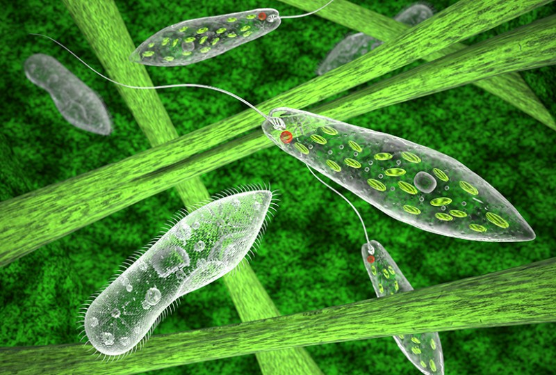
Euglena is one of the most visually striking microorganisms in any pond-water sample — a bright green, spindle-shaped protist that photosynthesizes like a plant yet swims like an animal. Its vivid chloroplasts, a distinctive orange-red eyespot, and two entirely different locomotion modes make it identifiable under a microscope even before you’ve studied the biology. This guide covers what you’ll see at each magnification, how to prepare your sample, and how to distinguish Euglena from every other common pond protist.
Shape, Size, and Color: Your First Look
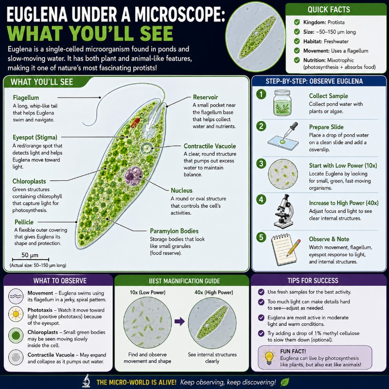
When you view Euglena under a microscope, you’ll see a bright green, elongated spindle- or teardrop-shaped cell roughly 50–100 micrometers long — small enough that you’d fit several side by side on a human hair. It’s one of the most visually distinctive microorganisms you’ll encounter in pond water. The vivid grass-green color (from real chlorophyll), a small but striking orange-red eyespot near the front end, and a swimming motion ranging from smooth gliding to an unmistakable accordion-like squirm set it apart from every other common protist. No other common pond protist combines all three of those features.

The table below summarizes what becomes visible at each magnification tier:
| Magnification | Objective | What You Can See |
|---|---|---|
| 100x | 10x | Green spindle shape, swimming movement, general cell outline |
| 400x | 40x | Eyespot (red dot), chloroplasts, euglenoid flexing, flagellar motion |
| 1000x | 100x oil | Pellicle striations, contractile vacuole, nucleus (with stain), paramylon granules |
How Much Magnification Do You Need to See Euglena?
You can spot Euglena with as little as 40x total magnification if your sample is dense, but you won’t resolve any meaningful detail at that power. Here’s what each tier actually gives you:
At 100x — Finding and Identifying Cells
At 100x (10x eyepiece × 10x objective), individual Euglena cells are visible as small green dashes moving across the field. You can confirm the color and see the elongated body shape, and the swimming motion is obvious. Internal structures like the eyespot are present but not clearly resolvable. This magnification is ideal for scanning a pond-water sample and confirming you have Euglena before switching to higher power. To understand how 40x and 100x objective lenses compare, or if you need to calculate total magnification for your setup, we have guides on both.
At 400x — The Best Working Magnification
Four-hundred times is the sweet spot for Euglena. At this power you can clearly resolve the red eyespot near the anterior end, the arrangement of green chloroplasts through the cell body, the flagellum’s movement effect (even if the flagellum itself is difficult to see), and — most rewarding — the characteristic body-flexing known as euglenoid movement. This is the magnification most biology labs and hobbyists use for ID and behavioral observation.

At 1000x — Fine Internal Detail
Oil-immersion at 1000x pushes the limits of what a standard light microscope can show. You may be able to make out faint spiral striations on the pellicle, the contractile vacuole near the anterior reservoir as it fills and collapses, and refractile paramylon granules (the cell’s carbohydrate storage, which look like colorless rings or lumps). The nucleus is usually only resolvable once the cell is stained — it then appears as a denser, rounded body in the central-to-posterior region. In an unstained live cell at 1000x you may at best glimpse a faint denser zone, but clear nucleus detail requires a stain. The catch: living cells are almost impossible to keep in frame at this power without immobilizing agents. Observe motility first at 400x, then prepare a stained slide if you want to study fine structure.
Key Structures You Can See
Chloroplasts and the Green Color
Euglena‘s vivid green color comes from its chloroplasts, which contain both chlorophyll a and chlorophyll b — the same pigments found in land plants. Under a bright-field microscope the color is genuine and unmistakable; you don’t need any staining to see it. The chloroplasts appear as green disc-shaped or ribbon-like bodies scattered throughout the cell. In Euglena gracilis the chloroplasts tend to have a star-like arrangement around a central pyrenoid, though this detail requires 400x or higher to appreciate. The presence of chloroplasts is one of the fastest visual IDs: if your cell is green, it’s not Paramecium or Amoeba.

Euglena is a mixotroph — in bright light it photosynthesizes like a plant; in darkness it can absorb dissolved organic nutrients heterotrophically. This dual lifestyle is what makes it the classic “neither plant nor animal” teaching example (more on that below).
The Eyespot (Stigma) — What the Red Spot Really Is
The small orange-red dot near the front of the cell is called the stigma or eyespot, and it’s one of the most eye-catching features at 400x. A common misconception is that it “sees” — it doesn’t. The eyespot is a shading device made of carotenoid pigment granules. Its job is to intermittently block light from reaching the paraflagellar body, a light-sensitive swelling at the base of the flagellum that is the actual photoreceptor. As the cell rotates while swimming, this light/shadow cycling tells the cell which direction light is coming from, enabling phototaxis — the ability to swim toward optimal light for photosynthesis.
At 400x the eyespot appears as a crisp, dense red-orange dot, usually near the anterior (flagellar) end. Its contrast against the green background makes it one of the clearest distinguishing markers for Euglena in a mixed pond-water sample.

The Flagellum (and Why It’s Hard to See)
Euglena technically has two flagella, but only one emerges from the anterior reservoir and is long enough to use for swimming. The emergent flagellum beats in a pulling, whip-like motion and is responsible for the smooth forward-rotating swim. Despite being the cell’s main engine, it’s surprisingly hard to see in standard bright-field because it is extremely thin and nearly transparent.
Tips for making the flagellum visible:
- Reduce the condenser aperture: Partially closing the iris diaphragm increases contrast on transparent structures.
- Use dark-field illumination: Dark-field microscopy makes the flagellum glow against a black background and is the most effective way to see it in living cells.
- Slow the cells: Add a drop of methylcellulose or Proto-Slo to reduce swimming speed without killing the cell.
- Stain: Iodine or methylene blue will reveal the flagellum but typically immobilizes or kills the cell — do this after observing living movement.
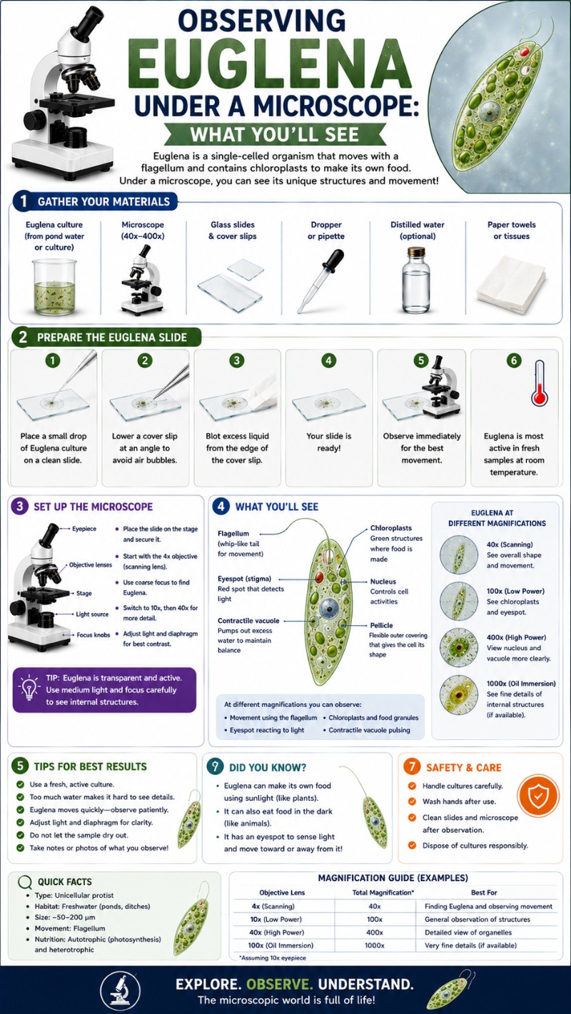
Pellicle, Contractile Vacuole, and Paramylon Granules
Instead of a rigid cell wall, Euglena has a pellicle — a flexible protein-strip layer just beneath the cell membrane. The pellicle gives the cell a defined shape while still allowing it to flex and contort. At 1000x you may be able to see faint spiral striations on its surface, though this detail is at the limit of standard bright-field optics.
The contractile vacuole sits near the anterior reservoir and acts like a bilge pump, collecting excess water and periodically contracting to expel it (osmoregulation). Watch for a clear bubble that expands and then suddenly collapses — that’s it. Best observed at 400–1000x in a living cell.
Euglena stores carbohydrates not as starch but as paramylon, a β-1,3-glucan unique to euglenoids. Paramylon granules appear as colorless, refractile lumps or rings scattered through the cytoplasm, distinct from the green chloroplasts. This biochemical difference from plants is a reliable identification and exam point.
How Euglena Moves — Flagellar Swimming vs. Euglenoid Movement
Euglena uses two completely different locomotion styles, and being able to distinguish them is one of the best diagnostic tests for identifying it in a sample.
Flagellar swimming is the default mode: the emergent flagellum beats in a helical pattern, pulling the cell forward while rotating it around its long axis. The result is smooth, directed movement — the cell travels purposefully across the field, often toward the light source if your microscope lamp is positioned to one side. Under the microscope this looks like a small green torpedo gliding steadily in one direction, sometimes briefly reversing.
Euglenoid movement (also called metaboly) is entirely different and immediately recognizable once you’ve seen it. When Euglena is attached to a surface, moving through a tight space, or simply “choosing” to, it shifts into a slow, peristaltic contraction-and-extension of the whole cell body. The cell bunches up at one end, then elongates, then bunches again — like squeezing a tube of toothpaste from one end. This is only possible because the pellicle is flexible rather than rigid. Paramecium and Amoeba, which lack this pellicle structure, cannot do it. Spotting metaboly is close to a definitive field ID for Euglena.
In practice, you’ll often see a single cell alternate between both modes: gliding smoothly, then slowing to a crawl and squirming, then resuming swimming.
Euglena vs. Paramecium vs. Amoeba
These three organisms frequently co-exist in pond-water samples and are the most common source of student confusion. The table below gives the fastest visual tells:
| Feature | Euglena | Paramecium | Amoeba |
|---|---|---|---|
| Color | Bright green | Colorless / grayish | Colorless |
| Shape | Spindle / teardrop | Slipper (fixed) | Irregular, no fixed shape |
| Typical size | 50–100 µm | 100–330 µm | 250–750 µm |
| Locomotion | Flagellum + metaboly | Cilia (fast, directional) | Pseudopodia (slow crawl) |
| Eyespot | Yes (red/orange dot) | No | No |
| Chloroplasts | Yes (green) | No | No |
| Distinguishing tell | Green + red dot + metaboly | Ciliated edge, oral groove, fast | Flowing pseudopods |
In a mixed sample at 100x, the quickest rule is: green cell = Euglena; large grayish fast-mover with fuzzy edges = Paramecium; slow colorless blob = Amoeba. Switch to 400x to confirm the eyespot on your Euglena candidate. You may also encounter Volvox, another green pond protist, but Volvox forms large spherical colonies made of hundreds of cells — easy to distinguish from a solitary Euglena once you know what to look for.
How to Find and Prepare a Euglena Sample
Where to Collect
Euglena thrives in still, organically rich freshwater — ponds, ditches, slow streams, birdbaths, and rain barrels are all good sources. Because Euglena is phototactic, it migrates toward the surface in daylight. The best samples come from the top centimeter of water, especially in areas where the water has a greenish tint — that bloom is often a massive population of Euglena or related algae. Scoop your sample with a dropper or pipette and transfer it to a sealed container. For more on observing pond water under a microscope, including what else you’re likely to find, see our full guide.
Slide Preparation
A simple wet mount is all you need. Place one small drop of your sample in the center of a clean glass slide, lower a coverslip at an angle to avoid bubbles, and you’re ready. To learn the full technique, follow our guide on how to make a wet mount slide or prepare your microscope slide for live specimens.
If the cells are swimming too fast to observe at 400x (a common frustration), try these slowing techniques:
- Add a small drop of methylcellulose or a commercial slowing agent like Proto-Slo to the edge of the coverslip and let it diffuse in.
- Gently press the coverslip to reduce the water depth — less room to swim means slower apparent movement.
- Place a single strand of lens-cleaning tissue fiber on the slide before the coverslip; Euglena will slow down in the fibrous barrier. You can also find ready-made mounting medium with fibers from microscopy suppliers.
For what bacteria look like vs. protists like Euglena, see our comparison — the scale difference alone (bacteria are 1–10 µm; Euglena is 50–100 µm) makes them easy to separate at even moderate magnification.
Lighting Tips
A standard bright-field microscope works well, but lighting setup matters more than most beginners expect:
- Partially close the iris diaphragm: A fully open aperture washes out the transparent structures of a live cell. Close it down to about 50–70% to increase contrast. You can always open it back up if the image is too dark.
- Dark-field: If your microscope has a dark-field condenser (or a dark-field stop), use it. The flagellum, pellicle, and cytoplasmic granules all glow against the black background and become far easier to see. The green chloroplasts look spectacular.
- Staining (last step): Apply iodine solution or methylene blue after you’ve finished observing live movement. Iodine will stain the paramylon granules and internal structure; methylene blue highlights the nucleus and contractile vacuole. Both solutions kill or immobilize the cell, so treat them as a final step.
Is Euglena a Plant or an Animal?
Euglena is neither — it’s a protist, and specifically a mixotroph: an organism that can switch between autotrophic (photosynthetic) and heterotrophic nutrition depending on environmental conditions. In bright light with adequate nutrients, Euglena photosynthesizes exactly like a plant, producing glucose from carbon dioxide and water using its chloroplasts. In the dark, or when nutrients are abundant and light is scarce, it absorbs dissolved organic compounds from its environment like an animal or fungus.
This dual strategy is one reason Euglena became historically contentious in taxonomy — early naturalists genuinely disagreed whether to classify it as an alga (plant) or a flagellate protozoan (animal). Modern classification places it in the phylum Euglenozoa — a lineage that branched early in eukaryotic evolution and is entirely separate from both the plant and animal kingdoms. The evolutionary distinctiveness of euglenoids is well documented in molecular phylogenetics. For comparison, single-celled yeast under the microscope is also neither plant nor animal — it’s a fungus — which shows how much eukaryotic diversity exists outside those two familiar kingdoms.
When your students ask “is Euglena a plant or an animal?”, the accurate answer is: Euglena is a protist that does both, which is exactly what makes it so useful as a teaching organism. It blurs the boundaries that simplified classification tries to draw, and that’s the lesson.
You might also spot Spirogyra algae in the same pond sample — a photosynthetic organism that, unlike Euglena, is a true alga and lacks any animal-like feeding mode. The comparison drives home the point nicely.
For a deeper dive into how scientists classify and observe diverse microorganisms, the Khan Academy phylogenetics resource is a solid starting point for students who want to understand the broader tree of life.
Frequently Asked Questions
How do you identify Euglena in pond water?
Look for a green, spindle-shaped cell about 50–100 µm long that swims with a rotating, gliding motion and occasionally squirms and contracts its body. At 400x, confirm the orange-red eyespot near the anterior end. No other common pond organism combines green color, an eyespot, and euglenoid movement.
What are the most common mistakes when first observing Euglena under a microscope?
The three usual culprits are too much light, too much water, and too much speed. A fully open iris diaphragm washes out the transparent cell, so close it to about 50–70% for contrast; a thick water layer lets cells swim out of focus, so press the coverslip gently to shallow the mount; and unrestrained cells dart out of frame, so add a drop of methylcellulose or place a cotton/lens-tissue fiber on the slide to corral them. Also resist jumping straight to 1000x — find and identify cells at 100x, then study them at 400x, which is the practical working magnification.
Can you keep a live Euglena culture at home or in a classroom?
Yes — Euglena is one of the easiest protists to maintain. Keep the culture at 16–22 °C in a well-lit spot out of direct sunlight (ordinary indirect daylight or fluorescent light is enough, since it photosynthesizes), and leave the cap loose so the cells get oxygen. Don’t refrigerate it. To keep a culture going long term, split it roughly every 8 weeks: move about half into fresh medium and discard or keep the rest as backup.
What is paramylon, and why does it matter as a teaching point?
Paramylon is Euglena‘s energy-storage carbohydrate — a β-1,3-glucan that it builds instead of the starch plants use or the glycogen animals use, which is why it’s a reliable exam and ID point. It collects as colorless, refractile granules in the cytoplasm, and the cell stockpiles far more of it in the dark (where it feeds heterotrophically) than in bright light, so paramylon load is a visible read-out of how the organism is currently getting its energy. That makes it a neat teaching bridge between structure and metabolism.
Does Euglena lose its green color if it’s kept in the dark?
It can. Euglena is mixotrophic — it photosynthesizes in light but can absorb dissolved organic nutrients in the dark — and under prolonged darkness its chloroplasts shrink into colorless proplastids and lose their chlorophyll, so the cells fade from grass-green toward pale. The change is reversible: returning the culture to light regenerates functional chloroplasts over roughly 1–3 days. This is exactly why a culture kept too long in a dark cupboard may look disappointingly colorless under the scope — move it into light for a couple of days before observing.
Conclusion
Euglena is one of the most rewarding organisms to observe under a microscope precisely because it rewards attention at every magnification tier. At 100x it’s a green dash in motion; at 400x it becomes a complex, behavior-rich cell with a vivid eyespot and visible internal architecture; at 1000x it reveals biochemically unique structures like paramylon granules that distinguish it from every other green organism in your sample. The twin locomotion modes — smooth flagellar swimming and the unmistakable metaboly squirm — make it instantly recognizable once you know what to look for, and its mixotrophic lifestyle makes it an ideal organism for teaching the limits of simple plant-vs.-animal classification.
Have you observed Euglena in a pond-water sample yourself? We’d love to know what magnification gave you the clearest view of the eyespot, or whether you’ve managed to catch metaboly on video. Share your experience or ask a question in the comments below.
