Put a spider web under a microscope and you’ll find far more than a sticky trap: smooth, glass-like dragline fibers radiating outward, and capture threads strung with evenly spaced glue droplets that look exactly like beads on a wire. Zoom in further — to the scanning electron microscope level — and those fibers reveal a hierarchical protein architecture that engineers are still working to replicate. This article walks you through how to view spider silk yourself, what to expect at each magnification level, and exactly why a strand thinner than a human hair can absorb more energy before breaking than a cable of high-grade steel.
How to Collect and Mount a Spider Web for the Microscope
Viewing a spider web under a microscope is one of the most accessible natural-materials projects for students and hobbyists — you need no special chemicals, and the sample is free.
What You’ll Need
- A fresh, intact spider web (orb-weaver webs found in gardens or window frames work best)
- A small piece of stiff cardboard or a plastic slide frame with a 1 cm center hole cut out
- Standard glass microscope slides and coverslips (optional, for compound viewing)
- Clear adhesive tape or white craft glue diluted 1:1 with water
- A stereo or compound microscope — either works for the first look; a stereo scope is easier for intact web fragments
Step-by-Step Collection and Mounting
- Find a fresh web. Early morning, when dew clings to the threads, makes the structure highly visible. Avoid webs that are tangled or dusty — the silk degrades and tangles quickly once disturbed.
- Make a cardboard frame mount. Hold the cardboard over the web and gently press the edges of the frame against the silk, letting the threads adhere across the opening. This keeps strands parallel and flat without crushing them.
- Optional: tape the edges. Run a strip of clear adhesive tape along each edge of the cardboard where the silk contacts it. This secures the frame threads and prevents the sample from contracting as it dries.
- Optional flat slide. For a compound scope, you can instead press a clean glass slide directly against a small web section and let the silk stick naturally. If you need a coverslip, make a wet mount slide using a very thin layer of glycerol to flatten the threads without crushing the glue droplets.
- Let it dry. Give the mount 5–10 minutes before placing it on the stage — fresh glue droplets are delicate and shift under airflow.
- Prepare your microscope slides and stage before you bring the sample over. You want to minimize handling time once the web is mounted. Learn more about how to prepare microscope slides properly before you start.
What a Spider Web Looks Like Under the Microscope

The visual payoff changes dramatically with each jump in magnification — here’s what to expect at each level.
At Low Power (Stereo / 10–40x)
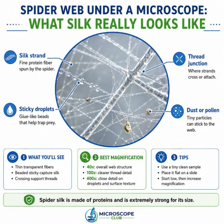
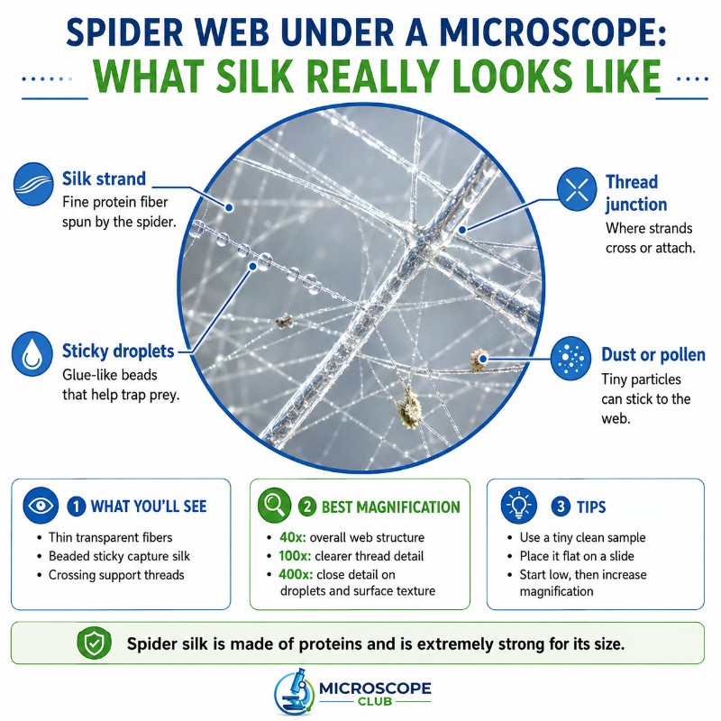
At 10–40x on a stereo or dissecting microscope, the web transforms from an invisible tangle into a precise geometric structure. The radial spokes — dragline silk — appear as straight, slightly reflective lines fanning outward. The spiral threads connecting them look noticeably different: thicker in apparent diameter, slightly more opaque, and strung with small, evenly spaced droplets. Those are the glue beads on the capture silk, and even at low power they’re unmistakable — like a string of translucent pearls. Where spokes meet spirals, you can see the piriform (attachment disc) silk forming dense anchor knots.
At Higher Power (Compound 100–400x)
A compound scope at 100–400x lets you measure and compare fiber types directly. Dragline fibers appear smooth, cylindrical, and almost colorless — their diameter falls in the 1–5 µm range for adult orb-weavers, far finer than human hair under a microscope (which runs 50–100 µm). Switch to a capture spiral strand and the difference is immediate: the axial core fiber is even finer, and the regularly spaced glue droplets are now clearly globular, surrounding the fiber like a series of miniature water drops frozen in place. Using objective lenses (40x, 100x), you can measure droplet spacing, compare radial versus spiral fiber width, and watch how the fiber bends differently under each — dragline threads are stiff; capture threads are visibly more elastic and sag slightly under their own droplet weight.
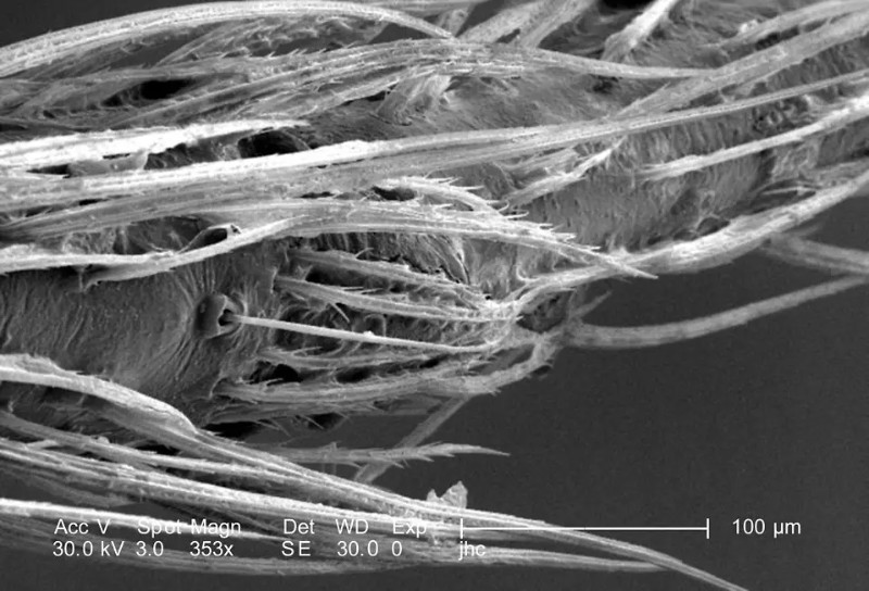
What SEM Reveals That Light Microscopes Can’t
The resolution limit of light microscopy is around 200 nm — enough to see fibers but not the surface texture or sub-fibril structure within them. That barrier is why understanding a light microscope versus an electron microscope matters here. Under a scanning electron microscope (SEM), dragline fibers show a smooth, almost glassy cylindrical surface — a single continuous strand, not obviously bundled. Capture silk imaged by SEM reveals how the aggregate glue coats the flagelliform core, and at very high magnification the periodic pinching of the fiber inside each droplet becomes visible — the structural signature of the “windlass” mechanism described in the next section. You can explore what electron microscope images of natural fibers look like to get a sense of the contrast before attempting SEM access through a university or makerspace facility.

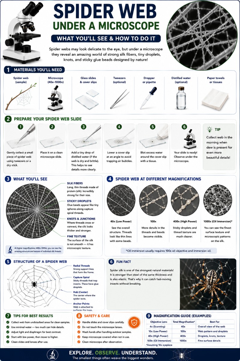
The Different Types of Spider Silk
A single orb-weaver spider can produce up to seven different silk types from separate glands, each tuned for a different mechanical role. The three most relevant to what you see under the microscope are listed in the comparison table below.
| Silk Type | Diameter (approx.) | Function | Visible under light microscope |
|---|---|---|---|
| Dragline (major ampullate) | 1–5 µm | Frame, radial spokes, lifeline | Smooth, straight, slightly reflective fibers |
| Capture spiral (flagelliform) | Sub-micron core | Stretchy sticky spiral — traps prey | Fine core thread strung with glue droplets |
| Aggregate glue | N/A (liquid coating) | Aqueous adhesive on capture threads | Evenly spaced beads (“beads-on-a-string”) |
Four additional types — minor ampullate (temporary scaffolding), aciniform (wrapping prey), piriform (anchor discs at junction points), and cylindriform (egg-case silk) — appear as dense, opaque knots or broad flat ribbons at lower magnifications. The Britannica overview of spider silk has a useful summary of all seven gland types if you want to map the full set.

One important misconception: not all spider silk is sticky. The radial dragline spokes carry no glue at all — that’s partly how the spider walks its own web without getting caught. Only the capture spiral threads carry the aggregate glue coating.
The Sticky Droplets: Spider Glue Under Magnification
The regularly spaced glue droplets on capture silk are one of the most striking things to see at 100–400x — and they do far more than simply make the thread adhesive. The droplets are composed of aggregate silk gland secretions: a water-based, hygroscopic adhesive that stays liquid and sticky even in dry conditions by pulling moisture from the air.
Under the compound scope, the spacing of the droplets along the fiber is remarkably consistent, typically 10–50 µm apart depending on the species. This regularity is not cosmetic — it reflects the “liquid wire” or windlass mechanism. When a capture thread is stretched, rather than the fiber simply going taut, the slack silk actually spools inward into each droplet, which acts as a tiny pulley or reel. The droplet prevents the thread from going limp and keeps tension distributed across the whole spiral. When you gently prod a capture spiral thread under the scope and watch it retract, you’re seeing this mechanism in action: the thread snaps back not just through elasticity but through the droplet geometry pulling the fiber back to resting length.

This makes the capture spiral a genuinely unusual engineering structure — a fiber that uses its own adhesive coating as a mechanical spring. Research published in leading journals like Nature’s spider silk coverage has described this as one of the most elegant passive tension-control systems found in biology.
Why Spider Silk Is So Strong: Inside the Nanostructure
The mechanical performance of dragline silk comes from its protein architecture — and while you can’t see the nanostructure directly with a light microscope, understanding it explains every visual feature you observe at higher magnifications.
Spider silk is built from proteins called spidroins. At the nanoscale, the fiber is a semi-crystalline composite. Tiny, tightly ordered beta-sheet crystallites — formed mainly from poly-alanine and glycine-alanine repeating sequences — are embedded in a softer, disordered amorphous protein matrix rich in glycine residues. Think of it like a fiber-reinforced composite material. The crystals act as the hard reinforcement phase, providing stiffness and tensile strength. The amorphous matrix provides the flexibility and energy absorption that prevents brittle fracture.
The result is a material that combines two properties that are normally in conflict: strength (resistance to breaking under load) and extensibility (ability to deform before breaking). Most strong materials — glass, ceramic, steel wire — are brittle at high load. Spider silk stretches significantly before failing. The measure that captures both properties is toughness — total energy absorbed per unit volume before fracture. By that measure, dragline silk outperforms both high-tensile steel and Kevlar per unit mass.
The American Museum of Natural History’s spider science resources offer accessible background on how this structure forms as the spider draws silk through its spinnerets — the protein actually self-assembles into its crystalline structure during extrusion.
Is Spider Silk Really Stronger Than Steel?
The comparison is real but needs a qualifier. The tensile strength of dragline silk is approximately 1–1.6 GPa — broadly comparable to high-grade steel wire at around 1–1.5 GPa in absolute terms. So in raw tensile strength, they’re roughly in the same range. The difference emerges when you factor in density: silk weighs around 1.3 g/cm³ versus steel’s roughly 7.8 g/cm³. That means for the same weight of material, silk delivers similar or greater tensile strength and dramatically superior toughness. By-weight (specific toughness), dragline silk exceeds both steel and Kevlar. The headline “stronger than steel” is a shorthand that holds true by weight — not in an absolute pound-for-pound sense.
Frequently Asked Questions
Can spider silk be harvested to make fabric or thread?
In theory yes, but spiders can’t be farmed like silkworms — they’re territorial and cannibalistic at high densities. A small amount of genuine spider silk fabric has been produced (most famously, a golden cape made from golden orb-weaver silk displayed at the Victoria and Albert Museum), but it required over a million spiders and years of effort. The practical path to spider silk textiles is synthetic: companies like Bolt Threads and Spiber express spidroin proteins in yeast or bacteria, then spin them into fiber. These bioengineered silks are now available at small commercial scale in specialty performance-wear applications.
What is spider silk made of?
Spider silk is a protein fiber built from spidroin proteins. At the molecular level it’s a semi-crystalline composite: hard beta-sheet crystallites (from poly-alanine amino acid sequences) embedded in a softer, glycine-rich amorphous protein matrix. This combination of hard and soft domains is what gives silk its unusual mix of strength and flexibility.
How thick is a strand of spider silk?
Dragline silk from an adult orb-weaver runs roughly 1–5 µm in diameter — most commonly around 3–4 µm. That’s 10–30 times thinner than a human hair. Capture spiral silk has an even finer axial core, often sub-micron, though the glue droplets make the overall thread appear thicker under a light microscope. Very small spider species can spin dragline silk below 1 µm in diameter.
Can you see spider silk with a light microscope, or do you need an electron microscope?
A basic stereo or compound light microscope is enough to see individual silk strands, distinguish dragline from capture silk, and observe the glue droplet pattern clearly. You do not need an electron microscope for the main visual features. What you can’t see optically are the surface texture of the fiber, the sub-fibril structure, and any detail below about 200 nm — for those features, SEM is required. Most hobbyists and students will get excellent results with a 40–400x compound scope or a stereo microscope.
Why aren’t the radial spokes of a spider web sticky?
The radial spokes are made of dragline silk from the major ampullate glands, which produces no adhesive coating. Only the spiral capture threads carry aggregate glue, secreted by a separate gland. This division is functional: the spider traverses its own web primarily along the non-sticky radial spokes. If the whole web were adhesive, the spider would trap itself.
Can I view spider silk at 1000x (oil immersion) on a compound scope?
You can, but the results are often disappointing. At 1000x with oil immersion, individual dragline fibers are so thin (3–4 µm) that the entire fiber width fills only a few pixels of the field, and the depth of focus becomes extremely shallow. You’ll lose the context of the web structure and gain little new information that 400x dry doesn’t already show. The most informative magnification range for a web sample is 40–400x. Oil immersion is better reserved for stained biological specimens where you need to resolve fine internal cell structures.
Conclusion
Spider silk is one of the most instructive materials you can put on a microscope slide — it bridges everyday biology and advanced materials science in a single thread. At low power you see the architectural logic of the web; at 400x you can measure fiber diameters and watch glue droplets hold their precise spacing; and even without SEM access, understanding the beta-sheet nanostructure explains every mechanical property you’ve just observed. The gap between what a spider produces effortlessly and what materials scientists can currently replicate is still enormous — and that gap starts with what you can see through your own eyepiece.
Have you mounted a spider web at home or in the classroom? We’d love to hear what species you sampled, what magnification gave you the best view of the glue droplets, or any tips you’ve found for keeping the threads flat — share it in the comments below.
