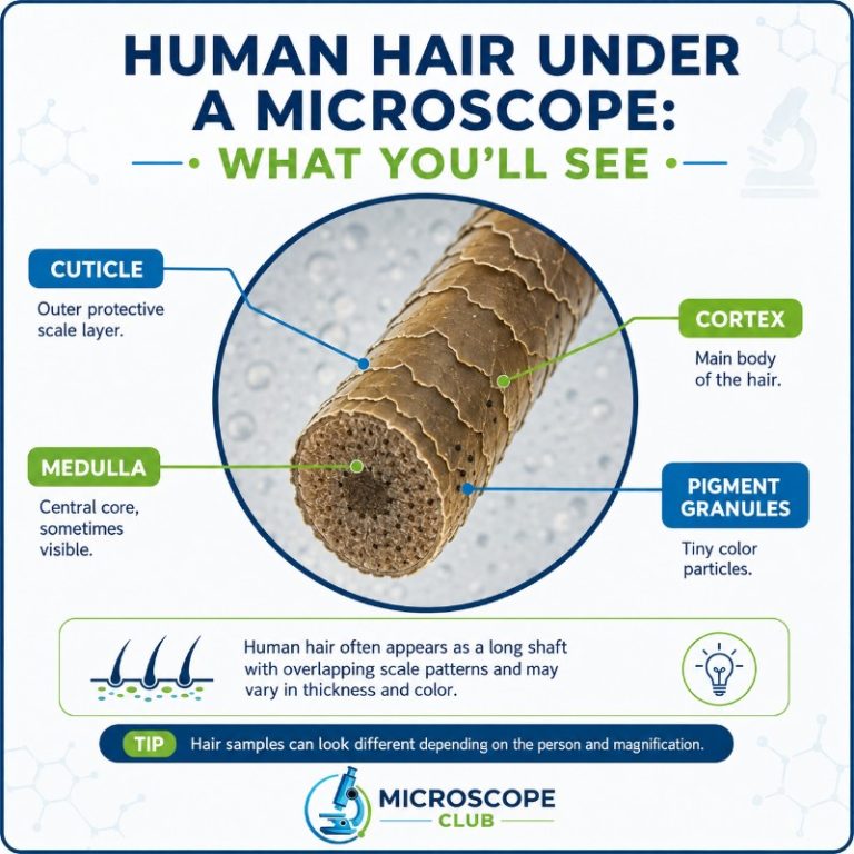
Human hair under a microscope reveals a semi-translucent rod with three concentric layers: the cuticle (an outer jacket of overlapping keratin scales), the cortex (the pigment-packed bulk of the shaft), and the medulla (a variable inner core that is often absent or fragmentary in fine hair). At low magnification you see its overall shape, colour, and diameter. Zoom in to 400× and the scaly cuticle pattern becomes the defining feature — telling you far more about health, damage, and species than the naked eye ever could.
What Does Human Hair Look Like Under a Microscope?
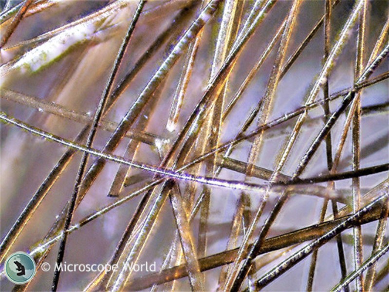
Place a single strand on a glass slide and the first thing you notice is how surprisingly thick it looks — a human scalp hair typically measures 50–100 micrometres (µm) across, thick enough to see with the naked eye but transformed by even a basic compound light microscope. The shaft is elongated and largely uniform, narrowing to a tapered tip (or a blunt cut tip if the hair has been trimmed). Its translucency means light passes through, revealing internal structure, while the outermost surface shows a faint but unmistakable scale-like texture.
For a broader look at hair of all kinds — including animal hair — see our general guide to hair under a microscope; this article focuses specifically on human hair.
The three layers tell a complete story about the hair’s biology, health, and origin — and each one becomes readable at a different magnification.
| Layer | Position | What it looks like under a microscope | Function |
|---|---|---|---|
| Cuticle | Outermost | Flat, overlapping transparent scales pointing toward the tip; like roof shingles or fish scales | Protects the cortex; waterproofing and physical barrier |
| Cortex | Middle (bulk) | Dense, pigmented zone; melanin granules give colour; tiny air spaces (cortical fusi) may appear as bright spots | Provides colour, strength, and elasticity |
| Medulla | Central core | A thin, often broken or absent channel; may appear as a pale or dark line running down the shaft | Not fully understood; may assist flexibility |
The Three Layers of a Human Hair
Cuticle — the Scaly Outer Shield
The cuticle is the first thing a microscopist notices, and for good reason: it is the most visually dramatic layer. Made of flat, transparent keratin scales arranged like overlapping roof shingles — always pointing from root toward tip — the cuticle is typically 6–10 layers thick. In reflected or oblique light, those scale edges catch the light and reveal a distinctive herringbone or imbricate pattern. The cuticle is colourless on its own; the colour you see through it comes from the cortex beneath.
Human hair cuticle scale pattern (described as “imbricate” — close-set, with narrow margins) is one of the primary features forensic analysts use to distinguish human hair from animal hair. Healthy cuticle scales lie flat and smooth; damaged ones lift, chip, or flake away entirely.

Cortex — Pigment, Colour and Strength
The cortex makes up the bulk of the hair shaft — roughly 80–90% of its diameter. It is packed with elongated keratin cells and, crucially, melanin pigment granules. Eumelanin (brown-black) and phaeomelanin (red-yellow) combine in different ratios to produce the full spectrum of human hair colour, from jet black through auburn to platinum blonde.
At 400×, the cortex appears as a grainy, densely structured zone. Look carefully and you may spot cortical fusi — tiny air spaces that appear as bright, oval voids scattered through the cortex. These are particularly common near the root end of pulled-out hair. Forensic analysts note the distribution and density of melanin granules as a racial/ancestral indicator (see Section 8 below).
Hair is approximately 95% keratin, a tough fibrous structural protein. The entire shaft is dead, keratinised tissue — no living cells remain in the part you mount on a slide.

Medulla — the Variable Inner Core
The medulla is the innermost layer and the one that most surprises first-time observers: in human hair, it is frequently absent altogether. When present, it may be continuous (a single unbroken channel), fragmented (intermittent dashes), or trace (barely detectable). Fine hair and blonde hair are especially likely to have no visible medulla at all.
This variability is a key human-vs-animal diagnostic. Most animal hairs have a prominent, continuous medulla — often patterned — that occupies more than half the total hair diameter. The medullary index (medulla width ÷ total hair width) is typically less than 1/3 in humans and greater than 1/2 in most animals. When you spot a strand with a thick, clearly-patterned central channel, you are most likely looking at animal hair, not human.
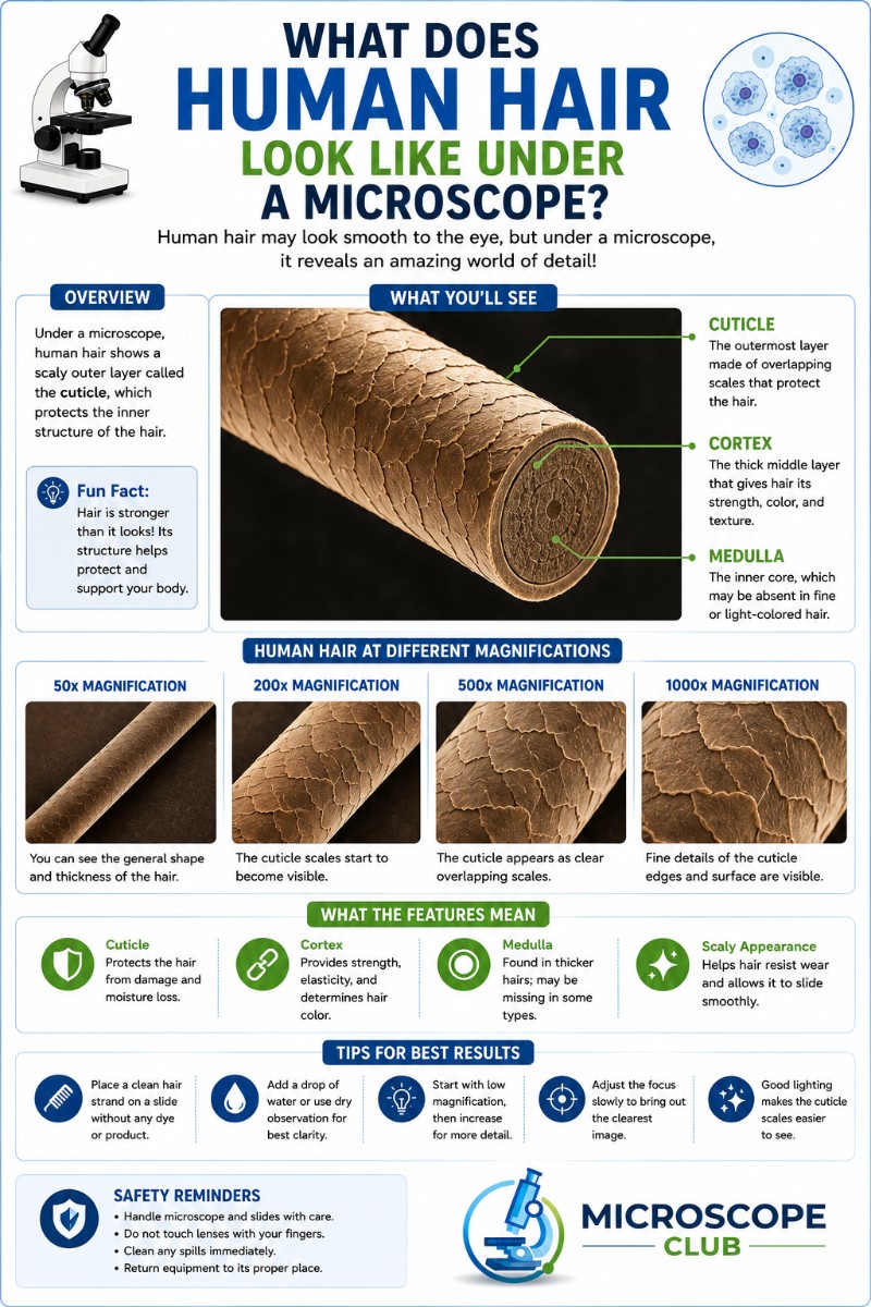
What You See at Each Magnification
The view changes dramatically as you increase magnification. A home compound light microscope and a laboratory scanning electron microscope (SEM) reveal completely different worlds — and it pays to know which features belong to which tool.
| Magnification | Objective | What becomes visible | Accessible with home scope? |
|---|---|---|---|
| 40× | 4× objective | Overall shape, approximate diameter, colour, gross damage (split ends), thickness comparison | Yes |
| 100× | 10× objective | Medulla presence and pattern, pigment distribution, general cuticle outline, cortical fusi | Yes |
| 400× | 40× objective | Cuticle scale edges and pattern, fine damage detail, cortical structure, trichoptilosis (split ends) | Yes — practical ceiling for most home/school scopes |
| 1000× (oil immersion) | 100× objective | Rarely useful for whole hair — the shaft is too thick to flatten for oil immersion | Possible but not recommended for hair |
| SEM (~500×–10,000×+) | Electron beam | Three-dimensional surface relief of individual cuticle scales, surface texture, nano-scale damage | No — laboratory instrument only |
The dramatic 3-D images of cuticle scales you see in textbooks and documentaries are almost always scanning electron microscope images. A home light microscope shows the pattern of scales — their edges and spacing — but not the raised, tile-like relief. That distinction matters when you are setting up a hair slide at home: what you see will still be genuinely informative, just different from the SEM imagery.
How to View Your Own Hair Under a Microscope at Home
Hair is one of the easiest specimens a beginner microscopist can mount — no staining is required, and you can collect the sample yourself without any equipment beyond a slide and cover slip. Before you start, check out our guide on how to prepare a microscope slide if you are new to wet mounts.
- Collect the sample. The shaft (the part above the skin) is what you want for structure. A plucked hair with the root bulb attached lets you see both shaft and follicle tissue — but handle the root gently. A cut hair works perfectly for cuticle and cortex study.
- Mount it. Lay the hair across the centre of a clean glass slide. Add one small drop of water (a wet mount). Lower a cover slip carefully at an angle to avoid bubbles.
- Start at low power. Begin at 40× to find and centre the hair, then step up to 100× and 400×. Adjust your compound light microscope’s condenser and iris diaphragm — reducing light slightly often improves contrast for transparent specimens like hair.
- Try oblique illumination. Angle your illumination slightly off-centre to cast shadows across the cuticle scales; this makes the scale pattern far more visible.
- Note the difference. Shaft = the elongated rod on the slide (dead, keratinised). Follicle/root/bulb = living tissue from beneath the skin. If you plucked the hair, you may see tissue attached at the base — that is where nuclear DNA is found, not in the shaft itself.
A first-time observer should expect a semi-transparent tube with a subtly textured surface. Do not expect the dramatic raised-scale SEM images; what you will actually see is the scale pattern — a faint herringbone on the surface, especially clear at 400×.
Healthy vs Damaged Hair Under the Microscope
The cuticle is a direct record of everything the hair has been through — heat styling, bleaching, UV exposure, mechanical friction, and age. Once you know what healthy looks like, damage becomes immediately obvious.
| Feature | Healthy hair | Damaged hair |
|---|---|---|
| Cuticle scales | Flat, smooth, lying close to the cortex | Lifted, chipped, cracked, or missing entirely |
| Surface texture | Even, uniform along the shaft | Rough, irregular, frayed sections |
| Diameter | Consistent along the length | May taper or narrow at damaged points |
| Tip | Tapered or cleanly cut | Split ends (trichoptilosis) — shaft fraying into two or more strands |
| Cortex exposure | Cortex fully protected by cuticle | “Weathered” zones where cuticle worn away, exposing cortex directly |
Heat is particularly destructive: a flat iron or repeated blow-drying lifts and eventually strips cuticle scales from the tip backward, leaving the cortex exposed and vulnerable. Bleaching dissolves melanin granules from the cortex as well as degrading cuticle protein — the combination creates the brittle, porous texture of heavily bleached hair that is obvious at 400× as a rough, irregular surface.
If the surface damage you observe is flaky rather than frayed, you may be looking at sebum and dead skin rather than damaged cuticle — see our guide to dandruff under the microscope to compare.
Do Different Human Hair Types Look Different?
Yes — and the most reliable indicator is the cross-section shape of the shaft rather than any feature of the surface (which looks similar across hair types at low magnification). Hair cross-section shape correlates directly with curl pattern:
| Hair type | Cross-section shape | Typical diameter |
|---|---|---|
| Straight (Type 1) | Roughly round | ~50–80 µm |
| Wavy (Type 2) | Oval | ~60–100 µm |
| Curly (Type 3) | Oval to flattened/elliptical | ~50–100 µm |
| Coily / tightly coiled (Type 4) | Strongly flattened, kidney-shaped | ~40–80 µm (often finer) |
The cross-section is the key: the more flattened or elliptical the shaft, the tighter the natural curl. Viewing a cross-section of hair (cut at a 90° angle and embedded in a medium) gives more structural information than a longitudinal view. On a longitudinal slide, curly hair may appear to undulate gently as you track along its length, while a coily strand may seem almost twisted. Overall shaft diameter across all human hair ranges from roughly 17–180 µm, though most scalp hair falls between 50–120 µm.
Human Hair vs Animal Hair Under a Microscope
The difference is immediately apparent once you know what to look for. The three key distinguishing features are medullary index, medulla pattern, and scale morphology:
- Medullary index: Human hair typically <1/3; most animal hairs >1/2. A strand where the central channel fills more than half the total width is almost certainly not human.
- Medulla pattern: Human medulla (when present) is simple — amorphous, fragmented, or a thin continuous channel. Many animal hairs have a patterned medulla (lattice, vacuolated, multiserial) that is visible at 100–400×.
- Scale morphology: Human cuticle scales are imbricate (narrow, tile-like, close-set). Many animal hairs have quite different scale shapes — pectinate (comb-like), coronal (crown-like in some fine mammal hairs), or diamond-shaped — which are visible in a scale cast or at high magnification.
It is worth noting that dog and cat hairs — the most common “unknown” hairs a hobbyist might encounter — typically have prominent, continuous medullas and distinctive scale patterns that look quite different from the faint, intermittent medulla of human hair.
What Forensic Scientists Look for in Hair
Forensic hair analysis was once far more prominent in criminal investigations than it is today, and understanding what microscopy can — and critically, cannot — determine is important context for anyone who has watched a forensic documentary and assumed hair is a definitive identifier.
What light microscopy can indicate:
- Species (human vs animal) — based on medullary index and scale pattern
- Broad ancestral/racial group — pigment granule distribution and density, medulla continuity, and cross-section shape offer population-level trends (not individual identification)
- Body area of origin — pubic, scalp, eyebrow, axillary, and beard hairs have characteristic features
- Whether the hair was forcibly removed — a root sheath or stretched root indicates traumatic removal
- Artificial treatment — a dye line (natural colour at root, dyed colour along shaft), bleach damage, or permanent-wave treatment leave visible traces
- General damage type — heat, UV, mechanical, or chemical damage are distinguishable
What microscopy alone cannot do: provide a positive individual identification. This point cannot be overstated. For decades, courtroom testimony claiming that microscopic hair “matches” were evidence of the same individual was presented far beyond what the science supported. A joint FBI/DOJ review found that microscopic hair comparison evidence had been overstated in hundreds of cases. Modern positive individual identification from hair requires nuclear DNA analysis from the follicle or root — not from the shaft itself.
The DNA distinction: Nuclear DNA (used for individual identification) is found in the living cells of the hair follicle and root bulb — below the skin. The shaft you mount on a slide is dead, keratinised tissue with no nuclear DNA. The shaft may yield mitochondrial DNA (inherited maternally, shared among relatives), but this cannot provide a unique individual match. If you pluck a hair with the root intact, that root tissue contains nuclear DNA; a shed hair (root shed naturally) may not.

Frequently Asked Questions
Does human hair have a medulla?
Not always. The medulla in human hair is described as “variable” — it may be continuous, fragmented, or completely absent. Fine hair and blonde hair are particularly likely to have no detectable medulla. This is one of the key differences from most animal hairs, which typically have a prominent, continuous medulla.
How should I store a hair sample so I can look at it again later?
For long-term storage, make a dry mount rather than a wet mount — hair is a dry specimen, so it sits well on a slide without water, and dry mounts can be kept indefinitely. Wet mounts are temporary: the water evaporates within hours and the sample shifts or degrades, so clean them off after use. If you want to keep a wet mount readable for longer, ring the edges of the cover slip with clear nail varnish or a smear of petroleum jelly to slow evaporation.
What are the most common mistakes when mounting hair on a slide?
The three usual culprits are air bubbles, too much light, and dust. Lower the cover slip slowly at about a 45° angle to push air out rather than trap it; if a bubble forms, a gentle tap nudges it to the edge. Because hair is transparent, flooding it with light washes out the cuticle pattern — close the iris diaphragm and lower the condenser to build contrast. Finally, wipe the slide and cover slip with ethanol or lens cleaner first, since stray fibres and dust are easily mistaken for hair detail.
How does chemically treated hair — dye, relaxer, or a perm — look different under the microscope?
Oxidative and alkaline treatments attack the same structures as heat and bleach, so the signs overlap: cuticle scales lift, chip, tear, or fragment, leaving a rough, uneven surface. Relaxers and permanent waves break and re-form the cortex’s bonds, which can leave voids, bubbles, or distortions visible inside the shaft at 400×. Permanent dye often shows a clear “dye line” — natural colour near the freshly grown root and treated colour along the older shaft.
Does a child’s hair look different from an adult’s or an elderly person’s under a microscope?
Yes. Fine infant and pre-pubertal (vellus) hair is much thinner — roughly 0.03 mm versus 0.06 mm or more for mature terminal hair — and usually has no visible medulla and little or no pigment. As hair ages, the most obvious microscope change is loss of melanin from the cortex: greying hair shows progressively fewer pigment granules until white hairs have almost none, because the colour-making cells in the root shut down.
Conclusion
Human hair is a deceptively complex structure. Under a microscope, what appears to the naked eye as a single strand resolves into three distinct layers — each telling its own story about pigment, health, ancestry, and biological history. The cuticle scale pattern is the most diagnostic feature a home microscopist can reliably observe at 400×, while the medulla (or its absence) is the quickest human-vs-animal tell. Understanding the difference between what a light microscope can show and what requires an SEM sets realistic expectations and prevents the common mistake of expecting dramatic 3-D surface images from a school scope. And knowing that the shaft holds no nuclear DNA — that individual identification requires the living root tissue — puts forensic hair science in its proper, honest context.
Have you mounted your own hair under the microscope? We would love to hear what you found — was your medulla visible, and did you spot any cuticle damage you were not expecting? Share your experience in the comments below.