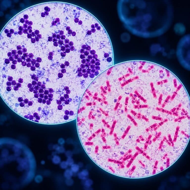
Gram staining step by step comes down to four reagents applied in a fixed order — crystal violet, iodine, alcohol, and safranin. The resulting bacterial smear turns either purple or pink, depending on how thick the cell wall underneath actually is. The whole run takes about five minutes on the bench, and exactly one of the four steps is judged by eye rather than by a clock. This guide walks through each step with its real timing, then explains why the color split happens and how to fix the run when it goes wrong.
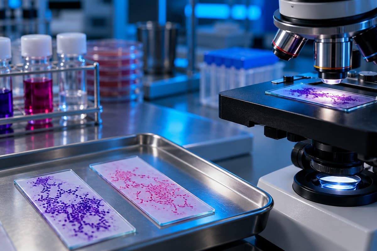
The Four Steps of a Gram Stain
A Gram stain runs through crystal violet, Gram’s iodine, an alcohol decolorizer, and safranin in that order, with a water rinse and a gentle blot between each one, following the standard ASM Gram stain protocol. Read the finished slide at 1000x under oil immersion. See OpenStax’s Gram stain explanation for additional context.
| Step | Reagent | Timing | What You Should See |
|---|---|---|---|
| 1. Primary stain | Crystal violet (0.5–2%) | ~1 minute | Smear turns solid deep purple, every cell included |
| 2. Mordant | Gram’s iodine | ~1 minute | No visible change — the dye is being locked inside the cell |
| 3. Decolorizer | 95% ethanol or acetone-alcohol | 3–15 seconds, judged by runoff | Purple runoff fades to clear; stop the instant it does |
| 4. Counterstain | Safranin (0.1–0.5%) | 30–60 seconds | Decolorized cells pick up pink/red |
Step 1: Crystal Violet (the Primary Stain)
Crystal violet floods the fixed smear for about a minute and stains every cell on the slide the same deep purple, Gram-positive and Gram-negative alike. It is an aggressive dye — it stains skin for days and leaves a permanent purple ring around the bottle cap, so gloves are not optional. At this point the slide tells you nothing yet; the differential part of the stain hasn’t happened.
Step 2: Gram’s Iodine (the Mordant)
Gram’s iodine acts as a mordant, binding the crystal violet already inside the cell into a larger, less soluble crystal violet–iodine complex. The reagent is amber-brown, like weak iced tea, with a faint antiseptic smell close to wound iodine. Skip this step and the crystal violet simply rinses back out during the next wash — there is no differential stain without the mordant.
Step 3: Decolorize — the Step That Decides Everything
The decolorizer is where the whole technique happens, judged visually, not by a stopwatch. Tilt the slide over a sink, drip the alcohol onto the top edge, and watch the purple ribbon running off the glass fade from deep violet to barely tinted. The instant the runoff stops carrying purple, hit the slide with water — on a thin smear that can be 3 to 5 seconds, well under the 10–15 seconds most guides quote for an average-thickness smear. Stopping too late is the single most common way a Gram stain fails.
Step 4: Safranin (the Counterstain)
Safranin is a thin, watery orange-pink — closer to diluted fruit punch than a dye — that stains the now-colorless Gram-negative cells for 30 to 60 seconds. Gram-positive cells are already saturated purple, so safranin makes no visible difference to them. Rinse, blot dry with bibulous paper (never rub), and the slide is ready to read.
Before You Stain: Preparing and Fixing the Smear
Smear preparation decides more Gram stain outcomes than the staining itself does, which is why most failures trace back here rather than to the reagents. Start with a culture that is 18 to 24 hours old — older cultures lose cell wall integrity and Gram-positive organisms start reading falsely Gram-negative or Gram-variable. If you’re still building basic slide-handling habits, preparing microscope slides properly is worth reviewing first. See OpenStax’s bacterial cell-wall overview for additional context.

Place a small drop of sterile saline on the slide, touch a loop to a single colony, and emulsify it into a thin, dime-sized film. A correct smear is thin enough that it looks like barely more than breath fog on the glass once dry. A visible white streak means the smear is too thick, which physically shields the cells at its center from the decolorizer. As a result, the middle of the slide reads falsely Gram-positive while the thin edge reads correctly.
Air dry the smear completely before fixing it — fixing a still-wet smear boils the cells and distorts them beyond recognition. Fix by passing the dry slide through a flame two or three times, or flood it with 95% methanol for about a minute. Methanol preserves cell morphology better for anything containing blood or host cells, since heat can lyse red cells. Decolorizer is also flammable, so keep it well away from the flame used for heat fixation — both reagents share the same bench.
Why Gram-Positive Cells Stay Purple and Gram-Negative Cells Turn Pink
Gram-positive and Gram-negative cells split apart at the decolorization step because their cell walls are built differently, not because one type resists dye and the other doesn’t. Crystal violet enters every cell equally, and iodine locks it into the crystal violet–iodine complex inside all of them — up to this point, both groups look identical. The decolorizer is what exposes the structural difference.

A Gram-positive cell wall is a thick, cross-linked peptidoglycan layer, 20 to 80 nm across and 40 to 90% of the wall’s dry weight, with teichoic acids threaded through it. Alcohol dehydrates that mesh and shrinks it, closing the pores and trapping the crystal violet–iodine complex inside — the cell stays purple. A Gram-negative cell wall carries only a thin peptidoglycan layer, 2 to 7 nm, sitting under a lipid-rich outer membrane built from lipopolysaccharide. Alcohol dissolves that outer membrane, and the complex washes straight out through the thin peptidoglycan underneath — the cell goes colorless and picks up safranin instead.
| Feature | Gram-Positive | Gram-Negative |
|---|---|---|
| Peptidoglycan thickness | 20–80 nm, multiple layers | 2–7 nm, one or a few layers |
| Outer membrane | Absent | Present, lipopolysaccharide-based |
| Teichoic acids | Present, threaded through the wall | Absent |
| Final color | Purple | Pink / red |
| Example organisms | Staphylococcus aureus, Bacillus subtilis | Escherichia coli, Salmonella enterica |
A common misconception is that Gram-negative bacteria have no cell wall — they actually have more layers, not fewer, since the thinner peptidoglycan sits under an extra outer membrane. Neither reaction means “safer” or “more dangerous”: the stain reports cell wall architecture, not virulence.
Reading the Slide at 1000x
Reading a finished Gram stain happens at 1000x under oil immersion — a 10x eyepiece paired with a 100x objective. Bacterial cells are too small to resolve reliably at any lower power. Gram-positive cocci read as a deep, jewel-toned royal purple, dense enough that a tight cluster looks nearly black at its center; they’re unmistakable. Gram-negative rods are a much fainter, disappointing salmon pink against a background that’s also faintly tinted, and beginners routinely scan right past them. Closing the condenser iris slightly brings up the contrast. What bacteria look like under a microscope more broadly is worth a look for the full range of shapes you’ll run into on a real slide.
Build in a control every time you stain: smear a known Gram-positive organism such as Staphylococcus aureus (the ATCC 25923 quality-control strain) on one end of the slide and a known Gram-negative organism on the other. Stain both together. If the Gram-positive control comes out pink, the run was over-decolorized — you know it without guessing at the unknown sample. It costs nothing beyond one extra smear and is the single most valuable habit a beginner can build.
Watch for stain precipitate too — scattered, hard-edged purple crystals that new students often mistake for Gram-positive bacilli. Precipitate isn’t alive; it sits at a different focal plane than actual cells.
What Gram-Positive and Gram-Negative Bacteria Look Like
Gram-positive organisms cover a recognizable set of shapes once you know the color to expect: Staphylococcus aureus forms purple cocci in grape-like clusters, Streptococcus pyogenes forms cocci in chains, and Bacillus subtilis forms large purple rods, occasionally showing an unstained endospore. Clostridium, Listeria, Enterococcus, and Lactobacillus round out the common examples.
Gram-negative organisms stain pink and generally show up as rods: E. coli under a microscope appears as short, plump pink rods, while Salmonella appears as pink rods with no way to ID it by sight alone. The two are morphologically indistinguishable on a Gram stain and need biochemical testing to tell apart. Pseudomonas aeruginosa forms slender pink rods, Neisseria gonorrhoeae forms kidney-bean-shaped diplococci in pairs, and Klebsiella pneumoniae often shows a clear capsule halo.
One organism trips up almost every beginner: yeast stains purple but is not a Gram-positive bacterium — it’s a much larger eukaryotic cell, five to ten microns and often budding, and new students routinely misread yeast under a microscope as an oversized coccus.
Troubleshooting: When the Stain Goes Wrong
Most failed Gram stains trace back to one of a handful of repeatable causes.
| Symptom | Likely Cause | Fix |
|---|---|---|
| Everything reads pink, including known Gram-positives | Over-decolorization | Decolorize drop-by-drop on a tilted slide, stop the instant the runoff clears |
| Everything reads purple, including known Gram-negatives | Under-decolorization, or smear too thick | Apply decolorizer a little longer; re-smear thinner next time and read the thin edge |
| Both colors appear on one field | Mixed culture, or an old/dying culture | Confirm culture purity and age; a mixed result on a pure culture usually means the culture is too old |
| Purple crystals or grit visible | Stain precipitate, not cells | Ignore — precipitate sits at a different focal plane and has hard, angular edges |
| Cells barely visible or too faint | Under-counterstaining, or reading at too low a magnification | Extend the safranin step slightly; read at 1000x oil immersion, never 400x |
A few habits prevent most of these outright. Keep the wash bottle in your off hand before decolorizing, since there’s no time to reach for it once the runoff starts. Aim the water stream above the smear rather than directly on it. And never let a reagent sit and dry between steps, since that produces heavy precipitate.
Bacteria the Gram Stain Cannot Identify
Several groups of bacteria fall outside what a Gram stain can resolve, according to the NCBI StatPearls entry on Gram staining. Mycoplasma species have no cell wall, so there’s nothing to retain or lose the stain. Mycobacterium tuberculosis and its relatives have a waxy, mycolic-acid wall that resists both dyes and need an acid-fast (Ziehl–Neelsen) stain instead. Spirochetes such as Treponema pallidum are too thin to resolve and are better read with dark field microscopy. Chlamydia and Rickettsia are too small and intracellular to stain this way. For suspected TB, labs switch to an acid-fast stain instead — our guide to what a sputum smear shows covers that slide.
Legionella, Haemophilus, and some anaerobes stain poorly with safranin, which is why some protocols substitute basic fuchsin instead. Some organisms are Gram-variable too: Actinomyces, Acinetobacter, and aging Bacillus or Clostridium cultures can read inconsistently between runs.
A Gram stain narrows bacteria into a shape-and-color category — it does not identify a species, and this article is educational, not diagnostic guidance. Clinical interpretation of a patient specimen belongs to a trained microbiologist working alongside culture and biochemical tests. Practice on safe, purchased teaching cultures or something like yogurt, never a throat swab or anything from a sick person. This technique is one part of the broader cell-staining toolkit worth knowing as a beginner microscopist.
Frequently Asked Questions
Can you Gram stain yeast or fungi?
Yeast picks up purple, but the result doesn’t mean the same thing as it does for bacteria. Yeast is a much larger eukaryotic cell that retains crystal violet for reasons unrelated to peptidoglycan, so a purple yeast cell is not a Gram-positive organism.
Who invented the Gram stain and when?
Hans Christian Gram, a Danish bacteriologist, developed the technique in 1884 in Berlin while trying to make bacteria visible in lung tissue from pneumonia patients. He published it believing it was imperfect and invited others to improve it — it has stayed in daily clinical use, largely unchanged, ever since.
Can you use tap water instead of distilled water for the rinses?
Distilled water is preferred because tap water’s mineral content can leave a fine residue on the slide that’s easy to mistake for stain precipitate. A quick rinse with tap water rarely ruins a stain, but if unexplained specks keep showing up under the scope, switch to distilled water before troubleshooting anything else.
How long does the whole Gram stain take?
The four staining steps together take roughly five minutes once the smear is fixed. Add smear preparation, drying, and fixation, and the full process runs under ten minutes for someone working efficiently.
Is safranin or basic fuchsin the better counterstain?
Safranin is the standard and works well for most organisms, but basic fuchsin gives better visibility for a specific group that stains poorly with safranin — Legionella, Haemophilus, and some anaerobes. Some published protocols specify basic fuchsin for exactly that reason.
Conclusion
Gram staining step by step is really just four reagents in a fixed order, but the decolorization step is where the real skill lives — everything else is mechanical. Keep the smear thin, the culture fresh, and the decolorizer timing judged by eye rather than by count, and the procedure will reliably split your slide into purple and pink exactly the way the cell wall chemistry predicts.
Have you run a Gram stain yourself, in a teaching lab or on a hobby scope at home? Tell us how your control slide turned out — and whether the decolorization step caught you off guard the first time — in the comments below.