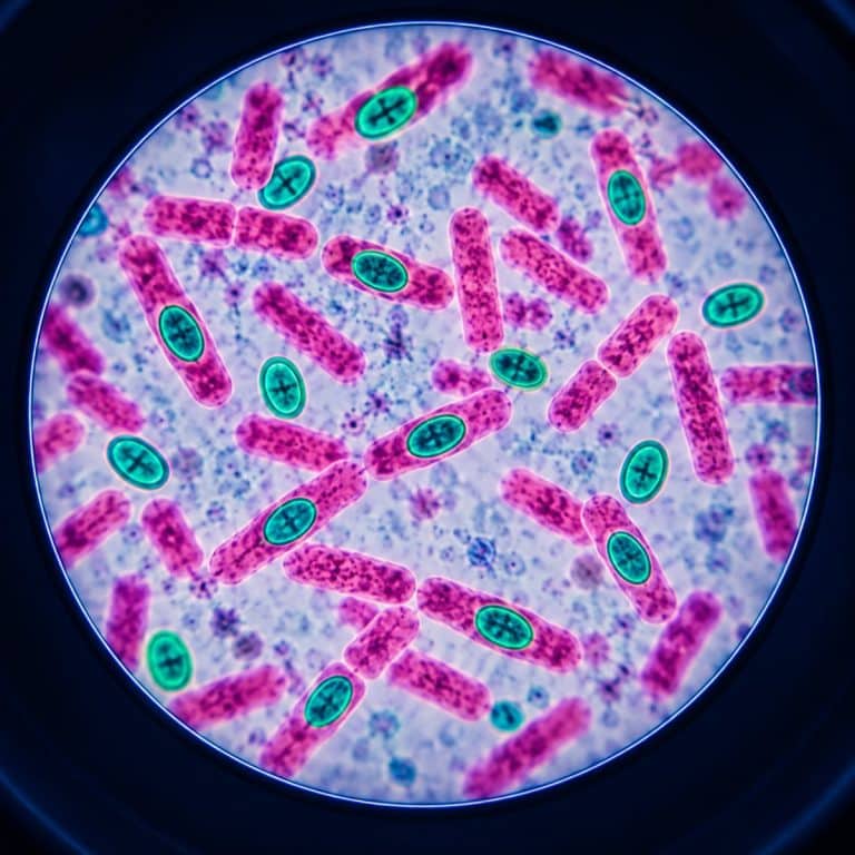
Endospore staining uses the Schaeffer-Fulton method to turn a dormant bacterial endospore green while the vegetative cell around it stains pink. It relies on malachite green as the primary dye, steam in place of a chemical mordant, and safranin as the counterstain. The whole procedure takes about 15 minutes including smear prep. It only works because heat — not iodine, not acid — is what forces dye through a spore coat that normally lets nothing in.
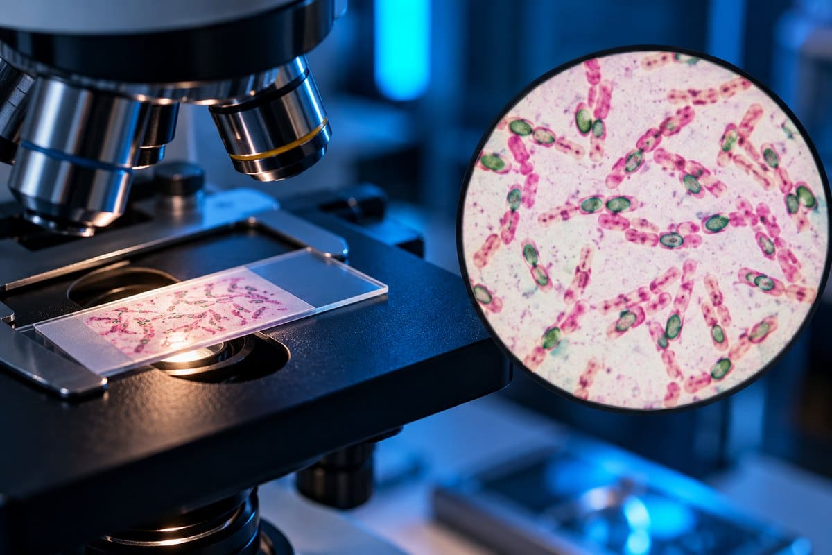
The Schaeffer-Fulton Method, Step by Step
Developed by Alice B. Schaeffer and MacDonald Fulton in the 1930s, this is the standard differential stain for bacterial endospores. Here is the full procedure at a glance before the step-by-step breakdown: See OpenStax’s endospore-stain explanation for additional context.
| Step | Reagent / Action | Timing | What You Should See |
|---|---|---|---|
| 1. Smear + heat fix | 48–72 hour culture, air dried, heat fixed | — | Thin, even smear on the slide |
| 2. Steam malachite green | Malachite green over blotting paper, steaming water bath | 5 minutes (some manuals extend to 15) | Smear turns near-black green |
| 3. Cool | Remove paper, let slide cool | 1–2 minutes | Slide returns to room temperature |
| 4. Rinse | Gentle stream of water | ~30 seconds | Green sheets off; smear fades to a pale wash |
| 5. Counterstain | Safranin | 1–2 minutes | Vegetative cells pick up pink/red |
| 6. Rinse + blot + read | Water rinse, bibulous paper, oil immersion | — | Green ovals inside pink rods, at 1000x |
Safety first: malachite green is a skin, eye, and respiratory irritant that stains everything it touches — wear gloves and eye protection. The slide comes off the steam bath genuinely hot, so use forceps or a clothespin. Keep the open flame you use for heat fixation away from loose paper or sleeves. Follow your lab’s standard aseptic technique for handling the culture and disposing of the slide afterward.
Step 1 — Prepare and Heat-Fix the Smear
Use a culture that is 48 to 72 hours old, not a fresh overnight one. Spread a thin smear on a clean slide, air dry it, then heat fix it by passing it through a flame two or three times. If you’re new to slide prep, preparing microscope slides properly covers smear thickness and heat-fixing technique in more detail. Set up your steaming water bath before you make the smear — waiting on a beaker to boil while a heat-fixed slide sits around is wasted time.
Step 2 — Steam the Malachite Green Through the Spore Coat
Lay a piece of blotting paper, cut slightly smaller than the slide, over the smear. Place the slide over the steaming water bath and flood the paper with malachite green. Steam for 5 minutes, adding more dye every minute or so — the paper should always look wet, never dry and curling at the edges. Some teaching manuals, including this endospore stain lab manual chapter, extend steaming to 15 minutes for older or stubborn cultures — either way, keep the paper wet throughout. Watch this step closely: a dried-out paper lets the dye crystallize on the smear instead of penetrating it.
Step 3 — Cool, Then Rinse With Water
Let the slide cool for 1 to 2 minutes before you touch water to it — dropping a hot slide straight into a cold rinse can crack it. The spore also needs that cooling window to reseal around the trapped dye anyway. Then rinse gently until the runoff is clear. A wide sheet of green will sluice off almost immediately; that is the stain working correctly, not the stain failing.
Step 4 — Counterstain With Safranin
Flood the slide with safranin for 1 to 2 minutes. It looks thin and watery next to the malachite green, almost too dilute to matter. But it only needs to color the now-colorless vegetative cells — it can’t displace dye that’s already trapped inside a resealed spore.

Step 5 — Blot and Read at 1000x
Rinse again, blot dry with bibulous paper (never rub — a rubbed smear takes the cells with it), and read the slide under oil immersion at 1000x. Getting familiar with microscope objective lenses and what each one resolves helps explain why this stain specifically needs the 100x oil lens. The spores are too small to read reliably at any lower magnification.
Why Heat Is the Mordant and Water Is the Decolorizer
A bacterial endospore is wrapped in a layered barrier — an exosporium in some species, a thick keratin-like spore coat, a peptidoglycan cortex, and an inner membrane with extremely low permeability. Inside that barrier, the spore’s core is deliberately dehydrated to roughly 10 to 30% water by weight, compared with 75 to 80% in a normal vegetative cell. It’s also packed with calcium dipicolinate. See OpenStax’s endospore overview for additional context.
That’s where heat comes in. In a Gram stain, iodine acts as a chemical mordant, binding to crystal violet and locking it inside the cell wall. Endospore staining has no chemical mordant at all — steam does the job physically instead, disrupting the spore’s permeability barrier just enough for malachite green to force its way in. As the slide cools, that barrier reseals with the dye trapped inside.

Water works as the decolorizer for the same reason a Gram stain needs alcohol instead. Malachite green is a weakly adherent, highly water-soluble dye with low affinity for cell material. So it rinses straight back out of any vegetative cell under plain water — no acid or alcohol required. It cannot rinse out of a spore, because the spore has already resealed around it. That single contrast — heat as the mordant, water as the decolorizer — is the entire mechanism of the stain in one sentence.
Reading the Slide: Green Spores in Pink Cells
A correctly stained slide shows a mix of three things: pink-only vegetative cells with no spore, pink cells with a green spore visible inside them, and loose green spores drifting free in the background. That last group appears where a mother cell has already lysed. Under oil at 1000x, the spores themselves are small, hard-edged, blue-green ovals or spheres — closer to tiny jade beads than to a diffuse stain. They sit inside noticeably paler pink rods. Free spores look like debris until you notice they’re all the same size and shape.
A pink cell with a green dot inside it is the diagnostic, textbook-perfect result, not a failure. Students who don’t expect it often report a “failed” stain the first time they see one.
Spore position is a genuine taxonomic clue, not decoration. A central spore sits in the middle of the cell, as in most Bacillus species. A subterminal spore sits between the center and the end, typical of Clostridium botulinum and Clostridium perfringens. A terminal spore sits right at the tip, and in Clostridium tetani it swells the cell wide enough to produce the classic “drumstick” or “tennis racket” shape. That’s a distending spore, unlike most Bacillus spores, which don’t widen the cell at all.
What an Endospore Actually Is (and Why It Resists Everything)
An endospore is a dormant, non-reproductive survival structure formed inside a bacterial vegetative cell — the ordinary, actively growing form covered in what bacteria look like under a microscope. It forms in response to nutrient depletion or environmental stress, and sporulation takes roughly 8 hours in Bacillus subtilis. This is the single biggest misconception to clear up: an endospore is not reproduction. One vegetative cell produces exactly one endospore, unlike fungal spores, which are reproductive structures made in large numbers — you can see that reproductive structure yourself in yeast under a microscope. A finished slide never has more spores than starting cells; it just has cells that have converted.
The resistance comes from that same layered structure, plus two chemical defenses. Calcium dipicolinate, 5 to 15% of the spore’s dry weight, drives core dehydration and heat resistance. Small acid-soluble proteins bind the spore’s DNA, shielding it from UV and heat damage. Together they let an endospore survive boiling at 100°C, freezing, desiccation, and most disinfectants, remaining viable for years to decades until it germinates back into a vegetative cell. The one process that reliably destroys them is autoclaving at 121°C, 15 psi, for 15 minutes — precisely why that’s the standard for sterilizing lab equipment.
Which Bacteria Form Endospores
Bacillus — an aerobic or facultative, rod-shaped genus including B. subtilis, B. cereus, and B. megaterium — is the genus most teaching labs hand you for this stain. It’s also a common find if you’ve ever looked at soil under a microscope. Clostridium and Clostridioides, obligate anaerobes, are the other major spore-forming genus, along with smaller groups like Paenibacillus, Sporosarcina, and Geobacillus. Every true endospore-former is Gram-positive or Gram-positive-derived — no Gram-negative organism makes one.
These genus names are context for the stain, not a diagnosis. An endospore stain tells you a spore is present and roughly where it sits in the cell. It does not identify a species and is not a clinical test on its own.
Endospore Stain vs Gram Stain
The two stains answer different questions with different chemistry. A Gram stain sorts bacteria into Gram-positive and Gram-negative, relying on how a Gram stain uses iodine as its mordant and alcohol as the decolorizer. An endospore stain detects a single structure using heat as the mordant and plain water as the decolorizer. You can actually spot the need for an endospore stain while looking at a Gram stain: spores show up there as unstained, refractile voids inside an otherwise purple or pink cell. That happens because crystal violet can’t get through the spore coat either. Compare that to something like E. coli under a microscope, which stains pink and forms no spores at all — there’s never a void to explain.
Troubleshooting: When the Endospore Stain Goes Wrong
| Symptom | Likely Cause | Fix |
|---|---|---|
| No green spores anywhere | Culture too young (12–18 hours) and hasn’t sporulated yet | Use a 48–72 hour culture |
| Green crystalline junk instead of oval spores | Blotting paper dried out during steaming | Keep the paper wet; add dye every minute |
| Clear, spore-shaped voids inside pink cells | Steaming too briefly — dye never crossed the coat | Steam the full 5 minutes; extend to 15 for older or stubborn cultures |
| Whole field reads green, no differentiation | Smear too thick, or rinsed before cooling | Thin the smear; cool 1–2 minutes before rinsing |
| Slide cracks during rinse | Thermal shock from a hot slide hitting cool water | Always let the slide cool first |
A negative control helps confirm which failure you’re looking at: smear a known non-spore-former, such as Staphylococcus or E. coli, on the same slide. If that control comes out green, the problem is under-rinsing, not the target culture. Reference protocols, including the version in ASM’s microbiology protocols collection, back the same core timings used here.
Frequently Asked Questions
How old should the culture be for an endospore stain?
Use a culture that is 48 to 72 hours old. A fresh 12 to 18 hour culture is often too young to have sporulated. You’ll end up with a slide of plain pink rods that looks like a failed stain when the organism simply hasn’t made spores yet.
Can you see endospores on a Gram stain?
You can see that a spore is there, but not as a color. Crystal violet can’t penetrate the spore coat any better than malachite green can at room temperature, so spores show up on a Gram stain as unstained, refractile voids inside an otherwise stained cell. That’s a clue that it’s worth running a dedicated endospore stain next.
What is the difference between the Schaeffer-Fulton and Dorner methods?
Both stain endospores by driving dye in with heat, and Schaeffer-Fulton is a modification of Dorner’s. Dorner published his method in 1922 using carbolfuchsin as the primary stain with nigrosin as a negative background stain, which leaves spores red against a dark field. Schaeffer and Fulton modified it in 1933, swapping in malachite green and speeding up the heating step, and theirs is the version taught in almost every lab today.
Is malachite green dangerous to handle?
It’s a skin, eye, and respiratory irritant that stains skin, clothing, and lab surfaces on contact, so gloves and eye protection are standard practice. It isn’t a serious hazard when handled with normal lab precautions, but it is genuinely hard to get off your fingers.
Do endospores stain with methylene blue or a simple stain?
No. A simple stain like methylene blue works at room temperature with no mordant, and the spore coat blocks it just as effectively as it blocks crystal violet or safranin. Getting dye into a spore requires the heat step — that’s the entire reason the Schaeffer-Fulton method exists as a separate technique.
Conclusion
Endospore staining comes down to one contrast worth remembering. Heat replaces the chemical mordant, and plain water is enough to decolorize because malachite green never gets a strong grip on a vegetative cell in the first place. Once that clicks, a pink cell with a green dot inside it stops looking like a mistake and starts looking like exactly what it is. It’s a spore that survived being nearly impossible to dye.
Have you run a Schaeffer-Fulton stain yourself, or seen the drumstick shape of a terminal spore under your own scope? Tell us what your slide looked like in the comments below.