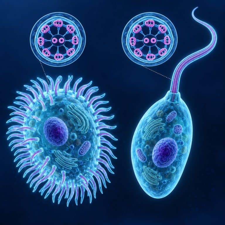
Cilia and flagella are both hair-like, motorized projections built on the same microtubule core, called the axoneme, but a cell doesn’t use them the same way. Cilia are short, cover a cell by the hundreds or thousands, and sweep in coordinated waves to move fluid past a cell that otherwise stays put. Flagella are long, usually just one or two per cell, and whip in an undulating wave to drive the whole cell forward.
Cilia vs Flagella at a Glance
| Feature | Cilia | Flagella |
|---|---|---|
| Length | Short, roughly 1–10 µm | Long, roughly 10–200 µm |
| Number per cell | Hundreds to thousands | Usually one or two |
| Movement pattern | Stiff power stroke, floppy recovery stroke, rolling metachronal wave | Symmetric, undulating wave along the length |
| Location | Covering the entire cell surface | One end of the cell, or a single tail |
| Primary function | Moves fluid or particles across a stationary cell, or propels small organisms | Propels the whole cell through fluid |
| Real-world example | Paramecium, cells lining the human airway | Euglena, human sperm |
Not every cell that moves relies on either structure — an amoeba crawls by extending pseudopods instead of beating a hair-like organelle at all. That’s a useful contrast to keep in mind as you read the rest of this comparison.
What Cilia and Flagella Have in Common: the 9+2 Axoneme
A eukaryotic cilium and a eukaryotic flagellum are built from the exact same core structure. Strip away the length and the count, and you’re looking at the same organelle deployed two different ways. The NCBI Bookshelf gives a detailed overview of the axoneme shared by cilia and flagella.
The 9+2 arrangement
The axoneme’s “9+2” name describes its cross-section: nine outer microtubule doublets arranged in a ring around two central single microtubules. This pattern is identical whether you’re looking at a motile cilium or a eukaryotic flagellum. It’s roughly 0.2–0.25 µm across — far too fine to resolve on a standard light microscope.
How dynein turns sliding into bending
Dynein arms, motor proteins that project from each outer doublet, generate the beat. Dynein “walks” along the neighboring doublet using ATP as fuel. Because nexin links and radial spokes tie the doublets together, that sliding motion gets converted into bending instead of the doublets simply separating. Stack enough of these bends together along the axoneme’s length and you get a beat or a wave. The NCBI Bookshelf’s chapter on cell movement in Molecular Biology of the Cell covers this sliding-to-bending mechanism in detail if you want the full molecular picture.
The video below walks through the shared 9+2 structure and how it powers movement in both cilia and flagella.
The basal body that anchors them
Every cilium and flagellum is rooted in a basal body, a short cylinder with a 9+0 triplet arrangement — nine microtubule triplets and no central pair. Structurally, a basal body is a centriole that has docked at the cell surface and templated an axoneme growing outward from it.

How Cilia Work — the Sweeping Stroke
Cilia move fluid by beating in two distinct phases. A stiff, extended power stroke pushes against the surrounding fluid, then a floppy recovery stroke drags back through less resistance so the cilium doesn’t undo its own work. A single cilium doing this wouldn’t accomplish much. A surface covered in thousands of them is different — each beats slightly out of phase with its neighbors, producing a rolling metachronal wave. It’s the same visual effect as wind rippling across a field of wheat. That coordinated wave is what moves mucus up a human airway, or pushes a Paramecium through pond water in a smooth, gliding line. Its companion chapter explains how dynein motors power ciliary and flagellar movement.
How Flagella Work — the Undulating Whip
A eukaryotic flagellum moves the opposite way a cilium does. Instead of a stroke-and-recovery cycle, a symmetric bending wave travels the length of the flagellum from base to tip (or tip to base), whipping side to side like a snake. There are only one or two flagella per cell rather than a coordinated field of thousands. So the whole cell gets pulled or pushed through the fluid, rather than fluid being swept past it. That’s how a human sperm cell’s tail drives the head forward in bursts. It’s also how a single flagellum tows a Euglena through water in the spiraling, tugging motion you’ll notice the first time you watch one under a scope.
The Bacterial Flagellum Is a Different Machine
A bacterial flagellum shares a name with a eukaryotic flagellum and almost nothing else. It contains no microtubules, no 9+2 axoneme, and no dynein. Instead it’s a thin, hollow, helical filament made of the protein flagellin, roughly a tenth the diameter of a eukaryotic flagellum. That filament attaches to a rotary motor embedded in the cell envelope, which spins it like a boat propeller rather than bending it like a whip. That motor runs on the proton motive force — a hydrogen-ion gradient across the membrane — instead of burning ATP directly the way dynein does. Biology LibreTexts has a detailed breakdown of that rotary motor if you want the ring-by-ring mechanics. It’s easy to see why students mix the two up, since “flagellum” gets used for both. But treating a bacterial flagellum as a small eukaryotic one is the single most common error on this topic. You can watch the effect of that rotary motor firsthand on E. coli swimming under rotary flagella, or more broadly on how bacteria use flagellin-based flagella to get around.

Primary (Non-Motile) Cilia: the Cell’s Antenna
Not every cilium beats. Most cells in the human body — including ones with no motile cilia at all — grow a single primary cilium with a 9+0 structure and no central pair. That means it can’t generate a power stroke. Instead of moving fluid, a primary cilium works as a sensory antenna, detecting chemical signals, fluid flow, or light depending on the cell type. Kidney cells use them to sense fluid flow through tubules, and photoreceptors in the retina rely on a modified primary cilium to house their light-detecting machinery. Olfactory neurons use them too, to catch scent molecules.
See Them for Yourself Under a Microscope
Cilia and flagella are two of the easiest organelles to observe in action, even though you’ll never resolve the axoneme itself on a school-grade scope. Unlike your own cheek cells sitting motionless on a slide, a live ciliate or flagellate powers itself across the field under its own steam. That motion alone tells you almost everything the comparison table above lists.
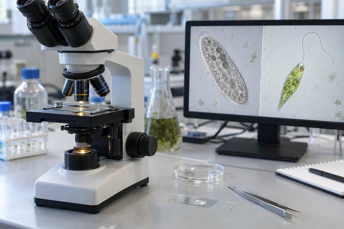
Start with a live Paramecium, the classic ciliated example. At around 400x you won’t see individual cilia as crisp hairs, but you will see a faint, shimmering, fur-like blur around the whole slipper-shaped body as it glides and spins. It reverses abruptly when it bumps something. For the flagellar side, watch the cilia beat give way to a completely different motion on Euglena, where the flagellum itself is often invisible. What you actually notice is the cell being tugged forward in a spiraling, jerky wobble. A drop of pond water is full of both ciliated and flagellated microbes in the same view. The beating corona on a rotifer is one of the more dramatic ciliary displays you can find on a hobbyist scope, looking almost like a spinning wheel.
Two mistakes trip up almost every beginner. First, don’t expect to see the 9+2 structure itself — the axoneme is roughly 0.2 µm across, well below what a light microscope can resolve. (Nikon’s MicroscopyU has a good explainer on resolution limits if you want the optics behind that ceiling.) What you’re actually seeing is the motion the axoneme produces, not the organelle itself. Second, don’t judge cilia versus flagella by length alone — also check the number (a fur coat versus one or two tails) and the beat pattern (a sweep versus a whip). Turning the light up full to see the “hairs” backfires too, since it washes out the low-contrast organelles entirely. Stopping the condenser diaphragm down improves contrast instead. Slowing the swimmers first — with a few teased cotton fibers, or a drop of methylcellulose — buys you time to actually watch the beat. Otherwise you’ll lose the organism off the edge of the field before you see anything. If you haven’t prepared one before, learning to prepare a wet mount slide properly is the first step to keeping your sample alive long enough to observe.
Frequently Asked Questions
Do humans have cilia and flagella?
Yes, both. Motile cilia line the respiratory tract and sweep mucus and trapped particles upward and out. A single flagellum, meanwhile, makes up the tail of a human sperm cell and drives it forward. Many other human cells also carry a non-motile primary cilium used for sensing rather than movement.
What is the difference between motile and primary (non-motile) cilia?
Motile cilia have the full 9+2 axoneme, usually appear by the hundreds per cell, and beat to move fluid. Primary cilia have a 9+0 structure with no central pair, appear as a single copy per cell, don’t beat, and function as a sensory antenna instead.
Can you see cilia and flagella under a light microscope?
You can see them move, but not their internal structure. The 9+2 axoneme is far below the resolving power of a light microscope. What you observe is the beating or whipping motion and its effect on the cell, not the microtubule arrangement itself. Resolving the axoneme requires an electron microscope.
How fast do cilia beat?
Motile cilia typically beat somewhere in the range of 10 to 20 times per second, though the exact rate varies by cell type and temperature. That speed is part of why individual beats blur together into the smooth, rolling metachronal wave you see under a scope rather than a visible stroke-by-stroke motion.
Are bacterial flagella always on one end of the cell?
No. Some bacteria have a single polar flagellum at one end, and others have a tuft of several flagella at one pole. Some, like many strains of E. coli, are peritrichous — covered in flagella distributed all over the cell surface that bundle together during forward swimming.
What happens if a person’s cilia don’t work properly?
A group of inherited conditions called primary ciliary dyskinesia impair normal cilia function. This most often leads to chronic respiratory infections, because mucus and trapped particles can no longer be cleared from the airway. MedlinePlus Genetics has more detail on the condition; this is background information, not medical advice.
Do plants have cilia or flagella?
Most plant cells have neither — flowering plants reproduce without swimming sperm cells at all. But a handful of older lineages — including ferns, cycads, and ginkgo — still produce flagellated sperm cells that swim to the egg. They use a structure built on the same 9+2 axoneme found in animal cilia and flagella.
Conclusion
Cilia and flagella turn out to be variations on one design rather than two unrelated structures. They share the same 9+2 axoneme, the same dynein-driven sliding-to-bending mechanism, and the same 9+0 basal body anchoring both. What separates them is deployment: cilia are short, numerous, and sweep fluid past a stationary cell, while flagella are long, few, and whip to drive the whole cell forward. Then there’s the one real trap in the topic — a bacterial flagellum borrows the name but none of the machinery.
Next time you get pond water under a scope, try picking out both movement types in the same drop. A Paramecium’s smooth glide next to a Euglena’s spiraling tug is one of the clearest side-by-side demonstrations biology offers. Have you spotted both in the same sample before? Tell us what you saw in the comments below.