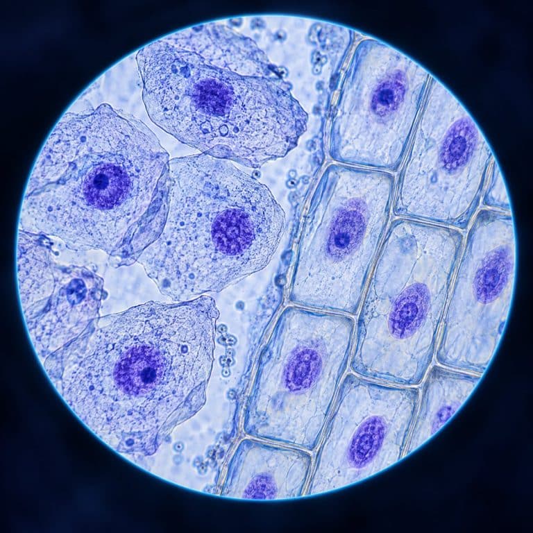
A cell nucleus under the microscope appears as a distinct, darker-stained round or oval body sitting inside an otherwise pale cell. It’s usually visible as a faint dot at 100x and clearly defined by 400x. You do not need an electron microscope to see it: a basic stain like methylene blue on a cheek cell or iodine on an onion cell is enough to make it jump out. This article walks through how to see it yourself, what each visible part is, and how roughly two meters of DNA fits inside a structure only about six micrometers wide.

How to See a Cell Nucleus Under the Microscope
The fastest way to see a nucleus at home or in a classroom is to start with an easy human cheek cell slide or an onion cell slide. Both are quick to prepare and give a reliable, high-contrast result at standard hobbyist magnification.
- Gently swab the inside of your cheek with a clean cotton swab or toothpick, or peel a thin, transparent layer from the inner surface of an onion.
- Smear the sample onto a clean slide and add a drop of water, then lower a coverslip at an angle to avoid trapping air bubbles. Follow the full wet mount slide steps if you’re new to this part.
- Add one small drop of methylene blue for a cheek cell, or iodine for an onion cell, at the edge of the coverslip and let capillary action pull it under. Blot any excess with the corner of a tissue — flooding the sample with stain washes out the contrast you’re trying to create.
- Start at 100x (10x eyepiece x 10x objective) to locate a single layer of cells rather than a thick clump, where overlapping cells hide individual nuclei.
- Switch to the 40x objective for 400x total magnification, then close the iris diaphragm slightly. Too much light washes out the faint difference between the nucleus and the surrounding cytoplasm — for background on this control, see how the iris diaphragm affects contrast.
- Rack the fine focus up and down slowly. The nucleus is the one spot that sharpens into a crisp, dense dot while the rest of the cell stays comparatively vague.
If your slides never quite look clean enough to spot this reliably, cleaner microscope slide preparation makes the nucleus far easier to find. The 400x figure above comes from calculating total magnification (objective times eyepiece). For more amateur staining technique, the Microbehunter Microscopy channel behind the video below is a solid reference.
What the Nucleus Looks Like at Each Magnification
The nucleus looks different at every step up in magnification, and knowing what to expect at each stop keeps you from second-guessing a normal, in-focus slide.
At 100x, you’re mostly hunting for cells rather than structures. A human cheek cell shows up as a pale, translucent shape a bit like a crumpled flat flake. The nucleus is just a faint central smudge you can barely separate from the rest of the cell. Switch to the 40x objective lens and that same cell resolves into a translucent, ghostly sheet of cytoplasm with one clearly darker-blue dot inside it. That dot usually sits near the center but is sometimes pushed off to one side. If you’re working with an onion epidermis mount instead, the cells form a tidy brick-wall grid, and the nucleus appears as a small denser oval sitting against one wall of each box. That’s easy to miss until you know to look for the single darker spot per cell. Push on to 1000x with oil immersion and, on a good slide, the nucleolus can show up as an even darker pinpoint inside the already-dark nucleus — a dot within a dot.
Two things ruin this view in practice. Mounts dry out under the lamp within a few minutes, shrinking the cells and distorting the nucleus. Work at a reasonable pace, or add a fresh drop of water if the edges start to curl. And an air bubble is not a cell — bubbles show up as thick, dark rings with nothing inside, while a real cell has visible internal texture and that one darker nucleus.
The Parts of the Nucleus You Can See
Under a light microscope, three structures make up what you’re actually looking at: the boundary, the DNA-containing material inside it, and a denser body within that material. The NCBI Bookshelf describes the internal organization of chromatin and the nucleolus.

| Part | Function | Appearance under the microscope |
|---|---|---|
| Nuclear envelope | Double membrane that separates the nucleus from the cytoplasm and controls what moves in and out | The sharp outer edge of the dark nucleus; the boundary itself, not something you can resolve in detail |
| Chromatin | DNA wound around histone proteins, holding the cell’s genome in its everyday, non-dividing state | The even, dark-blue fill of the nucleus, sometimes clumped darker near the edges |
| Nucleolus | Site of ribosomal RNA synthesis and ribosome subunit assembly | A smaller, even darker speck inside the nucleus, most visible at 400x to 1000x |
The Nuclear Envelope
The nuclear envelope is a double membrane — an inner and outer lipid bilayer — that encloses the nucleus and keeps its contents separate from the rest of the cell. The outer membrane is continuous with the rough endoplasmic reticulum. The whole envelope is perforated by nuclear pores, roughly 120 nanometers across, that regulate traffic like mRNA leaving the nucleus and proteins coming in. Those pores are far below what a light microscope can resolve, so under the scope the envelope simply reads as the crisp edge of the dark nucleus, not a structure you can pick apart on its own.
Chromatin vs. Condensed Chromosomes
Chromatin is what fills the nucleus during interphase — the normal, non-dividing state you’re looking at in any cheek or onion mount — and it’s DNA wound around histone proteins in a diffuse, spread-out form. Chromatin itself splits into two types: euchromatin, which is loosely packed, transcriptionally active, and stains lighter, and heterochromatin, which is tightly packed, inactive, and stains darker. This is why the edges or clumps inside a nucleus often look like the darkest part of the whole cell. Condensed chromosomes are a different, temporary state: only during mitosis or meiosis does that same chromatin coil tightly into the 46 discrete, X-shaped chromosomes most people picture from a textbook diagram. In a routine slide of resting cells, you’re seeing chromatin, not chromosomes — the two terms describe the same DNA at different points in the cell cycle, not two different substances. Pathologists lean on this distinction directly: they judge cancer cells partly by nuclei that look enlarged and irregular compared to the tidy, uniform nuclei of healthy tissue.
The Nucleolus
The nucleolus is a dense, roundish region inside the nucleus — most nuclei have one, though some have more than one. Unlike the nucleus itself, it has no membrane around it. It’s the site where ribosomal RNA is synthesized and packaged into ribosome subunits before they’re shipped out through the nuclear pores. Under the scope it appears as an even darker pinpoint within the already-dark nucleus. It resolves best once you’re at 400x to 1000x rather than the lower powers used just to find cells.

How the Nucleus Stores DNA
A single human cell holds about two meters of DNA in total, yet all of it fits inside a nucleus roughly six micrometers across. That’s a length-to-container ratio on the order of 330,000 to one. That’s only possible because DNA isn’t stored loose; it’s wound tightly at several levels. DNA first wraps about 1.7 turns around a histone octamer, a cluster of eight histone proteins, forming a nucleosome that holds roughly 147 base pairs. Strung together, nucleosomes look like beads on a string at around 10 nanometers, which then coils further into a roughly 30-nanometer fiber. That fiber loops into larger domains that make up the chromatin filling the nucleus. Each level of folding buys space. Without it, the DNA from every cell in your body would stretch across the solar system many times over, rather than sitting invisibly inside a structure smaller than the width of a human hair. OpenStax also outlines the nuclear envelope, chromatin and nucleolus in eukaryotic cells.
Why the Nucleus Stains Darker Than the Cytoplasm
The nucleus stains darker than the surrounding cytoplasm because it’s packed with DNA and RNA, and both have an acidic, negatively charged phosphate backbone. Basic, positively charged dyes — methylene blue, toluidine blue, and hematoxylin among them — bind readily to that negative charge. As a result, the nucleus soaks up far more dye than the protein-rich cytoplasm around it. This property has a name: basophilia. It’s the same mechanism at work in hematoxylin and eosin (H&E) staining used in clinical pathology labs, where hematoxylin turns nuclei blue-purple while eosin stains the cytoplasm pink. It’s also why a stain like methylene blue works so well on a home slide — you’re not adding color randomly, you’re exploiting real chemistry that concentrates dye exactly where the DNA is.
Cells Without a Visible Nucleus
Not every cell shows you a nucleus, and the exceptions help clarify what the nucleus actually does. Mature mammalian red blood cells are the clearest example: they eject their nucleus during maturation to make more room for hemoglobin. A stained red blood cell shows up as a pale, biconcave disc with no dark central dot at all. Bacteria go further still — as prokaryotes, they never had a true, membrane-bound nucleus to begin with. Their DNA sits loose in a region called the nucleoid, with no envelope around it. Between these two extremes sit cells with unusually shaped nuclei, like the lobed nuclei of white blood cells. That’s a useful reminder that the nucleus doesn’t have to be a plain circle to still be one continuous, membrane-bound compartment.
Frequently Asked Questions
Why is the nucleus sometimes off-center instead of in the middle of the cell?
Nuclear position shifts naturally with cell type and what else is packed inside — a large vacuole in a plant cell, for example, can push the nucleus flush against one wall. Off-center alone isn’t a warning sign; pathologists look at nuclear size, shape, and staining pattern together, not position, to flag something abnormal.
What stain makes the nucleus visible under a microscope?
Methylene blue works well for animal cells like cheek cells, while iodine is the standard choice for plant cells like onion epidermis. Both bind the acidic DNA in the nucleus and make it noticeably darker than the cytoplasm.
Is the nucleolus the same as the nucleus?
No. The nucleolus is a smaller, denser region located inside the nucleus, responsible for making ribosome components. It’s a structure within the nucleus, not a separate organelle or another name for the same thing.
Can you see the nucleus without a microscope?
No — at roughly 5 to 10 micrometers across, the nucleus is far smaller than the human eye can resolve; even the whole cell it sits inside is usually invisible without magnification.
Why does my nucleus look faint or blurry instead of sharp?
This is usually too little stain, a mount that’s starting to dry out, or the iris diaphragm left too wide open. Add a touch more stain, work quickly before the slide dries, and stop down the diaphragm to boost contrast rather than brightness.
Can I use iodine instead of methylene blue?
For plant cells like onion, yes — iodine is the standard choice there. For animal cells like cheek cells, methylene blue gives a cleaner result; iodine tends to stain starch and cell walls more than it stains a bare nucleus.
Is the nucleus visible in a live, unstained cell?
Sometimes as a faint outline under bright-field illumination, but rarely with any real definition — staining or techniques like phase contrast are what reliably reveal it on a standard light microscope.
Conclusion
The nucleus isn’t hidden behind a textbook diagram. With nothing more than a cheek swab, an onion peel, and a drop of stain, it’s one of the most reliably observable structures at ordinary hobbyist magnification. Once you know to look for a single darker dot at 400x, and can tell chromatin from a nucleolus from the envelope around them both, the idea of two meters of DNA folded into a six-micrometer sphere stops being an abstract number. It becomes something you’ve actually watched come into focus.
Have you managed to spot the nucleolus as that darker pinpoint inside the nucleus, or found a stain that works better for you than methylene blue or iodine? Share what you saw, and what magnification it took to get there, in the comments below.