
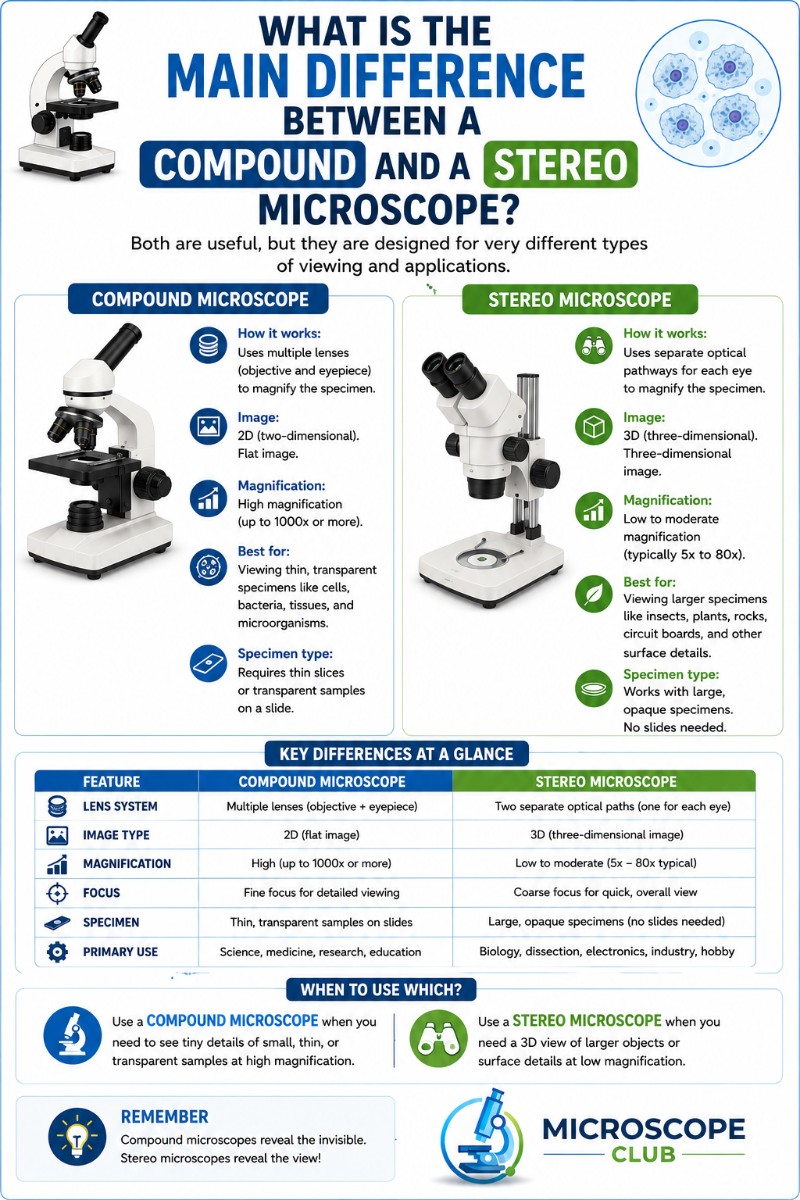
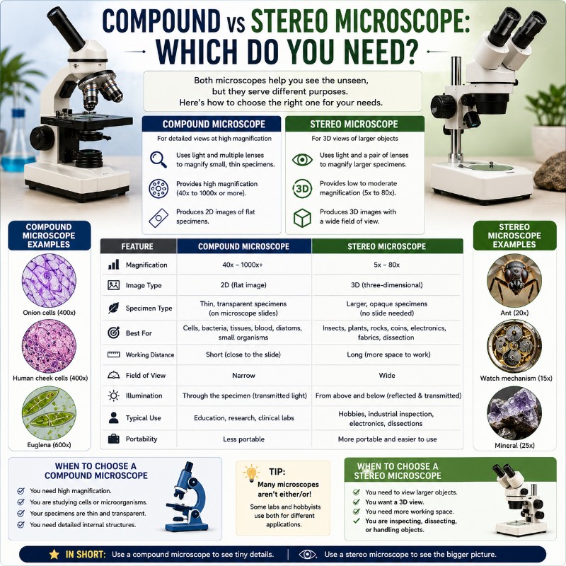
When choosing between a compound vs stereo microscope, the answer comes down to one question: are you looking through something thin on a slide, or at the surface of a solid object? Compound microscopes are built for cells, microbes, and slide-mounted samples at 40x–1000x magnification; stereo (dissecting) microscopes are built for 3D whole objects — insects, coins, circuit boards, flowers — at 10x–40x. Get this distinction right and the rest of the decision is easy.
Quick Comparison Table
| Feature | Compound Microscope | Stereo Microscope |
|---|---|---|
| Magnification range | 40x – 1000x | 10x – 40x (some to 80x) |
| Image type | 2D, flat, inverted | 3D, upright, non-reversed |
| Specimen type | Thin, translucent (on a slide) | Solid, opaque, whole objects |
| Lighting direction | Below (transmitted/diascopic) | Above (reflected/episcopic) |
| Working distance | Very short (0.2–0.6 mm at high mag) | Long (75–150 mm typical) |
| Slide required? | Yes | No |
| Typical price (entry) | $100 – $300 | $100 – $400 |
| Best for | Cells, microbes, biology slides | Dissection, inspection, repair, kids |

What Is a Compound Microscope?
How It Works
A compound microscope uses two lens stages in series: an objective lens close to the specimen and an eyepiece (ocular) at your eye. Total magnification equals objective power multiplied by eyepiece power — a 40x objective with a 10x eyepiece gives 400x. Light travels through the specimen from a source below the stage (transmitted illumination), which means your sample must be thin enough to let light pass through. The resulting image is flat, two-dimensional, and inverted. You can read more about the parts of a compound microscope if you want to go deeper on the optics.
The practical resolution ceiling of a light-based compound scope is about 0.2 micrometers (200 nm), set by the diffraction limit and the numerical aperture of the objective lens. Going beyond roughly 1000x produces “empty magnification” — the image gets bigger but no new detail appears, because you’ve hit the resolution wall, not because the lens is better. For more on this, see our guide on microscope resolution and numerical aperture. The Khan Academy microscopy primer also has a solid breakdown of how light microscopes work for students new to the concept.
What It’s Best For
Compound microscopes shine wherever you need to see structure at the cellular or sub-cellular level:
- Blood cells, bacteria, protozoa, pond microorganisms
- Onion cells, cheek cells, plant tissue cross-sections
- Prepared slides for school biology labs
- Yeast and mold spores at cellular detail
- Viewing blood cells and other stained tissue specimens
If your goal is to learn how to prepare microscope slides and explore the invisible world of cells, a compound scope is the tool for the job.
What Is a Stereo (Dissecting) Microscope?
How It Works
A stereo microscope — also called a dissecting microscope (same instrument, different name) — routes light through two completely separate optical paths, one per eyepiece, angled roughly 10–15° apart. Each eye receives a slightly different perspective on the same object, and your brain fuses those two views into a genuine 3D, stereoscopic image. The specimen sits in open air on a flat stage; light bounces off its surface from above (reflected illumination), though many models also have an optional bottom light for translucent items. The image is upright and non-reversed, which means you can use tweezers, a scalpel, or a soldering iron under the scope and your hands move exactly where you expect them to.
The depth of field is large and the working distance is generous — typically 75–150 mm — so a bumble bee, a circuit board, or a mineral specimen sits comfortably on the stage with room to work. The field of view is wide, which makes it easy to find and track objects.

What It’s Best For
Stereo microscopes are the go-to whenever the specimen is solid, opaque, or three-dimensional:
- Dissecting insects, flowers, small animals, seed pods
- Examining coins, stamps, minerals, and rocks
- Circuit board inspection, soldering, and electronics repair
- Watchmaking, jewelry setting, and gemstone grading
- Botany and entomology field specimens
- Young children exploring rocks, bugs, and leaves — no slide prep required
For a comprehensive look at stereo scopes specifically, our dissecting microscope guide covers everything from optics to accessories. The Nikon MicroscopyU introduction to stereomicroscopy is also an excellent technical reference if you want to understand the dual-optical-path design in more depth.

Key Differences Explained
Magnification: Compound scopes cover 40x–1000x; stereo scopes cover 10x–40x. Higher numbers don’t make a compound scope better for all tasks — they make it better only for specimens that need that resolution. A coin at 400x through a compound scope would be completely out of focus and useless.
Image dimensionality: Compound images are 2D and flat. Stereo images are genuinely 3D. This matters enormously for dissection or repair — you need depth cues to judge where your tool tip actually is.
Lighting direction: Compound scopes illuminate from below (light passes through the specimen). Stereo scopes illuminate from above (light reflects off the surface). This single difference explains most of the use-case split: if your specimen is opaque, transmitted light can’t reach your eyes and a compound scope won’t work at all.
Specimen preparation: Compound microscopy requires a thin, slide-mounted, often stained specimen. Stereo microscopy requires nothing — place the object on the stage and look. This makes stereo scopes far more approachable for kids and hobbyists.
Working distance and depth of field: At 400x on a compound scope, the objective lens is less than 1 mm from the slide; at 100x oil-immersion it’s 0.2 mm. Depth of field is razor-thin — a fraction of a micrometer. Stereo scopes have centimeters of working space and a large, forgiving depth of field.
The Magnification Myth: Why Higher Isn’t Always Better
The single most common buying mistake in microscopy is treating magnification as the primary quality metric. It isn’t. A 1000x compound microscope is completely useless for inspecting a coin or dissecting a flower — at that magnification, through-light optics, and a razor-thin focal plane, you’d see nothing useful. Conversely, a stereo scope’s 20x feels “low” on paper but is exactly right for seeing a tick’s mouthparts in full 3D detail.
The real limit of a light microscope is resolution — the smallest separation between two points that the scope can distinguish. The physics ceiling is about 0.2 micrometers, governed by the wavelength of light and the numerical aperture of the lens (Abbe’s equation: d = λ / 2NA). Magnification beyond the resolution limit is called “empty magnification”: the image gets bigger and blurrier, but no new information appears. A 2000x setting on a light microscope adds no detail — it just magnifies the blur. This is well-documented in cell biology reference literature on optical microscopy limits. For a deeper look at where optical limits actually sit, see our article on highest magnification and resolution in light microscopy.
The right question is never “which has more magnification?” It’s “which magnification range, image type, and lighting setup fits my specimen?” Answer that and you’ll pick the right scope every time.
Which Microscope Should You Buy? (Decision Guide)
Use-Case Decision Table
| Task / Use Case | Right Scope |
|---|---|
| Blood cells, bacteria, protozoa, pond microbes | Compound |
| Onion cells, cheek cells, plant tissue slides | Compound |
| School biology lab (prepared slides) | Compound |
| Yeast, mold spores at cellular level | Compound |
| Dissecting insects, flowers, small animals | Stereo |
| Coins, stamps, minerals, rocks | Stereo |
| Circuit board repair, soldering, electronics | Stereo |
| Watchmaking, jewelry, gemstone inspection | Stereo |
| Botany / entomology — whole specimens | Stereo |
| Examining mold colony surface / texture | Stereo |
| Young kids exploring rocks, bugs, leaves | Stereo |
For Students and Schools
If the goal is the standard biology curriculum — cell theory, mitosis, protists, bacteria — a compound light microscope is what every school biology lab uses and what every standardized curriculum assumes. Budget $150–$250 for a solid entry monocular or binocular compound scope. Binocular microscopes are more comfortable for extended lab sessions. If the school also does dissection, a separate stereo scope for that station is ideal — they serve different functions and trying to do both with one tool means compromising on both.
For Hobbyists and DIY / Repair
Electronics repair techs, watchmakers, jewelers, and model builders almost universally use stereo scopes. The 3D view, long working distance, and upright image make hands-on work practical. Hobbyist stereo scopes run $150–$400; professional trinocular models with camera mounts — see our guide to trinocular microscopes — run $400–$1,000+. If you’re also interested in pond-water biology or microbiology as a side hobby, you’ll want a compound scope separately — the two tools don’t overlap in function.
For Kids and Beginners
A stereo scope is almost always the better first microscope for children under 12. No slide preparation, instant 3D results, and a forgiving depth of field mean a kid can drop a beetle on the stage and immediately see something amazing. If the child is specifically interested in biology class and prepared slides, a simple compound scope works — but expect a steeper learning curve.
Can’t Decide? The Dual-Purpose Option
A handful of entry-level “combination” or “duo” microscopes offer both compound and stereo modes in one unit. The dual-purpose Duo-Scope microscope from My First Lab is the best-known example and a reasonable compromise for undecided beginners — you get a taste of both worlds without two price tags. The tradeoff: optical quality in each mode is somewhat lower than a dedicated single-purpose scope at the same price point. If you know your primary use case, buy dedicated. If you genuinely don’t know yet, the combo scope buys you time to figure it out.
For a broader view of your options, our overview of different types of microscopes covers the full landscape beyond just compound and stereo.
Frequently Asked Questions
What is the main difference between a compound and a stereo microscope?
A compound microscope uses transmitted light that passes through a thin slide-mounted specimen to produce a 2D image at 40x–1000x. A stereo microscope uses reflected light off solid objects and two separate optical paths to produce a 3D image at 10x–40x. One is for cells and slides; the other is for whole objects and hands-on work.
Which is better, a compound or a stereo microscope?
Neither is universally better — they do completely different jobs. A compound scope is better for viewing cells, bacteria, and slide-mounted specimens. A stereo scope is better for examining solid objects, dissecting specimens, and any task where you need to work with tools under the lens. “Better” only makes sense relative to what you’re trying to see.
Can a stereo microscope see cells?
Generally no. At 10x–40x, a stereo microscope shows surface texture and structure, but individual cells (typically 10–100 micrometers) require the higher magnification and resolution of a compound scope. You might see a mass of cells as a tissue layer, but you won’t resolve individual cell walls or nuclei.
What magnification do compound and stereo microscopes have?
Compound microscopes typically range from 40x to 1000x, with 100x oil-immersion being the practical optical ceiling for light microscopy. Stereo microscopes typically range from 10x to 40x, with some zoom models reaching 50x–80x. The stereo scope’s lower magnification is a feature — it delivers a wide field of view, depth of field, and working distance that high-magnification optics physically cannot.
Is a dissecting microscope the same as a stereo microscope?
Yes — they are exactly the same instrument. “Dissecting microscope” describes the most common use (dissecting specimens in a biology lab), while “stereo microscope” describes the optical design (two separate light paths creating a stereoscopic 3D image). Both terms refer to the same tool.
Do you need slides for a stereo microscope?
No. That’s one of the stereo scope’s biggest advantages. You place the whole object — an insect, a coin, a circuit board, a rock — directly on the stage and view it as-is. No mounting, no staining, no cover slip required. This makes stereo scopes dramatically faster and easier to use for solid specimens.
Which microscope is best for a beginner or child?
For young children (under 10–12), a stereo microscope is almost always the better choice — no slide prep, instant 3D results, and nothing to break or stain. For a teenager or adult beginning biology or microbiology study, a compound microscope is more aligned with what they’ll encounter in school and gives access to the cell-level world. If you’re truly undecided, a combination duo-scope lets you try both.
Conclusion
The compound vs stereo microscope question has a clear answer once you know what you’re trying to see. Compound scopes belong in any setting where the goal is cells, microbes, or slide-based biology at high magnification. Stereo scopes belong wherever the specimen is solid, three-dimensional, or needs to be worked on with your hands. Neither is a universal tool, and the magnification number on the box tells you far less than you’d think — what matters is lighting direction, image dimensionality, and specimen type.
Have you tried both types, or are you deciding between them for the first time? Drop a comment below and tell us what you’re hoping to look at — we’re happy to point you toward the right setup for your specific situation.