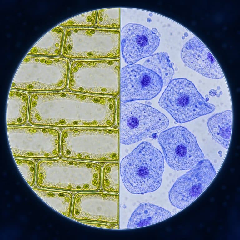
A plant cell vs animal cell comparison comes down to eight structural differences. Plant cells have a rigid cellulose cell wall, one large central vacuole, and chloroplasts, while animal cells have none of these but do have centrioles and a flexible outer membrane. Both are eukaryotic cells that share a nucleus, mitochondria, and several other organelles. The clearest way to see the split for yourself is under a light microscope, comparing a plant sample like onion with an animal sample like a cheek cell scrape.
Plant Cell vs Animal Cell at a Glance
The table below lines up the eight features that separate a plant cell from an animal cell, plus the two size/shape traits those features produce. OpenStax provides a detailed comparison of plant and animal cell organelles.
| Feature | Plant Cell | Animal Cell |
|---|---|---|
| Cell wall | Yes (cellulose) | No |
| Central vacuole | One large vacuole, up to 80–90% of volume | Small or none |
| Chloroplasts | Yes, in photosynthetic tissue | No |
| Plasmodesmata | Yes, channels between cells | No |
| Centrioles | Generally absent in higher plants | Yes |
| Lysosomes | Rare; vacuole handles digestion | Yes, common |
| Shape | Fixed, boxy/rectangular | Irregular, rounded |
| Typical size | 10–100 micrometers | 10–30 micrometers |
What Plant and Animal Cells Have in Common
Plant and animal cells are both eukaryotic, meaning each has a true, membrane-bound nucleus and a set of membrane-bound organelles doing the same basic jobs. That shared foundation matters: the differences below are variations on one cell plan, not two unrelated designs. Both cell types have a nucleus (with a nucleolus and chromatin inside), a plasma membrane, mitochondria, and cytoplasm. Both also have ribosomes, rough and smooth endoplasmic reticulum, a Golgi apparatus (called a dictyosome in plants), and a cytoskeleton. Mitochondria are worth flagging specifically, since it’s a common mix-up: plant cells have mitochondria too, and use them for respiration exactly like animal cells do. Chloroplasts are an addition, not a replacement. Its companion overview summarizes the shared structures and key differences of eukaryotic plant and animal cells.
Structures Only Plant Cells Have
Four structures show up in plant cells and not in animal cells: the cell wall, the central vacuole, chloroplasts, and plasmodesmata.
The Cell Wall
A plant cell’s cell wall sits outside the plasma membrane and is built from cellulose, a rigid fiber that animal cells never produce. That wall is what gives a plant cell its fixed, boxy shape and its structural strength. It’s roughly 0.1 to 10 micrometers thick — thick enough to show up as a crisp dark outline under a light microscope. An animal cell has no equivalent layer; only the flexible membrane holds it together.
The Large Central Vacuole
A mature plant cell has one dominant central vacuole, a fluid-filled sac that can take up 80 to 90 percent of the cell’s total volume. That volume pushes the nucleus and the rest of the cytoplasm out toward the wall. That vacuole stores water and dissolved ions and maintains turgor pressure — the internal push against the cell wall that keeps plant tissue rigid. Lose that pressure through water loss and the plant wilts, which is a direct, visible consequence of this one organelle. Animal cells may have a few small vacuoles or none prominent, but never one giant central reservoir.
Chloroplasts
Chloroplasts are the organelles that contain chlorophyll and carry out photosynthesis, converting light into chemical energy the plant can use. It’s worth correcting a common assumption here: not every plant cell looks green under a microscope. Onion bulb cells, for example, grow underground and typically carry few or no chloroplasts, so an onion slide looks clear or faintly colorless rather than green. Chlorophyll shows up in above-ground, light-exposed tissue like the chloroplast-packed cells inside a leaf, not in a root vegetable. Animal cells never have chloroplasts; they get their energy strictly from respiration in the mitochondria.

One more plant-only structure rounds out this group: plasmodesmata, tiny channels that run through the cell wall and connect one plant cell directly to its neighbors. They let water and small molecules pass cell to cell. Animal cells have no equivalent — their neighboring cells stay physically separate, connected only by looser junctions between membranes.
Structures Only Animal Cells Have
Animal cells carry their own set of structures that plant cells lack or rarely use in the same way.
- Centrioles. Paired cylindrical structures that organize the spindle fibers during cell division. Higher plant cells generally don’t have them and divide without this particular machinery.
- Lysosomes. The main digestive organelles in an animal cell, breaking down waste and worn-out cell parts. Plant cells can show similar digestive activity, but the large central vacuole absorbs most of that job, so lysosomes are the classic animal-cell marker.
- A flexible plasma membrane as the only outer boundary. With no wall on the outside, an animal cell’s edge can flex and deform, which is exactly why animal cells end up irregular and rounded instead of boxy.
- Smaller vacuoles, if any. Where a plant cell has one dominant central vacuole, an animal cell has several small ones or none prominent at all.
Shape and Size — Why Plant Cells Look Like Boxes
A plant cell’s shape is fixed and rectangular because the cellulose cell wall acts like a rigid box the membrane can’t push past. The cell grows into that shape and stays there. An animal cell’s shape is irregular and rounded because it has only the flexible plasma membrane holding it in. It deforms to fit against its neighbors or floats as a loose blob when unattached. Size follows the same logic on a general scale: plant cells typically run about 10 to 100 micrometers, while animal cells typically run about 10 to 30 micrometers. Both ranges vary a lot by cell type and species. Put a plant sample and an animal sample side by side under a microscope and this shape difference is the fastest way to tell them apart. You’ll spot it before you’ve identified a single named organelle.

Seeing the Difference Yourself: Onion vs Cheek Cells
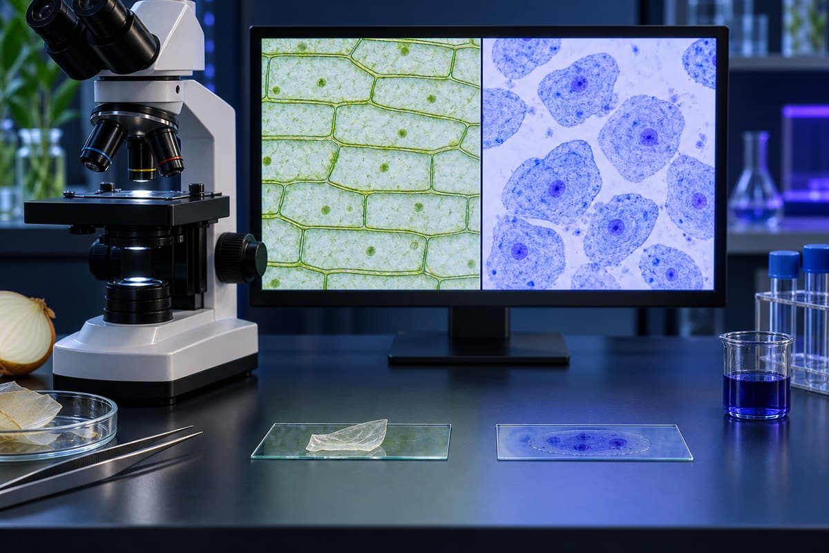
The fastest way to make this comparison concrete is to mount both samples yourself. An onion peel gives you a plant cell in seconds; a cheek scrape gives you an animal cell just as fast, and the two slides look nothing alike.
What onion (plant) cells look like under the microscope
Peel the thin, transparent inner membrane from an onion layer — not the thick fleshy scale — and you already have a sample one cell thick. Under the scope, what onion cells look like under a microscope is a clean tiled grid: long, brick-shaped cells packed edge to edge like a stone wall. Each one is outlined in a crisp dark line where the cell wall sits. Add a drop of iodine and a small dense dot appears — the nucleus — pushed off to one side rather than centered, because the large vacuole shoves it against the wall. The rest of the interior looks empty and clear; that emptiness is the vacuole itself. A common beginner mistake here is expecting green cells and assuming a colorless slide means failure — onion bulb cells have no chloroplasts, so clear is correct, not a mistake. A second mistake is viewing the thick, folded middle of the peel instead of its thin edge, which just produces a dark, unreadable pile under the stain.
What cheek (animal) cells look like under the microscope
A cheek scrape looks like almost nothing until it’s stained. Unstained, you’ll see faint, pale, rounded blobs drifting singly or in loose clumps, with no straight edges and no grid. Nothing is gluing them together the way a cell wall would. Add methylene blue and a round nucleus appears roughly centered in each blob; the edges stay soft and irregular, closer to a fried egg gone wrong than a geometric shape. That’s human cheek cells under a microscope in a nutshell. It’s worth comparing against another animal sample like human blood cells under the microscope to see the same rounded, wall-free pattern repeat. The single biggest beginner error is skipping the stain and concluding there’s nothing on the slide. Unstained cheek cells are nearly transparent, so the fix is simple: prepare and stain a microscope slide properly before judging what you see.
A few practical notes apply to both samples. Work quickly, since drying out shrivels cells and distorts their edges before you get a clean look. Too much light washes out the already low-contrast cheek cells, so stop down the condenser or iris rather than cranking up brightness. And watch for air bubbles — a perfect black circle with a thick ring is a bubble, not a nucleus. Start at 40x or 100x to find the overall pattern, then move to 400x once you’ve located a cell worth a closer look. If you want to build this technique from scratch, make a wet mount slide first — it’s the same basic prep both the onion and cheek samples use. For a historical bonus, the rigid, boxy look of plant cell walls is exactly what led Robert Hooke to coin the word “cell” in the first place. That coinage traces back to the cork cells Robert Hooke first drew in 1665.
Frequently Asked Questions
Do animal cells have a cell wall?
No. Animal cells have only a flexible plasma membrane as their outer boundary — the rigid cellulose cell wall is unique to plant cells.
Do plant cells have mitochondria?
Yes. This is a common misconception — plant cells have mitochondria and use them for respiration just like animal cells do; chloroplasts are an addition to that, not a substitute for it.
Do plant cells have a nucleus?
Yes. Both plant and animal cells are eukaryotic, which means both have a true, membrane-bound nucleus. Confusing this is a common beginner error, since some assume only animal cells have one.
Which is bigger, a plant cell or an animal cell?
Plant cells are generally larger, typically 10 to 100 micrometers, compared to roughly 10 to 30 micrometers for animal cells — though both ranges vary widely by species and cell type.
Do plant cells have lysosomes?
Generally no — or only rarely. In a plant cell the large central vacuole handles most of the waste breakdown that lysosomes do in an animal cell, which is why lysosomes are treated as a classic animal-cell marker rather than a shared organelle.
Conclusion
Once you know the eight features that separate them, a plant cell and an animal cell stop looking like a memorization exercise. They start looking like two logical variations on the same eukaryotic blueprint. The wall, the central vacuole, and the chloroplasts explain everything about a plant cell’s fixed, boxy shape and self-feeding lifestyle. The flexible membrane, centrioles, and lysosomes explain an animal cell’s rounded, mobile, digest-what-you-eat design just as fully. And the long list of shared organelles is the reminder that these are close relatives, not opposites.
The quickest way to make all of this stick is to look for yourself: mount an onion peel, mount a cheek scrape, and watch the grid-versus-blob difference appear under your own scope. Have you tried this comparison yourself? Tell us what you found in the comments below.