Cyanobacteria Under a Microscope: What You’ll See
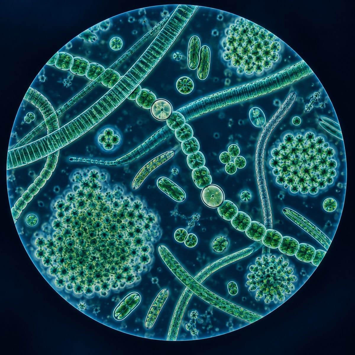
Recognize filamentous, colonial and single-celled cyanobacteria, identify heterocysts and prepare a pond-water wet mount safely.
Dive into the fascinating Microscopic World with stunning images and captivating discoveries. Our collection of microscopic images showcases bacteria, cells, crystals, pollen, and more, revealing the hidden beauty of the world around us. Be amazed by what’s invisible to the naked eye.
Recognize filamentous, colonial and single-celled cyanobacteria, identify heterocysts and prepare a pond-water wet mount safely.
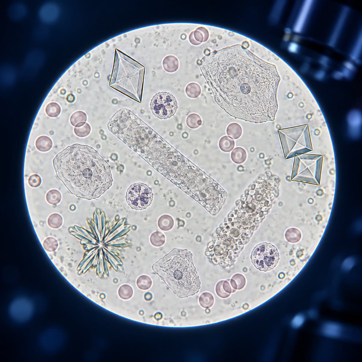
Learn how urine sediment is prepared for microscopy and what cells, casts, crystals and microorganisms may look like during urinalysis.
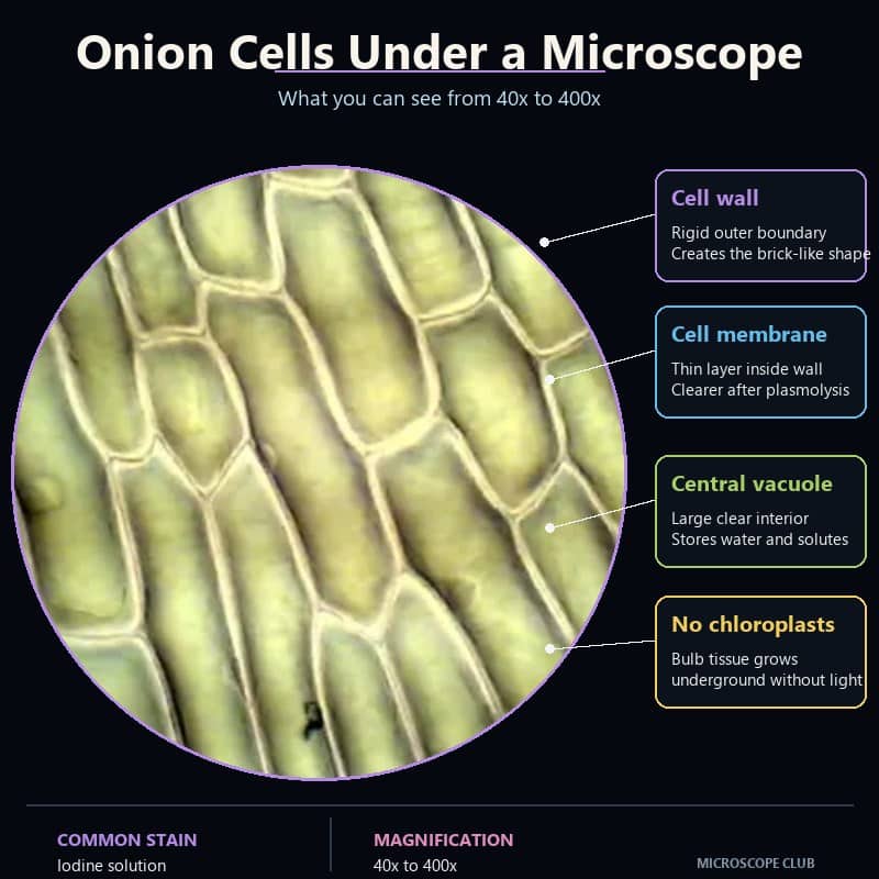
Onion epidermal cells (Allium cepa) appear under a compound light microscope as a tight, regular grid of elongated rectangles — like pale bricks mortared edge-to-edge with almost no gap. The moment you find focus at 40x, the pattern is unmistakable: dozens of identical cells tessellating across the field with a geometric precision you simply do …

Understand why ordinary light microscopes cannot resolve most viruses, how resolution differs from magnification and what electron microscopes reveal.

See how individual fungal hyphae form a mycelium and learn to prepare, stain and distinguish septate from coenocytic threads.
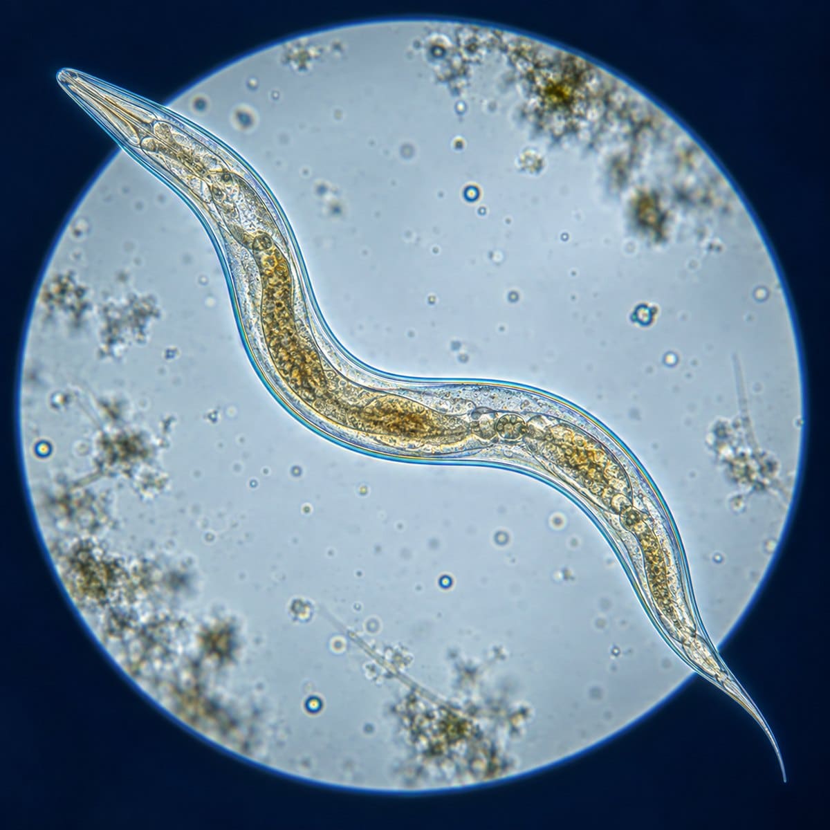
Learn where to collect free-living nematodes, prepare a wet mount and recognize their tapered body, internal gut and whip-like movement.
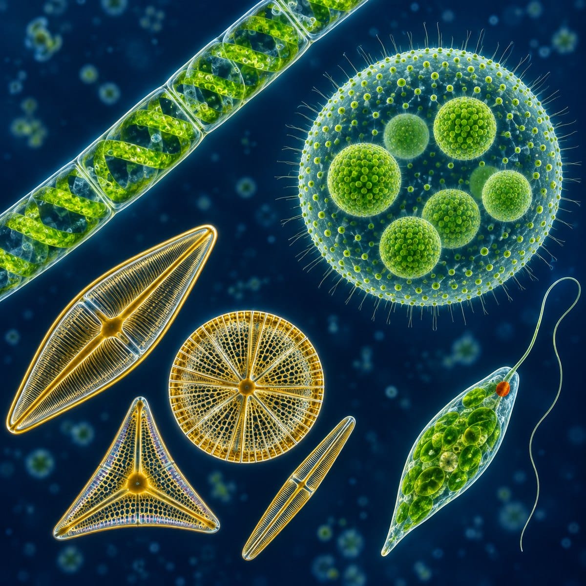
Explore the diversity of algae and learn how to collect, mount and recognize Spirogyra, Volvox, diatoms and Euglena under a microscope.

Compare the four traditional protozoan movement types and learn how to recognize Amoeba, Paramecium and Euglena in pond water.
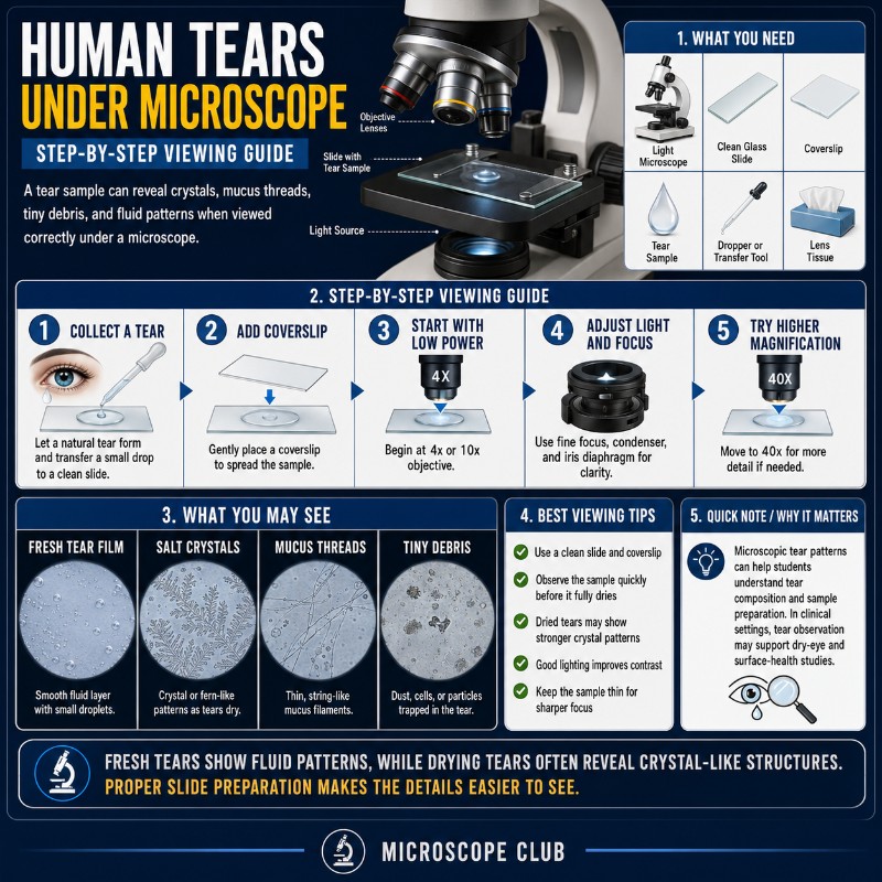
Dried human tears under a microscope look nothing like what you’d expect from a salty drop of liquid — they form branching, frost-like crystal landscapes that change shape from one tear to the next. Each drop leaves behind a map of salt, proteins, and lipids as the water evaporates, and no two ever look identical. …
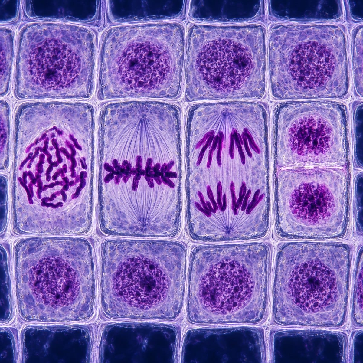
Learn how to prepare and examine an onion root tip slide, find the dividing zone and identify the visible stages of mitosis.