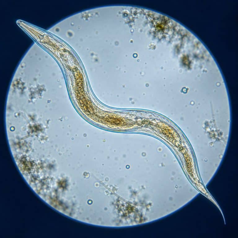
Nematodes under the microscope look like thin, glassy threads that snap into a tight curve and spring back before you even register their tapered shape. These unsegmented roundworms turn up in garden soil, pond water, and even a jar of raw vinegar. A basic student microscope is all it takes to watch one thrash across the field of view. This guide covers what to look for and where to collect a sample that actually has nematodes in it, plus how to mount a slide so they hold still long enough to study.

What Nematodes Look Like Under the Microscope
A nematode’s body shape is the giveaway before anything else about it: a smooth, unsegmented, round-in-cross-section tube that tapers at one end and looks blunt at the other. There is no visible banding, no legs, no shell — just a clear-to-milky thread that seems to appear out of nowhere in a drop of pond water, then suddenly moves. At low power, the motion catches your eye before the shape does; the body itself can be almost invisible against the background until it whips into a C and snaps back straight. OpenStax diagrams the pseudocoelomate body plan of nematodes.
Body Shape and the Tapered Tail
The tapered tail is the fastest identification cue a beginner can use. One end of the worm is rounded and blunt (the head), and the other draws down to a fine point (the tail). No other common pond or soil organism narrows quite that way. A stray cotton fiber or eyelash can look similar at first glance, but a fiber stays perfectly still and doesn’t taper evenly. An air bubble, by contrast, has a hard black rim instead of a soft, tapered edge. If it tapers to a point and moves, it’s a nematode.
What’s Translucent Inside: Gut, Pseudocoelom, and Eggs
A nematode’s translucent cuticle turns the whole animal into a living viewing window. Down the middle of the body runs a darker line — the intestine — visibly packed with granular contents that shift as the worm bends. Surrounding that gut tube is the pseudocoelom, a fluid-filled body cavity that sits between the gut and the outer body wall. It’s what gives the whole animal its slightly swollen, glassy look. In a well-fed, gravid (egg-carrying) female, you can often make out a neat row of oval eggs lined up like beads inside a tube. It’s one of the clearest “aha” moments of the whole exercise.
The Whip-Like Movement (and Why They Move That Way)
A nematode moves by thrashing side to side in a whip-like, S-shaped wave rather than crawling or inching forward. That single fact explains the entire motion: nematodes only have longitudinal muscles running the length of the body, with no circular muscles to squeeze and push the way an earthworm does. Unable to contract in a ring, the worm can only bend against the elasticity of its own cuticle, producing the sinusoidal thrash you see rather than a smooth peristaltic crawl. It’s also a useful behavioral tell — small bacteria-feeding nematodes lash frantically and barely travel anywhere, while larger predatory species glide more deliberately across the field of view.
Where to Find Nematodes: 4 Reliable Sources
Free-living nematodes are among the most abundant animals on the planet, and a single handful of healthy garden soil can hold thousands of them. The real challenge is sampling the right spot, not finding a rare organism. These four sources cover the range from “almost always works” to “guaranteed.”
Soil and Compost
Rich, moist garden or compost soil is thick with nematodes, most of them harmless bacterial or fungal feeders working through decaying organic matter — the same busy community you’d meet examining soil under a microscope more broadly. A simple home method: soak a soil sample in water and let it settle for 20–30 minutes. Then pipette from just above the sediment layer, rather than the clear water on top — that’s where the migrating worms collect. For a cleaner catch, use the Baermann funnel method described below.

Pond Water and Sediment
A drop of pond water pulled from just above the sediment, rather than the clear surface layer, is one of the most reliable places to find a nematode. Look there and you’ll often see one gliding past. Nematodes frequently share the same drop as rotifers, so don’t be surprised if both show up on the same slide.
Moss and Decaying Plant Matter
Squeezing a clump of rehydrated moss or lichen over a slide reliably releases nematodes along with whatever else is living in the moisture film. It’s the same moss-soaking trick used to find tardigrades. Compost and rotting plant matter work the same way: soak, squeeze, and check the runoff.
Vinegar Eels: The Guaranteed Beginner Source
Vinegar eels (Turbatrix aceti) are a small, harmless nematode that lives in unpasteurized vinegar. They are the single most reliable source for a classroom or a first attempt at home. Add a splash of raw, unfiltered apple cider vinegar — the kind labeled “with the mother” — to a jar of diluted vinegar and sugar water. A population will typically be teeming within a couple of weeks. Unlike a soil or pond sample that might come up empty, a jar of vinegar eels means live worms are available on demand. The hobbyist microscopy community at Micscape Magazine has documented this culture method for decades, and it remains the standard beginner recommendation.

How to Prepare a Nematode Slide
Preparing a nematode slide comes down to three steps. First, extract a sample rich in worms. Then mount it as a thin wet mount, and slow the worm down enough to actually study it.
The Soak-and-Pipette Method (Simple)
- Place a small sample of soil, moss, or compost in a jar and cover it with room-temperature water.
- Let it sit undisturbed for 20–30 minutes so nematodes migrate off the debris and settle toward the bottom.
- Use a pipette to draw liquid from just above the sediment layer, not the clear water on top.
- Place one drop on a clean glass slide and add a coverslip to complete the wet mount.
The Baermann Funnel (Higher Yield)
A Baermann funnel extracts a much larger, cleaner concentration of live nematodes from a soil sample than simple soaking. To build one:
- Line a funnel with a single layer of tissue or fine cheesecloth and fill it with water.
- Suspend a loose soil sample on the tissue so it’s in contact with the water below.
- Attach a short length of tubing to the funnel’s stem and clamp it shut.
- Leave the setup for 12–24 hours; live nematodes swim down through the soil and water, then sink into the tubing.
- Open the clamp and drain a few drops into a collection dish — this is your most concentrated sample.
Making the Wet Mount and Slowing the Worm Down
Use a small drop of liquid rather than a large flooded one. Too much water lets the worm swim out of the microscope’s thin depth of focus, while a slightly crowded mount pins it in one plane where you can actually track it. Work quickly, because a mount can dry out at the coverslip edge within minutes, stopping the worm along with it. Center and follow the worm at 40x first before stepping up to higher magnification; cranking straight to 400x makes it easy to lose the worm entirely. A wide-open condenser also washes out a translucent specimen — stopping down the iris a little gives the glassy body more contrast against the field.
What You See at Each Magnification
Total magnification equals eyepiece power multiplied by objective power. With a standard 10x eyepiece, switching between your 4x, 10x, and 40x objective lenses takes you from 40x up to 400x total.
| Magnification | What You See |
|---|---|
| 40x (4x objective) | The whole worm as a thread shape, tapered ends, and the gross whip-like thrashing motion |
| 100x (10x objective) | The translucent body wall, the darker central gut line, and internal contents shifting as it moves |
| 400x (40x objective) | Cuticle surface detail, the pharynx and its pumping bulb, and in gravid females, individual developing eggs |
Nematode Anatomy Through the Cuticle
A nematode’s cuticle is a tough, flexible, acellular (non-cellular) outer layer secreted by the hypodermis (the tissue layer just beneath it), and the worm molts this cuticle as it grows. Because the cuticle is translucent, most of the internal anatomy is visible without any staining. At the head end sits the mouth, or buccal cavity; some predatory and plant-parasitic species carry a spear-like stylet there, used to pierce prey or plant roots. Just behind the mouth is the pharynx, a muscular pumping tube that often ends in a visible bulb. In a live specimen at 400x, you can watch this bulb rhythmically contract as the worm feeds. The intestine runs as a dark, granular tube through most of the body length, suspended within the pseudocoelom. In a gravid female, the reproductive tract shows as a row of developing eggs near the rear third of the body. Feeding style tracks with mouthpart type: bacterial feeders (simple tube mouths) tend to thrash fast and erratically. Stylet-bearing plant parasites and larger predators move with a slower, more deliberate glide.
Friend or Foe? Free-Living vs. Parasitic Nematodes
Most nematodes you’ll find in soil, pond water, or vinegar are free-living and completely harmless to humans. They feed on bacteria, fungi, or decaying organic matter rather than on people, pets, or crops. A minority of species within the phylum are parasites — hookworm, Ascaris, and pinworm among them. But these are specific species with specific life cycles, not a trait of nematodes as a group. This guide is an educational observation resource, not medical advice, and it’s still good practice to wash your hands after handling soil or pond samples. For anyone concerned about parasitic species specifically, the CDC’s parasitic disease pages cover human health risks in detail. The taxonomy of the group as a whole is well summarized in Britannica’s roundworm entry. The nematodes you’re likely to pull from your backyard soil or a jar of vinegar are not the ones on those pages. Its invertebrate overview compares free-living and parasitic members of Nematoda.
Frequently Asked Questions
Are the nematodes in soil harmful to humans?
No, the vast majority of soil nematodes are free-living bacterial or fungal feeders that pose no risk to humans. The handful of parasitic nematode species that do affect people, such as hookworm, have specific transmission routes. They are not what you’ll typically find in a garden soil sample or compost pile.
What’s the difference between a nematode and a rotifer or other pond critter?
A nematode is a smooth, unsegmented thread with a tapered tail and no visible internal wheel-like structures. A rotifer, by contrast, has a distinctive ciliated “wheel organ” at its head that beats to create feeding currents, often paired with a segmented-looking foot. Nematodes also lack the fixed, boxy shape of many rotifers, moving instead in a loose whip-like thrash.
Can you see nematodes with the naked eye?
Larger free-living nematodes, at 1–3 mm long, can appear as barely visible pale threads to the naked eye in good light. You won’t see any real detail, though — body shape, the tapered tail, and internal structures all require at least 40x magnification.
How do you keep nematodes alive on the slide long enough to watch?
A trace of methyl cellulose stirred into the drop is the standard trick — it thickens the water just enough to slow a fast-moving nematode without harming it, which is what lets you actually watch details like the pharynx pumping. Even without it, a plain water mount usually holds up for five to ten minutes before evaporation at the coverslip edge stops the worm for good, so work through your observations in that window rather than saving the best magnification for last.
Conclusion
A nematode under the microscope is one of the easiest wins in hobby microscopy: a translucent, tapered thread that thrashes into view the moment you find the right sample. Between garden soil, pond sediment, damp moss, and a jar of raw vinegar, at least one of these sources will almost always turn up a live specimen. A simple soak-and-pipette mount is enough to see the tapered tail, the gut line, and that unmistakable whip-like motion.
Have you tried culturing vinegar eels or running a Baermann funnel on your own soil? Tell us what turned up in the comments below — and if you spotted anything else sharing the slide, we’d love to hear about that too.