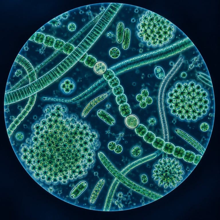
Cyanobacteria under a microscope appear as blue-green filaments, tangled colonies, or single rod-shaped cells drifting through pond water — and the first thing you’ll notice is what’s missing. Unlike the true algae swimming in the same sample, a cyanobacterium has no nucleus and no discrete chloroplast to find, because it isn’t a plant-like eukaryote at all. It’s a photosynthetic bacterium, and that single fact changes how you should read everything else on the slide.
The Three Growth Forms: Filaments, Colonies, and Single Cells
Cyanobacteria organize themselves into three basic growth forms, and sorting a sample into one of the three is the fastest way to start identifying what’s on your slide. Some species live as long threads called trichomes, others clump into jelly-bound colonies, and a few just float as isolated cells. OpenStax places cyanobacteria within the diversity of prokaryotic life.
Filamentous Forms: Oscillatoria, Anabaena, and Spirulina
Oscillatoria forms a long, straight, blue-green thread that looks like a stack of thin coins laid end to end, with the cross-walls between its disc-shaped cells barely visible. Watch one filament against the grid of your eyepiece reticle for a minute and it slowly slides and sways — a real glide, not the frantic thrashing of a flagellated protist. Cyanobacteria have no flagella at all. Anabaena, by contrast, looks like a string of green beads, with an occasional bead that’s larger, rounder, and washed-out pale — that’s a heterocyst, covered in the next section. Spirulina (technically Arthrospira) coils into an open corkscrew, a shape that’s easy to recognize even at low power.
The video above shows exactly what that Oscillatoria glide looks like in real time — a detail a still photo can’t capture, and one beginners often mistake for “nothing happening” because it’s so slow.
Colonial and Unicellular Forms: Microcystis and Merismopedia
Microcystis grows as a ragged clump of tiny spheres suspended in clear mucilage, peppered with darker specks that are gas vesicles. It looks like ground pepper spilled into a smear of green jelly. Merismopedia is easier to recognize at a glance: its cells sit in a flat, orderly grid, like a tiny sheet of graph paper. Unicellular genera such as Synechocystis and Synechococcus are smaller still, appearing as isolated spheres or short rods with no colony structure at all. Across all three forms, the overall color reads as olive to teal blue-green, spread evenly through the cell. That’s not the bright grass-green ribbon you’d see in a true green alga like Spirogyra.
Bacteria, Not Algae: Why That Changes What You See
Cyanobacteria are prokaryotes, placed in the domain Bacteria. That surprises most people meeting the name “blue-green algae” for the first time, because that name is a genuine misnomer. True algae, including green algae, diatoms, and Euglena, are eukaryotes with a nucleus and membrane-bound chloroplasts. You can usually spot the nucleus as a distinct round body inside the cell. A cyanobacterium has neither. Its DNA sits loose in a region called the nucleoid, and it has no mitochondria or other membrane-bound organelles. On a slide, the absence of a visible nucleus or a discrete green chloroplast blob is itself the identifying feature — not a failure to find one. Its biology guide explains the bacterial cell organization and photosynthetic membranes of cyanobacteria.
Despite lacking a chloroplast, cyanobacteria still perform oxygenic photosynthesis — the same water-splitting, oxygen-releasing chemistry used by plants and true algae like Volvox. The relationship runs deeper than a shared process. The chloroplasts inside every plant and algal cell descend from an ancient cyanobacterium that was engulfed and retained by a larger cell — a process called endosymbiosis. Cyanobacteria are credited with the Great Oxidation Event roughly 2.4 billion years ago, when their photosynthesis first filled Earth’s atmosphere with oxygen, according to UC Berkeley’s Museum of Paleontology.

Heterocysts and Akinetes: The Specialized Cells to Spot
A heterocyst is a larger, thicker-walled, pale or clear cell spaced along a filament in genera like Anabaena, Nostoc, and Cylindrospermum. It’s the single best identification feature in filamentous cyanobacteria. Heterocysts are where nitrogen fixation happens — they carry the enzyme nitrogenase, which is destroyed by oxygen. So the heterocyst’s thick wall and lack of active photosynthesis keep its interior oxygen-free. Under the scope it looks noticeably emptier and lighter than the green vegetative cells on either side of it.
An akinete is easy to confuse with a heterocyst at first glance, but the two look opposite. An akinete is larger, thick-walled, dark, and granular — a dormant resting cell built to survive cold or drought. It often sits right next to a heterocyst along the same filament. The quick rule: pale and clear means heterocyst, dark and grainy means akinete.
Why Cyanobacteria Are Blue-Green
Phycocyanin, a blue pigment, combines with ordinary chlorophyll a to give cyanobacteria their signature blue-green cast. Some species also carry phycoerythrin, a red pigment, which can shift the color toward olive or even reddish-brown. Unlike in a true alga, these pigments sit on thylakoid membranes that lie loose in the cytoplasm rather than packaged inside a chloroplast. That’s exactly why the color looks diffuse and even across the whole cell, instead of concentrated in a bright green blob or ribbon.

How to Find a Cyanobacteria Sample
Pond surface scum is the most reliable source: skim a sample from the thin, often iridescent film sitting on top of still water, rather than scooping clear water beneath it. The cyanobacteria live concentrated in the mat itself. Aquarium keepers usually find it as a dark, slimy film across the glass or substrate, commonly called “blue-green algae” or “slime algae” in the hobby. That earthy, musty smell is geosmin, a compound cyanobacteria produce — and it’s a strong clue you’re looking at Oscillatoria rather than a plant. Damp soil and sidewalk cracks after rain often turn up Nostoc, which forms small, jelly-like blobs, and it’s worth checking a damp soil sample the same way. Birdbaths and neglected plant saucers work in a pinch, and if none of those are available, a pinch of Spirulina powder from a health food store rehydrated in water gives you living filaments to examine.
How to View Cyanobacteria Under a Microscope (Step by Step)
- Collect from the mat or scum itself, not the clear water beside it — that’s where the cell density actually is.
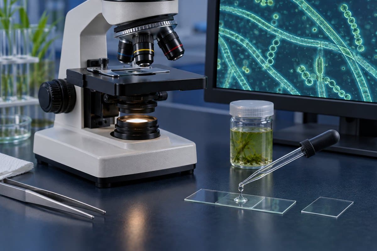
- Prepare a standard wet mount slide: a drop of sample under a coverslip, no stain required.
- Start at 100x on your compound light microscope to locate filaments or colonies before zooming in.
- Switch to the 40x objective for roughly 400x total magnification to resolve individual cells, heterocysts, and akinetes.
- Lower the condenser or close the iris diaphragm — the pale blue-green pigment washes out under full brightness, and reducing the light restores contrast.
- Give any Oscillatoria filament 30 to 60 seconds before deciding it isn’t moving; the glide is real but slow.
Skipping straight to 400x is the most common beginner mistake here — you’ll see nothing but a green blur because you never located the filaments at low power first. A wet mount also dries out fast. If you want to watch the gliding motion for more than a minute or two, seal the coverslip edge with a thin ring of petroleum jelly.
Cyanobacteria vs. True Algae at a Glance
| Feature | Cyanobacteria | True Algae |
|---|---|---|
| Domain / cell type | Bacteria (prokaryote) | Eukaryote |
| Nucleus | None (DNA in nucleoid) | Present |
| Chloroplast | None — free thylakoids | Present, membrane-bound |
| Pigments | Chlorophyll a + phycocyanin | Chlorophyll a/b, carotenoids |
| Typical cell size | 1–10 µm | 10–100+ µm |
| Movement | Gliding (no flagella) | Often flagellated |
| Example genera | Oscillatoria, Anabaena, Microcystis | Spirogyra, Volvox, diatoms |
A Safety Note on Blue-Green Algae Blooms
Some cyanobacteria produce cyanotoxins — including microcystins, anatoxin-a, cylindrospermopsin, and saxitoxins — during dense blooms in warm, nutrient-rich water. These harmful algal blooms can sicken people, dogs, and livestock that contact or swallow the water, according to the EPA and the CDC. A microscope cannot tell you whether a specific bloom is toxic — only laboratory testing can make that call. If you’re sampling pond, lake, or aquarium scum, wear gloves, wash your hands afterward, don’t taste the water, and keep pets away from any visible bloom or scummy film. This is educational information, not medical or water-safety advice; contact your state or local health department about a suspected harmful bloom.
Frequently Asked Questions
How can you tell Anabaena apart from Nostoc under the microscope?
Anabaena grows as a single free-floating filament that looks like a string of green beads with an occasional pale heterocyst. Nostoc packs many tangled filaments into a shared jelly-like ball or sheet you can often see with the naked eye. On a slide, check colony structure first, then confirm the beaded filaments and heterocysts inside it.
Do you need to stain a cyanobacteria sample?
No. The natural blue-green pigment provides enough contrast on its own — unlike onion or cheek cells, staining isn’t necessary.
Does boiling pond water remove cyanotoxins if a bloom is present?
No. Boiling can rupture cyanobacteria cells and release more toxin into the water, and it doesn’t break down the toxins already dissolved there. Standard filtration and typical water treatment aren’t reliable either — if a bloom is suspected, avoid the water entirely rather than trying to treat it yourself.
Can you see cyanobacteria without a microscope?
Yes, as mats, scum, or a slimy film — but you’re seeing millions of cells at once. Individual cells and structures like heterocysts only become visible around 400x.
Is the Spirulina powder sold as a health food the same thing as pond cyanobacteria?
Yes — Spirulina (Arthrospira) is a cyanobacterium, and food-grade powder rehydrated in water will give you living, helix-shaped filaments to examine.
Will a phone-camera microscope adapter show cyanobacteria detail?
It can show filaments and colonies at low magnification, but resolving individual cells and heterocysts really needs a compound microscope at 400x with proper objective lenses and controlled lighting.
Conclusion
Cyanobacteria reward a slow look: the three growth forms, the pale heterocysts strung along a filament, the diffuse blue-green cast with no chloroplast in sight. Every one of those details ties back to the same fact — that you’re looking at a photosynthetic bacterium rather than a true alga. Once that clicks, the rest of pond and aquarium samples start sorting themselves into “has a nucleus” and “doesn’t,” which is one of the more useful shortcuts in amateur microscopy.
Have you spotted Oscillatoria gliding across your slide, or found a heterocyst hiding in a string of Anabaena beads? Tell us what you found — and what magnification it took to see it — in the comments below.