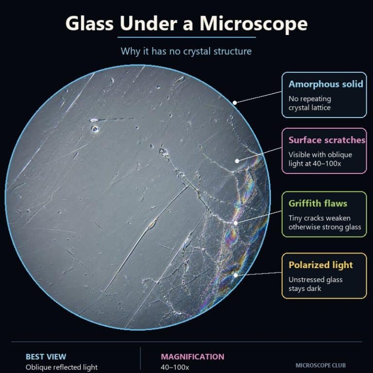
Glass under a microscope reveals a striking truth: this everyday material has no crystal structure whatsoever — it is an amorphous solid, its atoms locked in frozen disorder, its surface covered in scratches, pits, and microscopic cracks that the naked eye will never detect. It is also one of the most beginner-friendly specimens you can work with, because it needs no staining, no wet-mount slide, and no chemicals — just a piece of glass and the right angle of light.
Short-Range Order Without a Lattice: How Glass Is Built
Glass is an amorphous solid. That single phrase explains every unusual behavior glass has — the way it fractures in smooth curves, the way it has no sharp melting point, and the reason it looks structureless under the microscope.
Amorphous vs. Crystalline — What “No Lattice” Really Means
At the atomic level, all solid materials fall into one of two categories. In a crystalline solid such as a snowflake, atoms stack in a strict, repeating three-dimensional grid called a lattice. In an amorphous solid like glass, that repeating grid is absent. Each silicon atom still bonds to roughly four oxygen atoms, forming a local tetrahedron — that is the short-range order. But those tetrahedra angle off in different directions rather than snapping into parallel rows, so there is no long-range order and no lattice.
The practical result at the microscope: clear flat glass under transmitted light gives you almost nothing — a bright, featureless field. There is no grain boundary, no crystal face, no cleavage to hint at an underlying structure. The interesting material is entirely at the surface and the edges.
Everyday window glass (soda-lime glass) begins as a molten blend of roughly 72% silica (SiO₂), 14% soda (Na₂O) — a flux that drops the working temperature from pure silica’s punishing ~1,700 °C to a practical range — and 9% lime (CaO) as a stabilizer, as detailed in Britannica’s overview of glass composition. When that melt cools quickly enough, the silicon-oxygen network has no time to crystallize. It locks in place in its disordered state, producing an amorphous solid that looks like a frozen liquid — because structurally, that is exactly what it is.
The myth of flowing glass: Old cathedral windows are thicker at the bottom, which looks like evidence that glass slowly flows over centuries. The real explanation is manufacturing: crown glass was spun into discs of uneven thickness, and glaziers placed the heavier bottom edge downward by convention. At room temperature, glass is a rigid solid. It does not measurably flow on any human timescale.
Why Glass Stays Dark Under Polarized Light
Place a glass sample between two crossed polarizing filters and rotate the stage — the glass stays completely dark at every angle. That darkness is a diagnostic tool, and understanding it is the sharpest way to grasp what “amorphous” means in practice.

Glass is optically isotropic: light travels through it at the same speed in every direction. Crossed polarizers only transmit light that has been rotated away from the original polarization plane. Because glass does not rotate the plane of polarized light, none of it clears the second filter — the field stays dark, or “extinct” in petrographic terminology.
Crystalline minerals are typically anisotropic — light travels at different speeds along different crystal axes, which splits the beam and rotates the polarization. The result is bright interference colors (birefringence) that shift as you rotate the stage. This is why geologists use polarized light microscopy to identify minerals in rock thin-sections: a crystal lights up with color; glass does not. It is a genuine diagnostic technique used in forensic glass analysis.
One important exception: tempered or stressed glass shows stress birefringence — swirling, photoelastic color patterns visible under polarized light. This does not mean the glass has crystallized; it means locked-in internal strain has temporarily made the glass anisotropic. The same effect appears in some plastic screen protectors when viewed through polarized sunglasses.

The Surface — Scratches, Pits, and Griffith Cracks
Glass under a microscope comes alive when you move from the flat interior to the surface. Under transmitted light, clear flat glass looks like almost nothing — a bright, blank field. Move to a scratch, an edge, or a frosted patch, and the picture changes completely.
Scratches invisible to the naked eye appear at 40–100× as bright, hair-thin lines that flare with rainbow color where the groove catches the light at an angle. Those colors are thin-film interference from the scratch geometry itself — not pigment, just light bending through a shaped void.
More significant are Griffith flaws: microscopic surface cracks present on essentially every real-world piece of glass, even glass that looks flawless. These cracks concentrate mechanical stress at their tips, which is why glass fails at roughly 1/100 of its theoretical strength. Pristine glass should theoretically be stronger than most steel alloys by weight; the surface flaws accumulated during manufacturing and handling are what make it feel fragile. The Cambridge University DoITPOMS fracture mechanics module covers Griffith’s original analysis for anyone who wants to see the underlying mathematics.
Also visible on used glass surfaces:
- Pitting and hazing — hydrolysis (water slowly leaching surface alkalis) and repeated abrasion produce a finely cratered texture visible at 100×. A lens cleaned a thousand times shows this clearly.
- Abrasion marks — parallel lines from repeated wiping across a fixed grain; common on optical glass and display screens.
Handle broken or crushed glass specimens with care — shards cut. Use a cloth or gloves and never press samples directly with bare fingers.
Old Glass vs. New Glass Under the Microscope
The contrast between antique and modern glass is one of the most visually rewarding experiments a beginner can do. New float glass has a mechanically clean surface marked mainly by handling scratches — fine parallel lines and occasional pits from contact.
Old or excavated glass — antique bottles, window fragments from historic buildings — tells a completely different story. Decades of surface leaching pull alkali ions out of the glass, leaving a silica-rich skin that produces thin-film interference: an iridescent, oil-slick rainbow patina. Under the microscope, this appears as swirling, shifting color patches across an otherwise transparent material — like oil on water, but written into the surface by time and chemistry.
Deep weathering goes further and produces devitrification — the spontaneous formation of microscopic crystalline regions inside previously amorphous glass. Under polarized light, a devitrified patch will show birefringence (bright interference colors) even though the surrounding glass stays dark. It is a visible record of environmental exposure inscribed in the material itself.
Manufacturing defects appear in both old and new glass:
- Seeds — tiny trapped gas bubbles that appear under the microscope as perfect round black-rimmed circles with a bright center, like tiny portholes.
- Stones — unmelted solid particles, visible as opaque or cloudy inclusions.
- Cord / striae — streaks from uneven mixing during manufacture, visible as ripple-like variation in transparency or refractive index.
How to Look at Glass Under a Microscope (No Staining Needed)
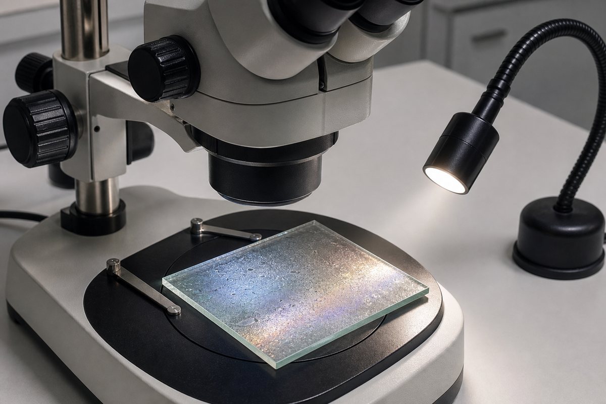
Glass is one of the most beginner-friendly microscope specimens because it needs no preparation. A standard compound light microscope works well for thin glass — slides, cover slips, or thin bottle glass placed directly on the stage. For thicker pieces and surface-feature work, a stereo microscope with top lighting is the better choice, because it sits the specimen on its stage without needing it to be thin or transparent.
- Pick the right piece. Frosted, colored, or antique glass gives you far more to see than a clean window pane. A used microscope slide, a broken bottle bottom, or a beach-worn piece of glass all work well.
- Clean the surface. Wipe with a lint-free cloth or isopropyl alcohol — fingerprints dominate the view if you skip this.
- Use reflected or oblique lighting. Transmitted bottom-light washes out shallow scratches on a flat surface. Angle a lamp at roughly 15–30° to the glass (oblique lighting) to make surface texture stand out dramatically.
- Start at 10–40×. Find a feature — scratch, edge, bubble, frosted patch — at low magnification before stepping up.
- Step up to 100× to examine individual Griffith crack networks, fine scratch lines, and seed bubbles in detail.
- Safety: If you break or crush glass to create viewable fragments, handle with care — shards are sharp. Use a cloth or gloves and never press samples with bare fingers.
What to View — Edges, Scratches, Frosted, Colored, or Crushed Glass
The most revealing beginner mistake is placing a clean, flat pane of clear glass under transmitted bottom-light and seeing almost nothing. The fix is immediate: move to an edge, a frosted section, or switch to oblique lighting. The edge of any glass piece shows conchoidal fracture — smooth, curved, shell-like ripples that radiate from the point of impact, looking like frozen ripples on a pond. This fracture pattern is the direct microscopic signature of the amorphous structure: with no cleavage planes and no lattice, glass has no preferred direction to break, so the crack curves wherever the stress releases fastest.
Frosted glass reveals a field of tiny craters and a dusty haze at 40×. Crushed glass shards from a broken bottle show both the curved conchoidal surfaces and fresh Griffith crack networks at the break edges. For related everyday-object comparisons, paper under a microscope or chalk are equally accessible no-prep specimens with very different structural stories — paper shows interlocked fibers, chalk shows compressed microfossils.
Glass vs. Quartz and Other Crystalline Solids
The sharpest way to understand glass is to compare it directly to quartz — a mineral with the identical chemical formula (SiO₂) but a completely different atomic arrangement.
| Property | Glass (amorphous SiO₂) | Quartz / crystalline solid |
|---|---|---|
| Atomic order | Short-range only (no lattice) | Long-range repeating lattice |
| Polarized light | Dark (extinct) at all angles | Bright birefringence and interference colors |
| Fracture | Conchoidal (smooth, curved) | Cleavage along flat crystal planes |
| Melting behavior | Gradual softening range | Sharp melting point (~1,713 °C for pure SiO₂) |
| Under microscope | Featureless in transmitted light; surface features with reflected light | Crystal grain boundaries and birefringence visible; crystal faces on single crystals |
The same contrast applies to other crystalline specimens. Unlike a snowflake — whose six-fold symmetry is the direct product of a hexagonal crystal lattice — glass has no geometry built into its structure. Unlike kidney stones under a microscope, which often display a visible crystalline lattice of calcium oxalate or uric acid, glass shows no internal pattern at all.
One memorable bridge concept: diatom shells are made of natural silica glass — amorphous opaline silica, not crystalline quartz. The same silicon-oxygen chemistry that makes your window glass a structureless blank under the microscope produces some of the most intricate microscopic architecture in the natural world. The glass is chemically the same; the organism built structure that the melt never achieved on its own.
For a practical decision on which microscope is right for glass surface work, our guide to choosing between a compound vs. stereo microscope covers when each tool is the right one for a given specimen type.
Frequently Asked Questions
What magnification do I need to see scratches and pits on glass?
Scratches and surface pits become clearly visible at 40–100× on either a stereo or compound microscope. For fine Griffith crack networks, 100× under oblique reflected lighting is most revealing. You do not need oil immersion or high-end optics — a basic 40× objective with a lamp angled at the surface shows significant detail on the first attempt.
Is glass a good specimen for microscope beginners?
Yes — it is arguably the single easiest specimen to start with. No staining, no wet mount, no timing, no chemicals. Pick up any frosted or colored glass piece, wipe it clean, place it under a stereo microscope with oblique lighting, and you will see something worth studying immediately. The one mistake beginners make is placing clear flat glass under transmitted bottom-light: it gives almost nothing. Switch to an edge, a frosted surface, or colored glass and the material comes alive at 40×.
Why is glass so much weaker than its chemistry would suggest?
The theoretical strength of silica glass — calculated from its bond energy — is roughly 10–14 GPa, which would put it in the range of high-performance structural steel. In practice, glass fails at around 50–100 MPa, about 1/100 of that figure. The culprit is Griffith flaws: microscopic surface cracks that form during manufacturing, handling, and environmental exposure. Each crack tip concentrates applied stress, multiplying the force at that point until the local bonds break. Controlling the surface flaw population — through polymer coatings, fire polishing, or controlled manufacturing environments — is the primary lever engineers use to make stronger glass products.
What does sea glass look like under a microscope?
Sea glass shows some of the most extreme surface weathering you can put under a microscope. Years of tumbling in salt water and abrasion against sand produce a thick frosted surface — a dense field of tiny craters and a cloudy haze from mechanical abrasion combined with hydrolysis. The original flat surfaces are completely replaced by rounded, pitted texture. On older or longer-tumbled pieces, the same iridescent patina visible on antique glass can appear. The edges lose their sharp conchoidal fracture detail and become rounded and smooth. It is glass that has visibly aged, and every year of ocean exposure is readable in the surface texture.
Can glass develop a crystal structure over time?
Yes — this is called devitrification, and it happens when glass is held at an elevated temperature long enough for the silicon-oxygen network to slowly reorganize into a crystalline arrangement. At room temperature over a human lifespan, it does not measurably occur. Under polarized light, devitrified regions show birefringence (bright interference colors) against the dark amorphous background, making them easy to identify. Glass-ceramics — materials like the original Corningware or telescope mirror blanks — are engineered to partially devitrify in a controlled way, producing a material that is part glass, part crystalline, with useful properties of both.
Is obsidian (volcanic glass) the same as regular glass under a microscope?
Structurally, yes — obsidian is natural glass: molten silica-rich lava that cooled too quickly to crystallize. Under the microscope it shares all the defining traits of manufactured glass: featureless in transmitted light, dark under crossed polarized filters, and exhibiting conchoidal fracture at broken edges. The differences are in composition and inclusions. Obsidian often contains tiny mineral crystals (phenocrysts) caught mid-formation when the lava quenched, plus gas bubble voids and flow banding (striae from turbulent cooling). A fresh obsidian break edge shows the same smooth curved conchoidal ripples as a broken bottle — one of the clearest illustrations that the “glass” structure is determined by cooling rate, not by human manufacturing.
Conclusion
Glass under a microscope is an exercise in seeing what you thought you already understood. It has no crystal structure — just short-range atomic order locked in place when a melt cooled too quickly to crystallize. That single structural fact explains every macroscopic behavior: why it stays dark under polarized light, why it fractures in smooth curves rather than flat planes, why its surface cracks quietly undercut its theoretical strength, and why antique glass develops a rainbow patina over decades. The Corning Museum of Glass documents how these properties have driven material innovation across centuries — a reference worth exploring if you want the full manufacturing history alongside the structural science.
The practical upside is that glass is one of the most accessible specimens you will ever work with. No preparation, no chemicals — just frosted bottle glass or a worn slide under oblique light, and you are already seeing something a casual observer would miss entirely. Try it with a piece of antique glass if you can find one: the iridescent patina alone is worth the five minutes it takes to set up. Drop a note in the comments if you spotted something unexpected — seeds, striae, stress birefringence on tempered glass, or the first Griffith crack network you have seen with your own eyes.
Originally posted 2026-07-29 12:00:00.