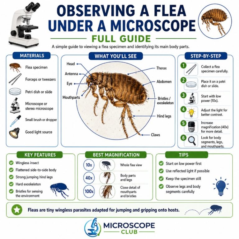
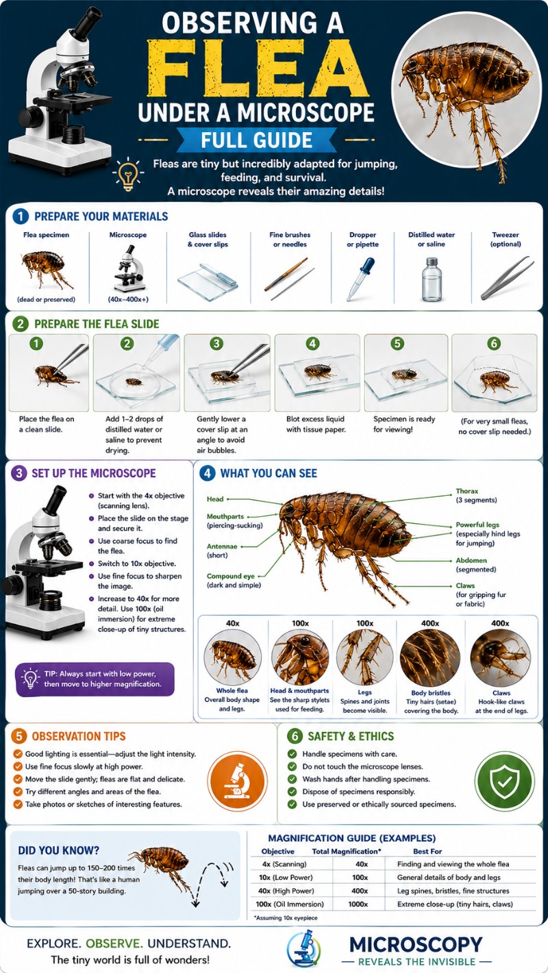
Observing a flea under a microscope reveals a surprisingly complex and well-engineered parasite. Under magnification, the flea appears as a wingless, reddish-brown insect with a body so laterally compressed it looks like it was squeezed in a vise — an adaptation that lets it slip through fur and resist being groomed away. Even at low power, you can see the rows of backward-pointing bristles, the oversized hind legs built for record-breaking jumps, and the dramatic comb-like spines called ctenidia that make flea identification unmistakable. This guide walks you through what you’ll see at every magnification level, which microscope works best, how to prepare a flea specimen at home or in the classroom, and what separates a flea from a louse or bed bug at a glance.
What Does a Flea Look Like Under a Microscope?
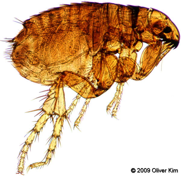
At low magnification, a flea looks like a tiny, armor-plated tank. The body is divided into the standard insect sections — head, thorax (three segments), and abdomen (ten segments) — but everything about its proportions screams specialization for a parasitic lifestyle. The body color ranges from reddish-brown to near-black depending on how recently it fed, and the hardened cuticle (exoskeleton) gives it a shiny, leathery appearance. No wings are present anywhere — fleas belong to the order Siphonaptera, literally “wingless siphon,” and that absence is one of the easiest ID points under the scope.
The most arresting features at first glance are the legs. The front and middle pairs are moderate in size; the hind pair is enormous relative to the body, folded up like a compressed spring ready to release. Rows of stiff spines and bristles cover the entire body, all angled backward to anchor the flea in host fur against grooming attempts. If you’re viewing a Ctenocephalides felis (cat flea, the species most commonly found on both cats and dogs in the US), you’ll spot two dramatic rows of comb-like spines — one along the lower edge of the head and one along the back of the first thoracic segment. Those combs alone are enough to make a flea unforgettable under the microscope.
Magnification Guide — What You See at Each Power
Choosing the right magnification matters. Here’s what resolves at each level — refer to this as your field checklist when you put a specimen on the stage. For a deeper breakdown of objective lenses, see our guide to 4x, 10x, 40x and 100x objective lenses.
| Magnification | What Becomes Visible | Best Scope Type |
|---|---|---|
| Naked eye | Dark speck, ~1.5–3.3 mm; jumps erratically | — |
| 10×–20× (stereo) | Body shape, color, leg proportions, bristles, general body outline; flea is clearly an insect | Stereo/dissecting microscope |
| 30×–40× (stereo) | Ctenidia (combs) on head and pronotum; tarsal claws; simple eye (ocellus); antennal groove | Stereo/dissecting microscope |
| 100× (compound) | Fine bristle texture, comb spine detail, body segment boundaries, flea dirt granules if present | Compound microscope (slide-mounted) |
| 400× (compound) | Mouthpart stylets (maxillary laciniae), setal patterns, resilin pads in legs, fine cuticle texture | Compound microscope (slide-mounted) |
To understand how to calculate total magnification for your specific scope setup, multiply the eyepiece power by the objective power.
Best Microscope for Viewing a Flea
For most people — students, hobbyists, pet owners — a stereo (dissecting) microscope at 10×–40× is the ideal tool. A stereo scope gives a three-dimensional view of the intact insect, provides enough working distance to reposition the specimen with a probe or forceps, and lights the surface from above (reflected light) so you can see color and texture, not just a silhouette. You can view a whole flea dry, without any mounting at all, which is especially convenient in a classroom.
A compound microscope works well for higher-magnification detail — the mouthparts, comb spine fine structure, and internal tissue (if cleared and stained) — but requires the flea to be flattened and slide-mounted, which destroys the 3D view. Use the compound scope as a second step after studying the intact specimen under the stereo. Learn more in our comparison of compound vs stereo microscope options and our full guide to the dissecting (stereo) microscope.

USB/digital microscopes at 20×–200× are a great budget option for classrooms and home use — they display the image on a screen or computer, which makes it easy to capture photos and share with students. They won’t match the optics of a dedicated stereo scope, but for a whole flea they get the job done. A budget microscope in the 20×–40× range is often enough to see ctenidia, legs, and body shape clearly.
Flea Anatomy Under the Microscope
Body Shape and Why Fleas Are Flat
The flea’s laterally compressed body — flattened side-to-side rather than top-to-bottom — is one of the most striking things you’ll notice under the scope. This isn’t a coincidence: lateral compression lets the flea orient itself edge-on to a hair shaft, giving it a profile so narrow that host grooming claws and teeth slide right past it. The hardened cuticle adds to this resilience — you can press a flea between your fingers and release it unharmed, which is why squeezing doesn’t kill them.

Compare this shape to a louse (dorsoventrally flattened — squashed top-to-bottom, like a tiny crab) or a bed bug (also dorsoventrally flattened, oval, no legs built for jumping). The flea’s side-to-side compression is specific to its lifestyle navigating dense fur and feathers. The entire body outline, from the smoothly arched back to the pointed hind end, is streamlined to minimize resistance as the flea moves forward through host hair.
Combs (Ctenidia)
The ctenidia are the flea’s most diagnostic microscope feature — once you’ve seen them, you’ll never mistake a flea for anything else. They are rows of rigid, dark, tooth-like spines that form neat combs on the body. The genal comb runs along the lower edge of the head (the “cheek” area), while the pronotal comb lines the posterior edge of the pronotum (the plate covering the first thoracic segment).
Ctenocephalides felis (cat flea) has both combs — typically 7–8 teeth in each. The dog flea (C. canis) has both as well, but the genal teeth are more vertically arranged. The human flea (Pulex irritans) lacks a genal comb entirely, making it easy to distinguish at 30× or higher. The ctenidia serve a mechanical function: they interlock with host hair, preventing the flea from being pulled backward during grooming. Under the scope at 30×–40×, the combs appear as a dense, dark rank of spines with a high-contrast, almost architectural regularity.
Mouthparts
Flea mouthparts are of the piercing-sucking type — the hallmark of an obligate blood feeder (the technical term is hematophagous ectoparasite). The functional cutting elements are the paired maxillary laciniae, two stylet-like blades that work together with the epipharynx to pierce skin and create a feeding channel. At 400× on a slide-mounted specimen, these appear as slender, needle-like projections beneath the head, darkly sclerotized and pointed.
When a flea feeds, it doesn’t simply poke a hole — the laciniae saw through tissue to reach a blood vessel, and saliva is injected to prevent clotting. That saliva contains proteins that trigger the immune response responsible for the characteristic itchy bite. The flea’s ability to detect a host uses CO₂, body heat, and vibration; the short, clubbed antennae that sit in sheltered grooves on the head are part of this sensory apparatus and are visible at 40× as stubby projections tucked behind the eye.
Legs and the Record-Breaking Jump
Under the stereo microscope, the size disproportion between the flea’s hind legs and the rest of its body is immediately obvious. The hind femora are thick and heavily muscled, and the tibiae are long and armed with stout spines. At the end of each leg, tarsal claws grip host hair with a mechanical lock that requires active muscle contraction to release — meaning a flea clinging to fur is passively anchored, not actively gripping.
Cat fleas can jump roughly 18 cm (about 7 inches) vertically and 33 cm (about 13 inches) horizontally — on the order of 100 times their own body length. The energy comes not from raw muscle power alone, but from a pad of the elastic protein resilin. The flea compresses resilin like a coiled spring using its muscles, then releases it in a fraction of a millisecond — achieving accelerations of around 100 g. Research by Sutton and Burrows using high-speed cameras showed that take-off force is transmitted through the tibia and tarsus (the foot), not by direct leverage of the femur as older models assumed. That finding overturned a decades-old assumption about flea jump mechanics. You can read the original research at The Journal of Experimental Biology.
Eyes, Antennae, and Bristles
Flea eyes are simple eyes (ocelli) — a single pair of small, convex lens-like eyespots, not the compound mosaic eyes of a fly or bee. Some cave-dwelling and deep-fur-dwelling flea species have reduced or absent eyes entirely, a clue that vision is less important to their lifestyle than chemical and mechanical sensing. Under the stereo scope at 30×–40×, the ocellus appears as a small, dark, smooth oval near the front of the head.
The backward-pointing bristles covering the body are visible even at 10× and are a key functional feature. Like the ctenidia, they resist backward motion through host fur — every spine is a one-way ratchet. The bristles are arranged in species-specific patterns, making them useful for taxonomists working on species ID under high magnification. For more on insect eyes under the microscope, including how simple and compound eyes compare, see our dedicated article on the subject.
How to Prepare and Mount a Flea Specimen
Preparing a flea for the microscope is straightforward and safe if you follow a few basic steps.
- Collect the flea. Run a fine-toothed flea comb through your pet’s fur over a white paper towel. Live fleas will jump; dead or sluggish specimens drop and can be collected with moistened forceps or a damp fingertip. Alternatively, place a shallow dish of soapy water under a lamp on the floor overnight — fleas are attracted to warmth and light, jump in, and the soap breaks surface tension so they drown.
- Euthanize. Drop the flea into a small vial of 70% isopropyl alcohol (rubbing alcohol). This kills it instantly and begins preservation. Alternatively, place the flea in a sealed container in the freezer for 30 minutes.
- Dry mount for stereo viewing. Remove the flea from alcohol, let it air-dry on a paper towel for a few minutes (or gently blot), then place it directly on the stereo microscope stage. No slide, no cover slip needed. Use a pin or probe to orient the body.
- Wet mount for compound viewing. Place the flea in a drop of water or glycerin on a glass slide. If you want to see internal detail (trachea, gut), you can clear the specimen first: soak in 10% potassium hydroxide (KOH) solution for several hours to dissolve soft tissue, then rinse and mount in glycerin. This step isn’t necessary for anatomy like combs and mouthparts, which are already heavily sclerotized and high-contrast. See our guides on how to make a wet mount slide and how to prepare microscope slides for more detail.
- Add a cover slip. Lower a cover slip gently at an angle to avoid bubbles. For a whole flea, you’ll probably crack the cover slip if you press hard — that’s fine for temporary viewing. Use modeling clay or cover-slip spacers to raise the cover slip if you want to avoid crushing.
Safety note: Fleas can carry bacteria including Yersinia pestis (plague) in rare endemic areas of the US, and Bartonella henselae (cat scratch disease). For classroom use, always work with specimens that have been thoroughly killed in alcohol first, wear gloves, and wash hands after handling. Do not handle live fleas from wild rodents.
Flea Life Cycle and Flea Dirt
Understanding the life cycle helps explain what else you might observe under the microscope when investigating a flea infestation. Fleas undergo complete metamorphosis: egg → larva → pupa → adult.
A female cat flea lays up to 40–50 eggs per day, depositing them on the host where they quickly fall into carpet, bedding, and furniture. The eggs are pearly-white smooth ovals, about 0.5 mm long — visible to the naked eye on dark fabric, and easy to see at 10× on a slide. Larvae are legless, worm-like, and roughly 2–5 mm long; they feed on organic debris and, critically, on flea dirt — the dried blood feces that adult fleas deposit in the host’s fur. The pupa spins a silk cocoon and can remain dormant for months, emerging only in response to host cues like vibration, elevated CO₂, and warmth. This is why moving into a vacant house with a previous pet can suddenly trigger a flea emergence. The whole cycle runs about 2–4 weeks under warm, humid conditions.
Flea dirt under the microscope appears as dark, comma- or crescent-shaped granules. The classic classroom test: collect some on damp white paper — genuine flea dirt dissolves to a reddish-brown color, confirming it contains digested blood. Ordinary dirt stays gray. At 40×–100× on a slide, the granules have an irregular, almost crystalline texture. The CDC’s flea information page is a useful reference for health context around fleas and the diseases they can carry.
Flea vs Louse vs Bed Bug Under the Microscope
These three ectoparasites are frequently confused by the people they bite. Under magnification, they’re actually very distinct.
| Feature | Flea | Louse | Bed Bug |
|---|---|---|---|
| Body compression | Lateral (side-to-side) | Dorsoventral (top-to-bottom) | Dorsoventral (top-to-bottom) |
| Legs | 3 pairs; hind pair hugely enlarged | 3 pairs; similar in size, claw-tipped | 3 pairs; similar in size |
| Wings | None | None | None (wing pads only) |
| Ctenidia (combs) | Present on most species | Absent | Absent |
| Jumps? | Yes — main dispersal method | No | No |
| Size | 1.5–3.3 mm | 1–4 mm | 4–7 mm (adult) |
| Shape | Narrow, streamlined oval | Flat, louse-shaped with claws gripping hair | Flat, broad oval; unfed = papery thin |
The single fastest field ID: if it jumps, it’s a flea. If it doesn’t jump and has a clamp-like grip on a hair shaft, it’s a louse. If it’s larger, brownish, and oval with no ctenidia or jumping ability, it’s likely a bed bug. For a classic insect anatomy comparison, see our article on ants under the microscope.
For additional insect anatomy reference, the University of Florida’s Featured Creatures database entry on cat fleas is one of the best freely available academic references, with photographs and taxonomy details.
Frequently Asked Questions
What diseases can fleas carry, and how do I handle specimens safely?
Fleas can transmit murine (flea-borne) typhus, caused by Rickettsia typhi and spread by infected cat fleas, Oriental rat fleas, or their feces — most US cases are reported from California, Texas, and Hawaii. They can also carry plague (Yersinia pestis) in rare endemic western US areas, Bartonella henselae (cat scratch disease), and tapeworm, which infects people who accidentally swallow an infected flea. Before viewing, kill specimens in 70% isopropyl alcohol, wear gloves, wash your hands afterward, and never handle live fleas from wild rodents.
How do I tell flea eggs, larvae, and pupae apart under magnification?
Eggs are smooth, pearly-white ovals under about 0.5 mm — a small pile looks like spilled salt, and unlike flat, sticky dandruff they are rounded and roll freely. Larvae are legless, eyeless, worm-like and 2–5 mm long, off-white to translucent with a ring of hairs on each segment, often curled into a C shape. Pupae are the hardest to spot: a sticky 3–4 mm silk cocoon coated in trapped dust and debris, so it usually just looks like a fleck of dirt.
What’s the easiest way to collect live fleas for viewing without getting bitten?
Set a passive light trap: place a shallow dish of soapy water on the floor under a desk lamp overnight — fleas jump toward the warmth and light, land in the water, and the soap breaks the surface tension so they drown without ever touching you. Alternatively, comb a pet over a sheet of white paper and let dislodged fleas drop straight into a vial of 70% alcohol. Either way you collect the specimen pre-killed, so there’s no live flea to bite or escape onto the bench.
What are the most common mistakes when mounting a flea, and how do I avoid them?
The biggest is crushing the specimen — a whole flea is thick, so pressing the cover slip flat shatters the legs and combs; use modeling-clay feet or cover-slip spacers to bridge over it. Trapped air bubbles are the next problem: lower the cover slip slowly at an angle onto the mounting drop rather than dropping it flat. Also avoid mounting a still-wet alcohol specimen (let it air-dry first for dry stereo viewing) and using too much fluid, which lets the flea drift out of focus.
Conclusion
Observing a flea under a microscope turns a familiar pest into an engineering case study. The laterally compressed body, the interlocking ctenidia, the resilin-powered jump, the piercing mouthparts — every feature maps directly to a specific survival problem that evolution solved with remarkable precision. A stereo microscope at 30×–40× is all you need to see the highlights: combs, bristles, oversized hind legs, and the simple eye. A slide-mounted specimen at 100×–400× on a compound scope takes you deeper into mouthpart and bristle detail. Either way, it’s one of the most rewarding specimens to study because so much anatomy is packed into 2–3 mm of insect.
Have you put a flea under your microscope — from a pet, a yard trap, or a classroom collection? We’d love to hear what you found most surprising. Drop your observations (and any photos if you’ve got them) in the comments below.