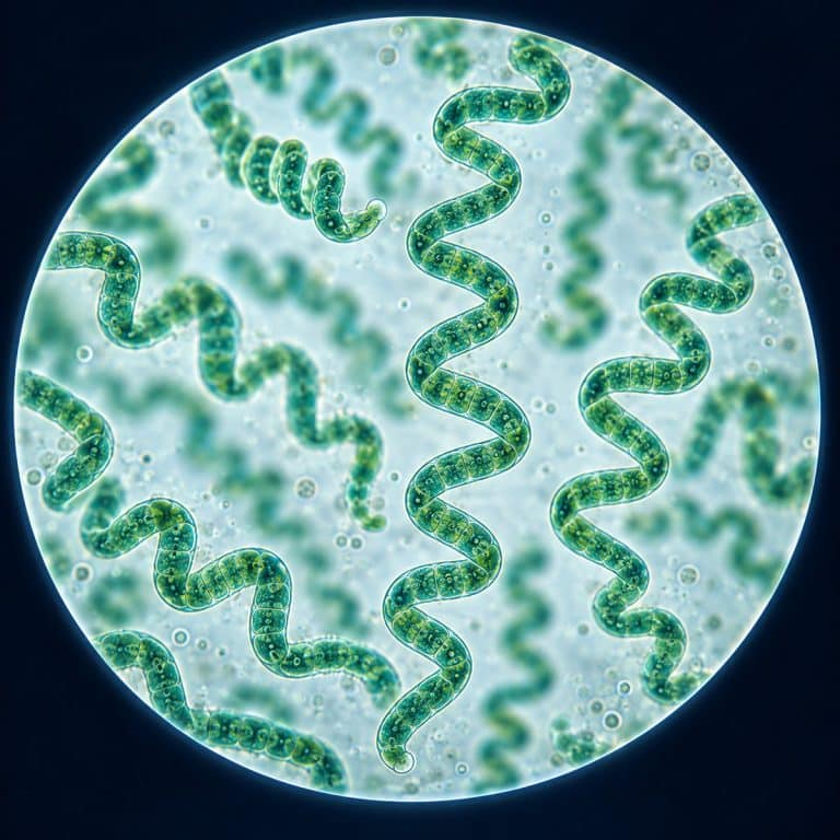
Spirulina under a microscope reveals a tangle of open, blue-green spiral filaments — corkscrew-shaped threads of the cyanobacterium Arthrospira coiled into a helix roughly 30 to 70 micrometers across. You don’t need a lab culture to see it. A pinch of the dried supplement powder already in your kitchen works. Rehydrate it in a drop of water, view it at 100x to 400x, and the same spirals appear on your own slide.
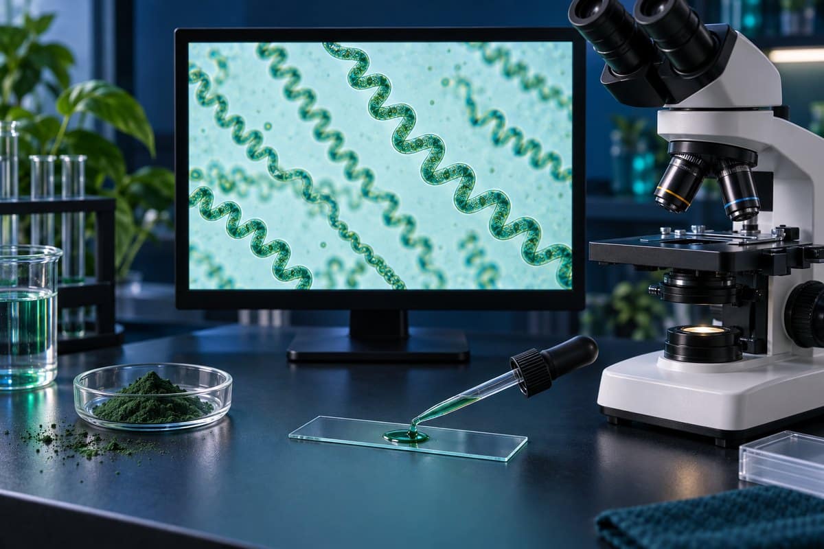
What Spirulina Looks Like Under a Microscope
A spirulina trichome is an unbranched, cylindrical filament twisted into an open left-handed helix — picture a tiny green bedspring floating in the water drop. The filament itself runs about 6 to 12 micrometers wide. The coil it forms is much larger, though, spanning roughly 30 to 70 micrometers from one side of the spiral to the other. Intact filaments can stretch up to 500 micrometers long, though what you’ll see from powder is usually shorter, broken fragments. The NCBI Bookshelf identifies Spirulina supplements with cyanobacteria in the genus Arthrospira.
| Feature | Typical Value |
|---|---|
| Magnification needed | 100x to locate, 400x for detail |
| Filament (trichome) width | ~6–12 µm |
| Helix (coil) diameter | ~30–70 µm |
| Filament length | up to ~500 µm (live); shorter fragments in powder |
| Color | blue-green to teal |
| Taxonomy | cyanobacterium, genus Arthrospira |
The spiral shape and size
Under the objective, each trichome reads as an open spring rather than a tightly wound coil — you can usually count the individual turns of the helix at 400x. The spiral isn’t a fixed feature of the organism: a handful of strains grow straight instead of coiled, and coiling can loosen under culture stress. So don’t be alarmed if a few filaments in your sample look more like gentle curves or short commas than full corkscrews.
Color — where the blue-green comes from
The color comes from two pigments working together: green chlorophyll a and phycocyanin, the blue accessory pigment. Phycocyanin is also extracted and sold on its own as a natural blue food coloring. Under brightfield illumination, healthy filaments look translucent teal-green; older or dried fragments can look duller and more olive.
How to See Spirulina Under a Microscope (From Powder or Culture)
Getting from a jar of supplement powder to a visible spiral takes five steps and a few minutes.
- Tap a very small pinch of spirulina powder — barely enough to tint the water — onto a clean slide, or crush a tiny corner of a tablet.
- Add one drop of water and let it sit for 30 to 60 seconds so the powder rehydrates and the filaments unfold.
- Lower a coverslip onto the drop at an angle to avoid trapping air bubbles, following the same technique used to make a wet mount slide.
- Start at 100x and scan the thin, spread-out edge of the drop rather than the dense center.
- Once you spot a filament, center it and step up to 400x to resolve the individual coils and faint cross-walls.
From a supplement tablet or powder
Powder-sourced spirulina is dead and fragmented, so expect shorter, static curls rather than long unbroken springs. That’s normal — the spiral shape survives drying and grinding even though the organism doesn’t.
From a live culture
If you’re growing spirulina rather than buying it dried, sample from a well-mixed, actively growing culture — it should look bright green, not settled and pale. Live filaments are longer, and you may catch one gliding slowly within its mucilage sheath.

Best magnification and settings
100x is enough to locate the filaments; 400x is where the coils and cross-walls actually resolve. A standard brightfield scope works fine, but if your filaments look pale and washed out, closing the substage iris diaphragm down helps. Either dark field microscopy or a phase contrast microscope will make the translucent spirals stand out much more clearly, as explained in Nikon’s introduction to phase contrast microscopy.
Is Spirulina a Bacterium or an Alga?
Spirulina is a bacterium, not a plant and not a true alga — the supplement name refers to the cyanobacterium genus Arthrospira, most commonly Arthrospira platensis. As a prokaryote it has no nucleus, no chloroplasts, and none of the membrane-bound organelles a true algal cell would have; its photosynthetic machinery sits directly in the cell’s cytoplasm. “Blue-green algae” is the legacy name that stuck from before scientists reclassified cyanobacteria as bacteria. That’s why supplement labels still call it algae even though it’s taxonomically closer to what bacteria look like under a microscope than to any plant or true algal cell. Details on the genus’s morphology and classification are documented in this Arthrospira platensis profile, and the broader cyanobacteria group is covered in Britannica’s entry on blue-green algae. OpenStax supplies broader context on cyanobacteria within prokaryotic diversity.
Light microscopy can’t resolve internal organelle detail in a cell this small and this simple. That level of structure (thylakoid membranes, ribosomes, DNA fibrils) only shows up under an electron microscope. What you can see, though, is the trichome’s segmented structure: faint transverse cross-walls dividing the filament into individual cells, visible as ladder-like lines running across a well-focused spiral at 400x.

Powder vs Live Spirulina Under the Microscope
Dried supplement powder and a living culture look related but distinctly different on the slide. Powder gives you the shape without the motion; live culture gives you both.
| Feature | Powder / Tablet | Live Culture |
|---|---|---|
| Filament state | Dead, often fragmented | Intact, long helices |
| Movement | None — static | Slow gliding/rotation possible |
| Color | Duller, sometimes olive | Bright teal-green |
| Typical length seen | Tens of µm (broken bits) | Up to ~500 µm |
If you want to compare the powder view against another well-known freshwater green organism, the green, motile euglena is a useful contrast — same color family, completely different structure and swimming behavior.
Troubleshooting — Why You Might Not See the Spirals
Most failed attempts trace back to one of a few easy-to-fix habits.
- Too much powder. A heavy pinch turns the drop into an opaque green sludge with no visible individual filaments. Use a tiny pinch — barely enough to tint the water.
- Looking at the clump instead of the edge. The thick center of a powder clump is too dense to resolve. Hunt the thin, spread-out edge of the drop where single filaments separate out.
- Jumping straight to 400x. Find a filament at 100x first, center it, then climb in magnification — searching at high power wastes time and narrows your field of view before you’ve found anything.
- Powder that won’t wet. Dry clumps sometimes float as little rafts instead of dispersing. Stir the drop gently or let it sit 30 to 60 seconds so the powder rehydrates properly.
- An over-bright field. Full brightfield illumination can wash out the pale, translucent filaments. Close the iris diaphragm down for contrast, or switch to dark field or phase contrast.
One source-related note: only view spirulina from a purchased supplement or a known, deliberately cultured sample. Never scoop wild pond scum to look for spirulina. Natural blue-green blooms can include genuinely toxic cyanobacteria species that are unsafe to handle or ingest, as the CDC’s overview of harmful algal blooms explains. If you want to explore wild aquatic samples safely, a sample of pond water is a better, lower-risk place to start. You’ll likely spot other organisms too. Check our guide to plankton under a microscope or the glass-shelled diatoms under the microscope for what else turns up in a drop of water. For slide technique generally, our guide to prepare your microscope slides covers the basics in more depth.
Frequently Asked Questions
Why is spirulina spiral-shaped?
The helix is simply how the trichome grows — an open left-handed coil is the default form for most Arthrospira strains. It isn’t fixed for life, though: a small number of strains grow straight, and coiling can loosen under culture stress, so shape alone isn’t a perfect identifier.
Can you see individual spirulina cells, or just the whole filament?
You can usually make out the filament’s segmented structure — faint cross-walls dividing it into individual cells — at 400x under good lighting. You won’t see internal cell detail like organelles, because spirulina is a prokaryote and that level of structure requires an electron microscope.
Is spirulina powder alive under the microscope?
No — dried supplement powder is dead. The filaments keep their spiral shape but sit completely still. Only a live culture shows movement, typically a slow gliding or rotation as the trichome moves within its mucilage sheath.
Is “spirulina” the same thing as the genus Arthrospira?
Close, but not exactly. The organism sold as spirulina is scientifically Arthrospira — taxonomists split it from the separate genus Spirulina years ago. The older common name stuck for marketing, though, and never got updated on supplement labels.
Will spirulina look different depending on which brand of powder I buy?
Slightly, but the core shape holds. Grinding, drying method, and how finely a brand mills its powder all affect how fragmented and clumped the filaments are on your slide. Every brand is still the same organism, though — you should see the same open spiral shape regardless of which jar it came from.
How much spirulina powder should I use for a slide?
Far less than seems intuitive — a pinch small enough to just barely tint a drop of water. Too much powder is the single most common reason beginners see a green smear instead of individual spirals.
Conclusion
Spirulina under a microscope turns out to be one of the easiest “aha” specimens to find. That’s precisely because most readers already own it as a kitchen supplement rather than needing to culture or collect anything. A tiny pinch of powder, a drop of water, and a few minutes of patience at 100x to 400x is all it takes to see the open blue-green spirals. And that’s enough to understand, first-hand, why this “algae” is really a bacterium wearing a corkscrew shape.
Have you tried rehydrating your own spirulina supplement under the microscope? Tell us what you found — how tight the coils looked, whether you caught any movement in a live sample, or what magnification worked best for you — in the comments below.