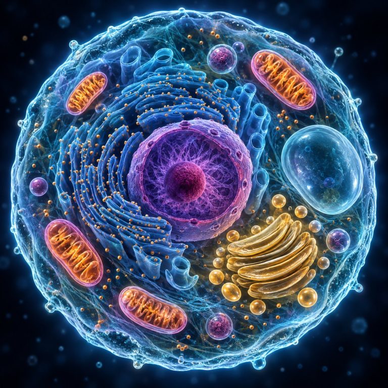
Cell organelles are specialized structures inside a cell that perform essential jobs such as storing DNA, building proteins, generating energy and packaging materials. This visual guide explains the major organelles, compares their roles in plant and animal cells, and shows which structures you can actually see with a light microscope.
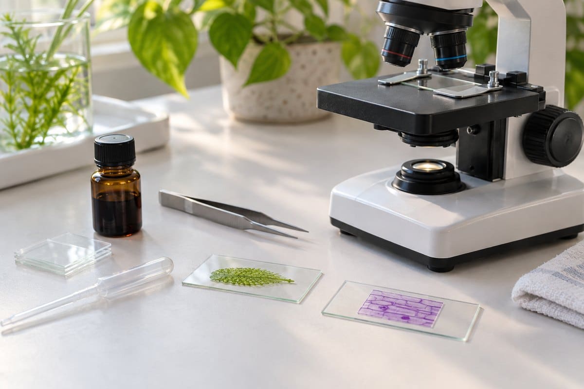
The Cell Organelles at a Glance
The table below maps every major organelle to its core job and to the cell types that carry it. The deep dive that follows breaks down structure, function, and microscope visibility one organelle at a time.
| Organelle | Function | Found in |
|---|---|---|
| Cell membrane | Controls what enters and leaves the cell | All cells |
| Cytoplasm | Fluid that holds organelles; site of many reactions | All cells |
| Nucleus | Houses DNA; directs the cell’s activity | Animal & plant |
| Nucleolus | Builds ribosomal subunits | Animal & plant |
| Ribosomes | Synthesize proteins | All cells, including bacteria |
| Rough ER | Builds and folds proteins for export | Animal & plant |
| Smooth ER | Makes lipids; detoxifies; stores calcium | Animal & plant |
| Golgi apparatus | Sorts and packages proteins into vesicles | Animal & plant |
| Mitochondria | Produce ATP via aerobic respiration | Animal & plant |
| Chloroplast | Carries out photosynthesis | Plant only |
| Vacuole | Storage; turgor pressure (plants) | Animal & plant |
| Lysosome | Breaks down waste and worn-out organelles | Mainly animal |
| Peroxisome | Breaks down fatty acids; neutralizes peroxide | Animal & plant |
| Cytoskeleton | Shape, support, internal transport | Animal & plant |
| Centrioles | Organize the spindle during cell division | Mainly animal |
| Cell wall | Rigid outer support | Plant, fungi, bacteria |
The Organelles, One by One
Nucleus and Nucleolus
The nucleus is the cell’s control center: a double-membraned compartment that houses the DNA as chromatin and directs protein synthesis by dispatching mRNA out through pores in its envelope. Inside it sits the nucleolus, a denser region that isn’t its own organelle but a workshop for assembling ribosomal subunits. Under a light microscope, the nucleus is the one organelle you can count on seeing reliably. It shows up as a small, dense dot in onion cells or human cheek cells, especially once a stain is added. Learn more about how the nucleus fits into overall cell structure.
Ribosomes
Ribosomes are the sites of protein synthesis, translating mRNA into amino acid chains. They’re made of rRNA and protein, and they either float free in the cytosol or stud the rough endoplasmic reticulum. Ribosomes are the one organelle every cell has, bacteria included — though a bacterial 70S ribosome is smaller than a eukaryotic 80S one. At roughly 20-30 nanometers, a ribosome is about ten times smaller than a light microscope can resolve; you will never see one without an electron microscope.
Endoplasmic Reticulum: Rough and Smooth
The endoplasmic reticulum is a folded membrane network that comes in two flavors. Rough ER is studded with ribosomes and builds proteins destined for membranes or secretion. Smooth ER has no ribosomes and instead handles lipid and steroid synthesis, detoxification (it’s abundant in liver cells), and calcium storage in muscle. Individual ER sheets are far too thin and low-contrast for a school scope — this is electron-microscope territory.
Golgi Apparatus
The Golgi apparatus works as the cell’s post office: its cis face receives proteins and lipids from the ER, then modifies and sorts them. Its trans face ships them out in vesicles. Like the ER, its individual stacked membranes (cisternae) sit below what a light microscope can resolve.

Mitochondria
Mitochondria generate the cell’s usable energy, producing ATP through aerobic respiration inside an inner membrane folded into cristae. They carry their own DNA, a leftover of their endosymbiotic origin as free-living bacteria billions of years ago. Their numbers also track a cell’s energy demand — a liver cell can hold 1,000 to 2,000 of them, while a mature red blood cell has none. Under a light microscope with Janus Green B stain, mitochondria show up as faint blue-green specks or short threads, not the labeled bean-with-cristae diagram; the internal folds are EM-only.
Chloroplasts
Chloroplasts are the plant cell’s solar panels, carrying out photosynthesis inside stacked thylakoid membranes (grana) suspended in a fluid called the stroma, using chlorophyll to capture light energy. Like mitochondria, they carry their own DNA from an endosymbiotic bacterial ancestor. They’re also the easiest organelle to actually see: in a fresh leaf or Elodea sample, chloroplasts appear as rows of bright-green ovals. In a warm, living leaf you can often watch them slowly circulate around the cell in a process called cytoplasmic streaming.
Vacuoles
A vacuole is a storage sac. Plant cells typically have one large central vacuole that can occupy 30 to 90 percent of the cell’s volume, maintaining turgor pressure and storing water, nutrients, and waste. Animal cells instead have several small, temporary vacuoles. The plant central vacuole is easy to spot under a light microscope — it’s the large, clear space that pushes the cytoplasm and organelles to the cell’s edge.

Lysosomes and Peroxisomes
Lysosomes are enzyme-filled recycling centers, mostly found in animal cells, that break down waste, worn-out organelles, and invading pathogens. Peroxisomes, present in both plant and animal cells, break down fatty acids and neutralize reactive oxygen species by producing and then destroying hydrogen peroxide. Both are typically 0.1 to 1 micron across — at or below the light microscope’s resolution limit, so they’re generally invisible without an electron microscope or specialized staining.
Cytoskeleton and Centrioles
The cytoskeleton is an internal scaffold of microfilaments, intermediate filaments, and microtubules that gives the cell its shape, supports it structurally, and moves cargo internally. Centrioles are paired microtubule cylinders, found mainly in animal cells, that organize the spindle during cell division; plants generally lack them. Both structures require an electron microscope to resolve their fine architecture.
Cell Membrane
The cell (plasma) membrane is a phospholipid bilayer that forms a selectively permeable boundary around every cell, controlling what enters and leaves. It’s universal — animal, plant, and bacterial cells all have one — but in cells without a rigid wall, like human cheek cells, the membrane is all that holds the cell’s shape. That’s why unstained cheek cells look like floppy, nearly transparent flakes rather than crisp bricks.
Cell Wall
The cell wall is a rigid structure that sits outside the cell membrane, built from cellulose in plants, chitin in fungi, and peptidoglycan in bacteria. Animal cells never have one. Under a light microscope it’s one of the clearest structures to see. The crisp, dark, brick-like outlines that define each cell in an onion epidermis sample are cell walls, not membranes.
Cytoplasm
Cytoplasm is the gel-like fluid that fills the cell and holds every organelle in place. It’s also where many metabolic reactions — including the first stage of glycolysis — actually happen. It’s not a discrete organelle so much as the medium everything else sits in, and under a microscope it appears as the translucent background field the visible organelles float in.
What You Can Actually See Under a Microscope
A standard light microscope’s resolution tops out around 200 nanometers, set by the diffraction limit of visible light. That’s also why cranking a school scope past roughly 1000x to 1500x just produces a bigger, blurrier image rather than more detail, an effect microscopists call empty magnification. Optical resolution limits like this one are why so much of the idealized textbook diagram is invisible at the eyepiece. The rule of thumb: if a structure is smaller than about 200 nanometers, or too low-contrast without a stain, a light microscope won’t show it.
| Organelle | Approx. size | Visible on light scope? | Needs staining? | EM-only? |
|---|---|---|---|---|
| Nucleus | 5-10 µm | Yes | Helps | No |
| Chloroplast | 3-10 µm | Yes | No | No |
| Central vacuole | Up to 90% of cell | Yes | No | No |
| Mitochondrion | 0.5-1 x 1-10 µm | As faint specks | Yes | Internal structure only |
| Lysosome / peroxisome | 0.1-1 µm | Rarely | Yes | Mostly |
| ER / Golgi | Membrane networks | No | N/A | Yes |
| Centriole | ~0.2 x 0.5 µm | No | N/A | Yes |
| Ribosome | 20-30 nm | No | N/A | Yes |
A transmission electron microscope resolves down to about 0.1 to 0.2 nanometers, which is what finally reveals mitochondrial cristae, ER sheets, and individual ribosomes. Between the two extremes sit techniques that push a light-based setup further without going electron. A phase contrast microscope brings up low-contrast, unstained organelles that a standard bright field microscope would miss, and super-resolution microscopy techniques break past the 200-nanometer diffraction limit altogether using fluorescent labeling.
Plant vs. Animal Cell Organelles
Plant and animal cells share most of the same core organelle set, but three structures are plant-only and two are effectively animal-only.
| Structure | Plant cell | Animal cell |
|---|---|---|
| Cell wall | Yes, cellulose | No |
| Chloroplasts | Yes | No |
| Central vacuole | One large, permanent | Several small, temporary |
| Centrioles | Generally absent | Yes |
| Lysosomes | Rare/minor role | Prominent |
The cell wall, chloroplasts, and the large central vacuole are the three structures a plant cell has that an animal cell simply doesn’t. Going the other way, centrioles and prominent lysosomes are essentially an animal-cell feature; plants handle cell division and waste breakdown through other mechanisms instead.
Do Bacteria Have Organelles?
Bacteria, as prokaryotes, lack membrane-bound organelles entirely — there’s no nucleus, no mitochondria, no ER or Golgi. Their DNA instead sits loose in a region called the nucleoid, with no membrane around it. That doesn’t mean a bacterial cell is empty: it still has a cell membrane, a cell wall, cytoplasm, and ribosomes. This is where the “no organelles” claim trips people up — ribosomes aren’t membrane-bound, so bacteria have them too. If you want to see this structure for yourself, a stained slide of bacteria under a microscope shows simple rods or spheres. None of the internal compartments a plant or animal cell displays are visible here.
How to See Cell Organelles Yourself
You don’t need lab-grade equipment to see several of these organelles firsthand — just the right sample and a stain.
- Onion epidermis — peel a thin layer from an onion’s inner surface, mount it in water, and add a drop of iodine to make the cell walls and nuclei stand out clearly.
- Elodea or a thin leaf edge — mount a fresh, warm leaf tip to see rows of green chloroplasts, and watch for cytoplasmic streaming on a live sample.
- Cheek cells — swab the inside of your cheek, mount, and stain with methylene blue to bring the nucleus into view against an otherwise near-invisible cell.
Two mistakes derail beginners every time: skipping the stain (an unstained nucleus is nearly invisible) and cranking the magnification too high. Past roughly 1000x it just adds blur, not detail — work at 400x and let staining and contrast do the work instead. Make a wet mount slide first, then review the basics of preparing and staining your slides. If you’re unsure your setup can even resolve what you’re after, check how to calculate your microscope’s total magnification. For a broader tour of how these structures fit together, the Genetic Science Learning Center’s cell walkthrough is a solid companion reference.
Frequently Asked Questions
What is the most important organelle in a cell?
There isn’t a single “most important” one — but the nucleus, mitochondria, and ribosomes are the three a eukaryotic cell can’t function without.
How many organelles are in a cell?
There’s no fixed number; it varies enormously by cell type. A single liver cell may hold 1,000 to 2,000 mitochondria alone, while a mature red blood cell has none at all.
What organelle is found only in plant cells?
The chloroplast is the cleanest answer — it’s exclusive to plant and algal cells. The cell wall and large central vacuole are also plant-associated, though a few other organisms share versions of them.
Is the nucleolus the same thing as the nucleus?
No. The nucleolus is a smaller, denser region located inside the nucleus, dedicated to assembling ribosomal subunits — it’s a sub-structure, not a separate organelle.
Is the cell wall the same as the cell membrane?
No. The cell membrane is a thin, universal boundary present in every cell. The cell wall is a separate, rigid outer layer found only in plants, fungi, and bacteria — animal cells never have one.
Can a cell survive if it loses a few mitochondria?
Usually, yes. Mitochondria counts vary widely and cells can function with fewer, though a severe loss cripples the cell’s ability to generate ATP. That’s why energy-hungry tissue like muscle and liver keep such large reserves.
Is it safe to use iodine or methylene blue stains at home?
Both are safe in the small quantities used for slide staining, but they do stain skin and fabric. Handle them over a tray, avoid contact with eyes, and wash your hands after mounting a slide.
Conclusion
Every cell is really a set of specialized compartments working together: a nucleus directing operations, mitochondria supplying energy, and ribosomes building proteins. A supporting cast of ER, Golgi, vacuoles, and more handles everything in between. Knowing the function of each organelle is only half the picture. Knowing which ones you can actually resolve at the eyepiece, and which are permanently electron-microscope territory, is what separates a real understanding of cell structure from the flat textbook diagram.
Have you managed to spot chloroplast streaming in a live leaf sample, or gotten a clean look at a stained nucleus in your own cheek cells? Tell us what you found — and what you couldn’t see — in the comments below.