
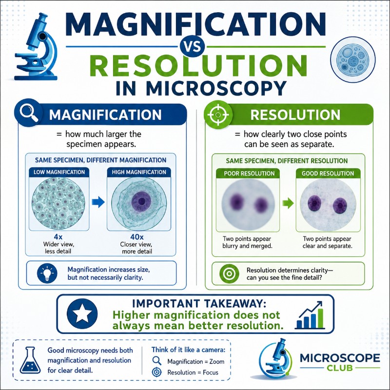
Magnification tells you how much larger an image appears compared to the real object. Resolution tells you how much fine detail you can actually distinguish. A microscope can magnify an image 1,000 times and still produce a blurry, useless picture if its resolution is poor. In microscopy, these two properties are independent — and understanding the difference is the single most important concept for anyone choosing or using a microscope.
Magnification vs Resolution — The Short Answer
Before going deeper, here is a direct side-by-side comparison:
| Magnification | Resolution | |
|---|---|---|
| What it means | How much larger the image is vs. the object | Smallest distance between two points still seen as separate |
| What controls it | Objective lens power × eyepiece power | Numerical aperture (NA) and wavelength of light |
| Units | Dimensionless ratio (e.g., 400×) | Distance (nanometers or micrometers) |
| Better when the number is… | Higher | Lower (smaller gap = more detail) |
| Can you improve it? | Yes — add a stronger eyepiece or objective | Yes — use a higher-NA objective, immersion oil, or shorter wavelength light |
The key takeaway: resolution is the true measure of a microscope’s power. Magnification without resolution is just a bigger blur.
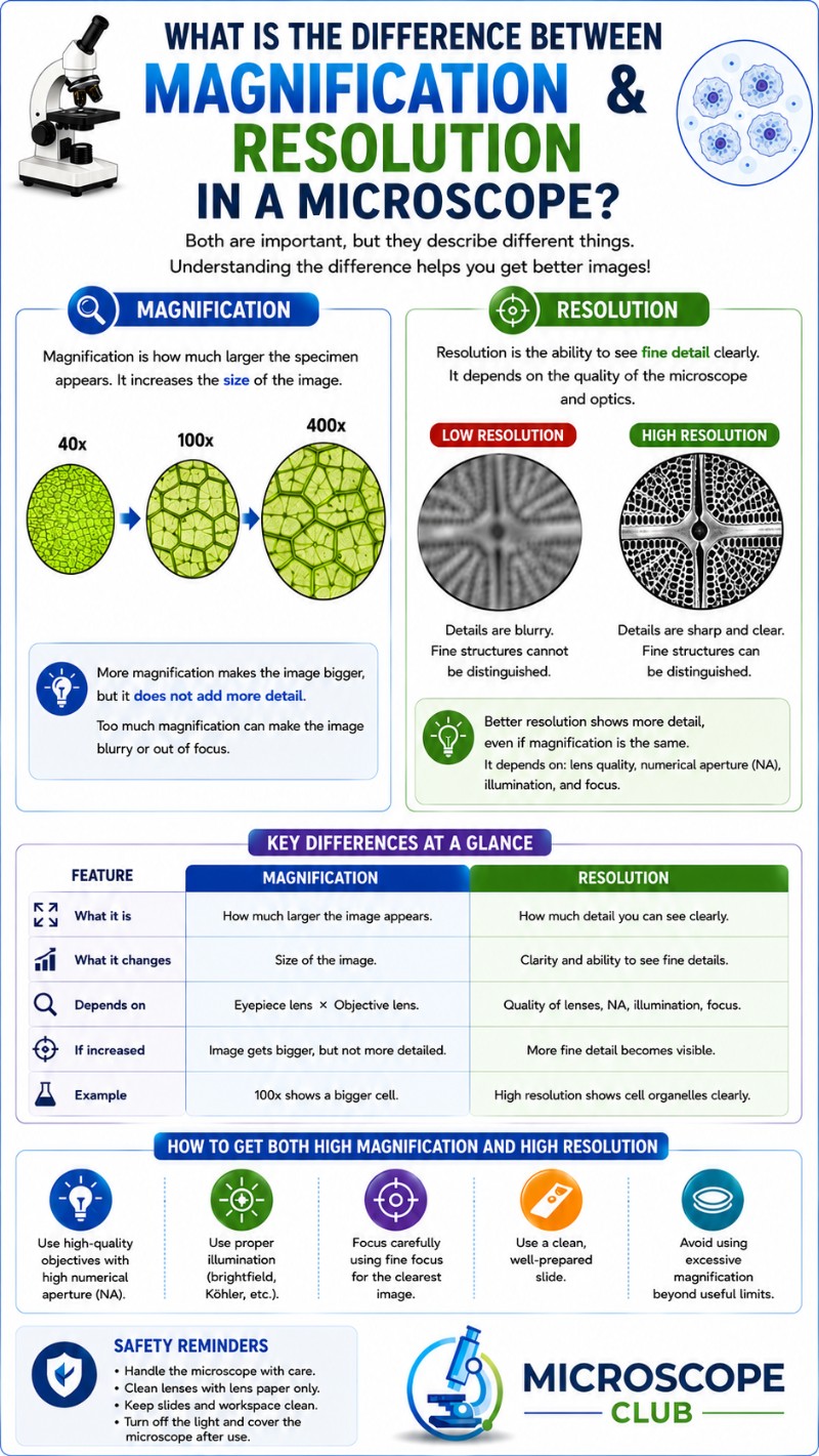
What Is Magnification?
Magnification is the ratio of an image’s apparent size to the real size of the specimen. In a compound light microscope, you calculate total magnification by multiplying the objective lens power by the eyepiece (ocular) power:
Total magnification = objective magnification × eyepiece magnification
For example, a 40× objective combined with a standard 10× eyepiece gives 400× total magnification. Switch to a 100× oil-immersion objective and you reach 1,000×. The calculation is simple arithmetic — knowing the parts of a compound microscope and their individual powers tells you exactly what magnification you will achieve.

Magnification is easy to increase. You can swap in a higher-power eyepiece or add a photo relay lens. The problem is that magnification has no opinion about image quality — it scales whatever is there, sharp details and blur alike.
What Is Resolution?
Resolution (also called resolving power) is the minimum distance between two points at which the microscope can still display them as two distinct points rather than a single merged blob. A microscope with a resolution of 0.2 micrometers (µm) can distinguish two structures that are 200 nanometers (nm) apart. One with a resolution of 2 µm cannot — those same two structures would blur into one.
Notice the counterintuitive direction: a lower resolution number means better resolving power. Resolving 0.2 µm is far superior to resolving 2 µm. This trips up beginners constantly, so make sure the number’s meaning is clear before reading any spec sheet.

For reference:
- The unaided human eye resolves about 0.1 mm (100 µm)
- A quality light microscope resolves about 0.2 µm (200 nm)
- A scanning electron microscope (SEM) resolves 0.5–1 nm
- A transmission electron microscope (TEM) resolves below 0.05 nm
Electron microscopes win because electrons have a far shorter wavelength than visible light — and wavelength is one of only two variables that determine resolution.
Numerical Aperture — The Key to Resolution
The other variable is the numerical aperture (NA) of the objective lens. NA is defined as:
NA = n × sin(θ)
Where n is the refractive index of the medium between the lens and the specimen (air = 1.0, immersion oil ≈ 1.52), and θ is the half-angle of the cone of light collected by the objective.
Higher NA means better resolution. This is why microscope resolution, numerical aperture, and light wavelength are always discussed together — they are the three levers that determine what your microscope can actually see.
It is also why immersion oil matters. By replacing air (n = 1.0) with oil (n = 1.52) between the objective and the slide, you raise the refractive index, which raises the NA, which improves resolution. The condenser plays a related role: it shapes the cone of light illuminating the specimen, and a properly adjusted condenser maximizes the effective NA of the system. Skimping on condenser adjustment is a common reason high-power objectives underperform.
The Abbe Diffraction Limit — Why Light Microscopes Stop at ~200 nm
In 1873, German physicist Ernst Abbe derived the mathematical limit of resolution for light microscopy. Known as the Abbe diffraction limit, it states:
d = λ / (2 × NA)
Where d is the minimum resolvable distance, λ is the wavelength of the illuminating light, and NA is the numerical aperture of the objective.
Worked example using green light (λ = 550 nm) and a high-quality oil-immersion objective (NA = 1.4):
d = 550 nm / (2 × 1.4) = 550 / 2.8 ≈ 196 nm ≈ 0.2 µm
That is the hard floor of conventional light microscopy. No matter how good the optics, no matter how high the magnification, a light microscope cannot resolve two points closer than roughly 200 nm. Physics will not allow it — the diffraction of light waves sets the ceiling.
You may also see the Rayleigh criterion, which uses the formula d = 0.61λ / NA. Applied to the same values: d = 0.61 × 550 / 1.4 ≈ 240 nm. Both formulas yield the same practical limit of ~200–250 nm, and both are cited in textbooks. The Abbe formula is more common in microscopy contexts.
According to Britannica’s overview of microscope resolution, this diffraction barrier remained the fundamental limit of light microscopy for over a century — until super-resolution techniques emerged in the 2000s.
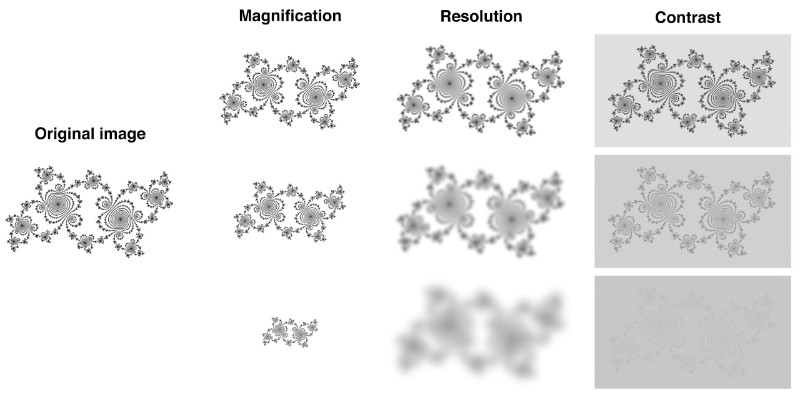
Empty Magnification — Bigger but Not Clearer
Once you understand the Abbe limit, empty magnification makes immediate sense. Empty magnification is the condition where you increase the image size beyond the point where any new detail can appear. You are enlarging existing blur, not revealing new structure.
The practical rule: useful magnification runs from 500 × NA to 1000 × NA. With the best oil-immersion objective (NA = 1.4), that ceiling is about 700×–1,400× total magnification. Most practitioners cite the practical upper limit of a light microscope as 1,000×–1,500×.
Any magnification above ~1,000 × NA is empty. Zoom past that threshold and the image simply gets larger without getting sharper.
How to spot empty magnification on a spec sheet:
- Check the objective NA, not just the power. A “100×” objective with NA 0.65 is mediocre; a “100×” objective with NA 1.25 oil is excellent.
- Apply the rule: useful max = 1000 × NA. NA 0.65 → 650× useful max. Pushing it to 1,000× is empty magnification.
- Any microscope advertising “2000×” in a consumer or toy category is almost certainly delivering empty magnification. The optics to back that claim cost hundreds of dollars in objective lenses alone.
Digital zoom compounds the problem. Cropping and enlarging a digital image is pure empty magnification — it adds no optical information and throws away field of view with nothing to show for it. Optical NA, not pixel count, sets real resolution. For more on how this plays out across instrument types, see which microscope achieves the highest magnification and resolution.
When Magnification Matters vs When Resolution Matters
Both parameters matter — the question is which one is the actual bottleneck for a given task.
When magnification is the bottleneck: You are viewing large structures — pond organisms, insect anatomy, plant cross-sections — that are big enough to resolve at your current NA, but too small to see comfortably. Here, adding magnification (up to the useful limit) directly improves the experience. A 4× or 10× objective may be all you need, and a lower NA is perfectly fine.
When resolution is the bottleneck: You are trying to see fine cellular detail — bacterial flagella (~20 nm wide, below the light limit), cell membrane structures, chromosome banding — where the detail itself is at or below the diffraction limit. More magnification does nothing. The fix is a higher-NA objective, immersion oil, blue/UV illumination (shorter wavelength), or a fundamentally different instrument (electron microscope, confocal).
Practical scenarios:
- Viewing pond water microorganisms (50–200 µm): Magnification is the limit. A 40× dry objective is plenty; worrying about NA at this scale is over-engineering.
- Resolving mitochondria in a cell (~1 µm): Resolution starts to matter. Use a 60× or 100× oil-immersion objective with NA ≥ 1.25.
- Resolving individual bacteria (~0.5–2 µm long, ~0.2–1 µm wide): You are at or near the diffraction limit. Oil immersion is non-negotiable; consider blue-light illumination.
- Imaging viruses (~20–300 nm): Beyond the light limit entirely. You need an electron microscope or a super-resolution fluorescence technique.
Also worth noting: resolution and depth of field trade against each other. Higher-NA objectives that improve lateral resolution simultaneously reduce depth of field — the slice of specimen that appears in focus gets thinner. This is not a flaw; it is physics. Plan your sample preparation accordingly.
Beyond the Light Limit — Electron and Super-Resolution Microscopy
The Abbe diffraction limit applies to visible light. Electrons have wavelengths thousands of times shorter (~0.002 nm at typical accelerating voltages). That is why a light microscope vs electron microscope comparison is not even close in resolution terms. TEMs resolve individual atoms; SEMs resolve surface topography at the nanometer scale. The tradeoff is that specimens must be prepared in vacuum, stained with heavy metals, or coated in conductive material — live cells are generally not compatible with electron microscopy.
Super-resolution fluorescence techniques (STED, STORM, PALM, and structured illumination) break the Abbe limit through a different approach: they use clever optical or molecular tricks — such as selectively switching fluorophores on and off — to extract spatial information beyond what classical diffraction theory allows. Resolutions of 20–50 nm are now routine in fluorescence super-resolution. The Nobel Prize Committee’s 2014 chemistry award recognized the development of super-resolved fluorescence microscopy. These techniques require specialized equipment and labeling protocols, but they represent the frontier of what light-based microscopy can achieve.
For a broader picture of where each instrument type sits on the resolution spectrum, see different types of microscopes and how electron microscopes image atoms.
For a grounded primer on the underlying physics, Khan Academy’s microscopy overview covers the core concepts clearly and is freely available.
Frequently Asked Questions
What is the difference between magnification and resolution in a microscope?
Magnification is how much larger the image appears compared to the real object. Resolution is the smallest distance between two points that the microscope can show as separate. A microscope can have high magnification and poor resolution — meaning it shows a large but blurry image with no additional detail.
Is higher magnification always better?
No. Magnification is only useful up to the point where the resolution of the objective can support it. Beyond that — roughly 1,000 × the numerical aperture — you get empty magnification: a bigger image with no new detail. For most biological work, 400×–1,000× is the practical sweet spot.
What is empty magnification?
Empty magnification occurs when total magnification exceeds the resolving power of the optics. The image gets larger but no new structural detail appears — you are simply enlarging existing blur. It typically begins above 1,000 × NA for the objective in use. Many inexpensive consumer microscopes advertise “2000×” power that is entirely empty magnification.
What determines the resolution of a microscope?
Two factors: the numerical aperture (NA) of the objective lens, and the wavelength of the illuminating light. Higher NA and shorter wavelength both improve resolution (lower the minimum resolvable distance). The relationship is captured in the Abbe formula: d = λ / (2 × NA).
What is the maximum useful magnification of a light microscope?
Approximately 1,000×–1,500× total magnification, depending on the objective NA. The useful range is 500 × NA to 1,000 × NA. With the best oil-immersion objectives (NA ≈ 1.4), that is roughly 700×–1,400×. Beyond that, additional magnification is empty and adds nothing useful.
What is the Abbe diffraction limit?
The Abbe diffraction limit is the theoretical minimum resolvable distance for a light microscope, derived by Ernst Abbe in 1873. The formula is d = λ / (2 × NA). For visible green light (550 nm) and a high-quality oil objective (NA 1.4), d ≈ 200 nm (0.2 µm). No conventional light microscope can resolve finer detail than this physical limit.
Does numerical aperture affect resolution or magnification?
Numerical aperture affects resolution directly — it is the single most important specification for resolving power. It does not affect magnification. Two objectives with the same power (e.g., 40×) but different NAs (0.65 vs 0.95) will magnify identically but resolve very differently.
Why does my microscope image look blurry at high magnification?
The most likely cause is that your magnification has outrun the resolving power of the objective — empty magnification. Other common causes include: the condenser is not aligned or stopped down, the objective NA is too low for that magnification, you are missing immersion oil on an oil-immersion objective, or the slide is too thick or dirty. Check NA first, then condenser alignment.
Conclusion
Magnification and resolution are not the same thing, and treating them as interchangeable is the most common mistake beginners make when evaluating or using a microscope. Resolution — governed by numerical aperture, light wavelength, and the hard ceiling of the Abbe diffraction limit — determines what detail is actually present in the image. Magnification simply scales that image up. Push magnification past the useful range and you get empty magnification: a larger blur, nothing more. The specification worth scrutinizing on any objective is the NA printed on its barrel, not the power.
Have you run into empty magnification in your own work — maybe a high-power eyepiece that made things worse instead of better, or a “2000×” microscope that disappointed? Tell us what you found in the comments below. It is one of those lessons that sticks best once you have seen it firsthand.