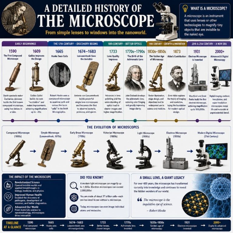
The history of the microscope is a 2,000-year relay race — passed from ancient glassworkers to Islamic scholars, Dutch lens-grinders, and 20th-century physicists pushing far beyond the limits of light itself. The microscope stands as one of the most consequential instruments in science, granting human eyes access to a world invisible to the naked eye, from the cells inside a cork sliver to the molecular scaffolding of a virus. What follows is a detailed account of that journey, corrected against the best current sources and enriched with what each instrument actually revealed when it was first turned on a specimen.
How the Idea of Magnification Was Born

Light refraction — the bending of a light ray as it passes from one medium to another — is the physical phenomenon that makes magnification possible. Claudius Ptolemy, the Greek-Egyptian mathematician and astronomer who lived c. 100–170 AD (the 2nd century AD, not BC as older accounts claim), described the bending of light passing through water in his treatise Optics. He noted that a rod partially submerged in water appears kinked at the surface — a direct observation of refraction. It was a perceptual puzzle that would take another nine centuries to ripen into a practical tool.
The crucial next step came from the Islamic world. Ibn al-Haytham — the Arab polymath known in Latin sources as Alhazen — authored the Book of Optics (Kitāb al-Manāẓir) around 1021 AD in Cairo. It was the first work to describe, in systematic mathematical terms, how a convex lens refracts light rays to a focal point and can produce a magnified image of an object placed closer than that focus. Ibn al-Haytham’s intromission theory — light travels from the object to the eye, not vice versa — was the correct model. When Latin translators carried his work into medieval Europe in the 12th and 13th centuries, it provided the optical theory that spectacle-makers and eventually microscope-builders would need.
Glass, Burning Lenses, and the First Spectacles (1st–14th Century)

Glass itself is far older than Rome — Egyptian and Mesopotamian craftsmen were making glass beads as early as 3500 BC. What changed in the 1st century AD was the Roman mastery of clear, blown glass thin enough to shape into lenses. Roman glassworkers, along with Egyptians and Greeks, ground lenses in the shape of a lentil bean — thick at the center, thin at the edges — which is precisely why we call them lenses today (from the Latin lenticula, “small lentil”). These so-called burning glasses could focus sunlight to ignite tinder and, the Greek physician Galen records, were used to cauterize wounds in surgery.
The step from burning glass to reading aid happened in Italy in the late 13th century. Spectacle-makers in Venice and Florence — their names are unfortunately lost — began grinding lenses and mounting them in frames around 1286–1300. By the early 14th century, craftsmen were also making simple hand-held magnifiers, producing magnifications of roughly 6×–10×. Because these lenses were frequently used to examine fleas and small insects, they became known informally as flea glasses — a nickname that reveals exactly what curious observers were doing with them.
The First Compound Microscope: Middelburg, c. 1590–1620 (Disputed)

The decisive conceptual leap was combining two lenses in a tube — one (the objective) to gather light from the specimen and form an intermediate image, a second (the eyepiece) to magnify that intermediate image further. This is the compound microscope. Its invention is usually credited to Dutch spectacle-makers in the city of Middelburg, the Netherlands, sometime between 1590 and around 1620, though the historical record is genuinely contested.

The names most often cited are Zacharias Janssen (born approximately 1585) and his father Hans Janssen, and separately Hans Lippershey, who is better known today as the claimant of the first telescope patent (1608). Because Zacharias would have been only about five years old in 1590, historians generally regard the instrument as a family workshop effort at the earliest, or place it somewhat later. The magnification of these first compound instruments is reported to have ranged from 3× to 9× — a respectable range but far short of what a good single lens could ultimately achieve — and the images were blurry enough that the devices were more novelty than scientific instrument.
Galileo Galilei also built compound optical instruments during this period, using them primarily to observe the sky. While he did turn a two-lens instrument toward small objects and called it the occhiolino (“little eye”), the telescope is Galileo’s instrument, not the microscope.
The Word “Microscope” and the Birth of a Discipline (17th Century)
The Latin term microscopium was coined in 1625 by the Italian botanist Giovanni Faber, in a letter to the mathematician Federico Cesi of the Accademia dei Lincei (the same learned society that had Galileo as a member). Faber assembled the term from the Greek mikros (small) and skopein (to look at). The word first described Galileo’s two-lens instrument, not Leeuwenhoek’s later single-lens scope — a reminder that naming and invention rarely happen simultaneously.

Robert Hooke and Micrographia (1665)
The publication that turned the compound microscope into a cultural event was Robert Hooke‘s Micrographia, published in January 1665 as the Royal Society’s first major scientific publication. [source: Royal Society — Micrographia] The diarist Samuel Pepys stayed up until 2 o’clock in the morning reading it and called it “the most ingenious book I have read in my life.” The reason: Hooke’s fold-out copper-plate engravings were revelatory. His rendering of a common flea filled an entire pull-out sheet and showed the creature’s legs equipped with barbs, its eyes faceted like cut gemstones, its body sheathed in overlapping armor-plates. Readers who had spent their whole lives swatting at fleas suddenly saw them as intricate machines.
More scientifically important was Hooke’s examination of a thin slice of cork. Through a three-lens compound microscope of his own design, he saw a regular pattern of empty box-like compartments separated by walls. He named them cells, from the Latin cella (small room), because they reminded him of the cells of a honeycomb or a monk’s chambers. The cork cells he saw were dead — only the cell walls remained — but the word stuck, and cell biology was born.
Antonie van Leeuwenhoek and the Simple Single-Lens Microscope

While Hooke was working with compound instruments, a Dutch draper in Delft was taking a radically different approach. Antonie van Leeuwenhoek (1632–1723) had no university education but exceptional manual skill. He learned to grind and polish small glass balls into near-spherical lenses with very short focal lengths — his finest surviving lens has been measured at approximately 266–270× magnification, far beyond anything the compound microscopes of his era could produce.
The key insight is why Leeuwenhoek’s single-lens (simple) microscope out-resolved compound scopes. Each additional lens in a compound instrument compounds the aberrations: chromatic aberration (each color of light bends by a slightly different angle, creating colored halos around edges) and spherical aberration (light passing through the edge of a lens focuses at a slightly different point than light through the center, blurring the image). Leeuwenhoek’s tiny, near-perfect single lens avoided that accumulation. The trade-off was that the lens was only about 1 mm in diameter and had to be held extremely close to both the specimen and the observer’s eye — uncomfortable, but optically superior to anything else available until the achromatic lens was invented.
What Leeuwenhoek first saw with his instrument in 1674 was a sample of green lake water from Berkelse Mere. He described tiny transparent creatures moving about with great agility — rotating, darting forward, standing still. These were protozoa (single-celled eukaryotes), which he called “animalcules.” He communicated this finding to the Royal Society of London on 7 September 1674. [source: Phil. Trans. Royal Soc. B — Leeuwenhoek’s “little animals”]
Bacteria came two years later. In 1676, Leeuwenhoek prepared a pepper-water infusion and observed far smaller organisms — true bacteria — which he reported to the Royal Society on 9 October 1676. This makes 1676 the correct date for the discovery of bacteria, not 1674, and the 1674 lake-water organisms were protozoa, not bacteria. For these discoveries, Leeuwenhoek is credited as the father of microbiology and was elected a Fellow of the Royal Society in 1680. [source: NCBI/PMC — Leeuwenhoek as Father of Microbiology]
Correcting Aberration: The 18th and 19th Centuries
The compound microscope’s weakness — aberration — motivated a century of optical engineering. The solution arrived in two stages.
Chromatic aberration makes lens images appear ringed with color, because different wavelengths of light refract by slightly different amounts through a simple glass lens. If you focused a 17th-century compound microscope on a sharp black line on white paper, you would see it surrounded by bands of violet and red — the lens was bending blue light more than red. Chester Moor Hall, an English barrister and amateur optician, discovered around 1729–1733 that pairing a converging crown-glass lens with a diverging flint-glass lens — an achromatic doublet — largely cancelled the chromatic difference. Hall kept his discovery private and had the two elements ground by different craftsmen to prevent anyone from deducing the formula. John Dollond independently arrived at the same design and patented it in 1758; courts later upheld Dollond’s patent on the grounds that Hall had never published or commercialized the idea. Both names deserve credit.
Spherical aberration — the residual blur that remained even in achromatic lenses — was addressed by Joseph Jackson Lister in 1830, who showed how to combine low-power lens elements at specific separations to cancel spherical aberration without losing sharpness.
The theoretical ceiling of light microscopy was defined by Ernst Abbe at Carl Zeiss in Jena in 1873. Abbe’s diffraction limit states that no light microscope, however perfectly built, can resolve two points separated by less than roughly half the wavelength of the illuminating light — approximately 200 nanometers under visible light. Abbe pioneered oil-immersion objectives (filling the gap between lens and coverslip with immersion oil to increase numerical aperture) and invented the Abbe condenser for controlled specimen illumination. His sine condition — a mathematical relationship between lens aperture and image quality — remains the foundation of microscope optics today.
Chemistry Joins the Story: Staining and Sample Preparation
Seeing depended as much on chemistry as on optics. Most biological specimens are nearly transparent and colorless under a microscope — early microscopists often could not distinguish meaningful structures in the fog. The breakthrough was dye chemistry. Synthetic aniline dyes, developed in the 1850s and 1860s from coal-tar derivatives, had an unexpected property: different dyes bound preferentially to different types of biological tissue. Suddenly researchers could stain a tissue slice and see nuclei in one color, cytoplasm in another.
The most consequential staining technique in microbiology was published in 1884 by the Danish physician Hans Christian Gram. The Gram stain separates bacteria into two groups — Gram-positive (which retain a crystal-violet dye and appear purple) and Gram-negative (which lose the stain and appear pink after a counterstain) — based on differences in their cell-wall structure. A century and a half later, Gram-positive and Gram-negative remain the first classification a clinician reaches for when identifying a bacterial pathogen. The stain works because of the microscope; the microscope became clinically useful because of the stain.
The 20th Century: Seeing Below the Wavelength of Light
By 1900, the compound light microscope had been developed as far as Abbe’s diffraction limit would allow. What came next were instruments that either bent that limit or abandoned visible light altogether.
Köhler illumination — a system for producing uniform, glare-free specimen illumination — was introduced by August Köhler of Carl Zeiss in 1893 and remains the standard setup used in research microscopes today. A decade later, in 1904, Köhler and Moritz von Rohr at Carl Zeiss built the first ultraviolet microscope, using a cadmium arc lamp and fused-quartz optics (ordinary glass is opaque to UV). During this UV work, Köhler noticed that some specimens glowed faintly when illuminated with ultraviolet light — a casual observation that would eventually blossom into fluorescence microscopy, now one of the most widely used techniques in cell biology.
Richard Zsigmondy invented the ultramicroscope in 1903, using oblique illumination to make particles far smaller than the resolution limit visible as bright spots against a dark background — the first instrument to detect nanoparticles. He received the Nobel Prize in Chemistry in 1925 for colloidal chemistry work enabled by this instrument.
The phase-contrast microscope was developed by Frits Zernike, a Dutch physicist, in the early 1930s. The problem it solved was fundamental: living, unstained biological cells are nearly transparent, and staining kills them. Zernike’s design converts tiny differences in the refractive index of cell structures — differences invisible to the eye — into differences in brightness, making the internal structures of living, unstained cells visible for the first time. Carl Zeiss brought the instrument to market in 1941. Zernike received the Nobel Prize in Physics in 1953.
The Electron Microscope (1931)
The hardest barrier in microscopy — Abbe’s 200-nm diffraction limit — was broken not by improving lenses but by replacing visible light with electrons. Electrons, accelerated through an electric field, behave as waves with wavelengths thousands of times shorter than visible light.
Ernst Ruska and Max Knoll built the first electron microscope prototype at the Technische Hochschule Berlin in 1931. [source: Britannica — Ernst Ruska] Ruska improved the instrument to roughly 12,000× magnification by 1933. The first commercial transmission electron microscope (TEM) was produced by Siemens in 1939. Ruska received the Nobel Prize in Physics in 1986 — 55 years after the invention — for this work. The electron microscope ultimately allowed researchers to photograph viruses, see the double-helix of DNA in crystallography, and resolve protein structures.
Scanning Probe Microscopes: Touching the Atomic Surface (1981–1986)
Gerd Binnig and Heinrich Rohrer at IBM Zürich invented the scanning tunneling microscope (STM) in 1981 (first published 1982). The STM works by bringing a metal tip to within a nanometer of a conducting surface and measuring the quantum-mechanical tunneling current that flows. As the tip scans across the surface, variations in current map the positions of individual atoms with sub-angstrom precision. The first STM images of the silicon surface (1983) showed individual atoms arranged in their crystal lattice — a photograph of matter at its most fundamental scale. Binnig and Rohrer received the Nobel Prize in Physics in 1986.
Binnig, together with Calvin Quate and Christoph Gerber, extended the principle in 1986 with the atomic force microscope (AFM), which works by measuring the deflection of a tiny cantilever as its sharp tip is dragged across a surface. Because AFM measures force rather than current, it works on non-conducting samples — biological membranes, polymer films, pharmaceutical crystals — making it indispensable in both materials science and cell biology.
Fluorescence and Super-Resolution: The 21st-Century Frontier
Fluorescence microscopy turned Köhler’s 1904 observation into the dominant imaging modality in modern cell biology. In a fluorescence microscope, molecules within the specimen — either naturally fluorescent or tagged with synthetic fluorescent dyes or genetically encoded fluorescent proteins (GFP, discovered in jellyfish and developed for biology in the 1990s) — are excited by a specific wavelength of light and emit a longer wavelength that is detected separately. The result is that a specific protein, organelle, or gene sequence can be lit up in one color inside an otherwise dark cell, with extraordinary specificity.
The theoretical problem with fluorescence microscopy was the same as ordinary light microscopy: Abbe’s 200-nm limit. Structures smaller than that — individual proteins, synaptic vesicles, virus capsids — blurred together. Three researchers independently developed ways around the diffraction limit, and collectively won the 2014 Nobel Prize in Chemistry: Eric Betzig (PALM), William Moerner (single-molecule detection), and Stefan Hell (STED microscopy, which depletes fluorescence outside a nanometer-scale spot using a second laser). These super-resolution microscopy techniques brought light microscopy resolution down to 20–30 nm — ten times below the classical limit — enabling researchers to watch individual proteins move inside living cells in real time.
Cryo-electron microscopy (cryo-EM) represents a second revolution. Rather than staining or chemically fixing a biological sample (which distorts its structure), cryo-EM vitrifies it — freezing it so rapidly (within milliseconds) that water molecules have no time to form ice crystals, trapping proteins in their native conformations in a glass-like water matrix. Jacques Dubochet, Joachim Frank, and Richard Henderson developed the methods that made this feasible, earning the 2017 Nobel Prize in Chemistry. [source: Nobel Prize Chemistry 2017] Cryo-EM can now determine protein structures at near-atomic resolution without crystallization — it was used to determine the structure of SARS-CoV-2’s spike protein within weeks of the virus being sequenced, accelerating vaccine development.
Today: Microscopes From the Classroom to the Molecular Scale
Microscopes of every type — from beginner to research-grade — are manufactured primarily in Germany, Japan, and China, and mass production has put functional optical microscopes within reach of science classrooms worldwide. The compound light microscope remains the standard classroom instrument: affordable, durable, and capable of resolving bacteria and cells at 400×–1000×. Digital microscopes connect to a computer screen or tablet, making real-time specimen capture accessible without an eyepiece.
At the research frontier, the electron microscope, AFM, STED, and cryo-EM are the tools resolving structures at the protein, membrane, and atomic level. The 500-year arc from Zacharias Janssen’s blurry 9× tube to cryo-EM resolving amino acid side-chains at 1.2 Å is a continuous story of one question: how much smaller can we see?
Microscope Magnification: How It Works and How to Calculate It
Microscope total magnification equals eyepiece magnification multiplied by objective magnification — that is the formula every microscopist uses.
Total magnification = eyepiece magnification × objective magnification
A worked example: if your microscope has a standard 10× eyepiece and you select the 40× objective, the total magnification is 10 × 40 = 400×. The specimen appears 400 times larger than its actual size.
| Eyepiece | Objective | Total magnification | Typical use |
|---|---|---|---|
| 10× | 4× | 40× | Scanning — get the full slide in view |
| 10× | 10× | 100× | Low power — tissue architecture |
| 10× | 40× | 400× | High dry — individual cells, bacteria |
| 10× | 100× (oil) | 1,000× | Oil immersion — bacteria detail, blood cells |
Important: magnification and resolution are not the same thing. Resolution is the smallest separation at which two points appear distinct rather than merged. Ernst Abbe’s diffraction limit fixes the resolution ceiling for a light microscope at roughly 200 nm (half the shortest wavelength of visible light). Increasing magnification beyond what the resolution allows simply makes a blurrier image bigger — what microscopists call “empty magnification.” The jump to electron and super-resolution microscopes was driven by the need to break through that 200-nm wall.
History of the Microscope: Timeline
| Date | Who | Milestone |
|---|---|---|
| c. 100–170 AD | Claudius Ptolemy | Describes light refraction in water (Optics) |
| c. 1021 | Ibn al-Haytham | Book of Optics — first mathematical description of magnifying convex lenses |
| c. 1286–1300 | Italian spectacle-makers | First eyeglasses; lens grinding becomes a trade |
| c. 1590–1620 (disputed) | Janssen / Lippershey, Middelburg | First compound microscope (3×–9×) |
| 1625 | Giovanni Faber | Coins the word microscopium |
| 1665 | Robert Hooke | Micrographia published; “cell” coined from cork |
| 1674 | Antonie van Leeuwenhoek | Discovers protozoa (“animalcules”) in lake water |
| 1676 | Antonie van Leeuwenhoek | Discovers bacteria in pepper-water infusion |
| c. 1729–1733 | Chester Moor Hall | Invents the achromatic doublet (lens); John Dollond patents it 1758 |
| 1830 | Joseph Jackson Lister | Corrects spherical aberration in compound microscopes |
| 1873 | Ernst Abbe | Defines the diffraction limit (~200 nm); pioneers oil-immersion objectives |
| 1884 | Hans Christian Gram | Gram stain — transforms clinical microbiology |
| 1893 | August Köhler | Köhler illumination — standard for research microscopes |
| 1903 | Richard Zsigmondy | Ultramicroscope — detects nanoparticles (Nobel 1925) |
| 1904 | Köhler & von Rohr (Zeiss) | First ultraviolet microscope; fluorescence serendipitously observed |
| 1931 | Ernst Ruska & Max Knoll | First electron microscope prototype (TEM) |
| Early 1930s | Frits Zernike | Phase-contrast microscope — live unstained cells visible; Nobel 1953 |
| 1939 | Siemens | First commercial transmission electron microscope |
| 1981 | Binnig & Rohrer (IBM Zürich) | Scanning tunneling microscope (STM) — images individual atoms; Nobel 1986 |
| 1986 | Binnig, Quate & Gerber | Atomic force microscope (AFM) — works on non-conducting surfaces |
| 2014 | Betzig, Hell, Moerner | Super-resolution fluorescence microscopy — Nobel Prize in Chemistry |
| 2017 | Dubochet, Frank, Henderson | Cryo-EM — near-atomic protein structures without crystals; Nobel Prize in Chemistry |
Frequently Asked Questions
Who invented the microscope?
The compound microscope is credited to Dutch spectacle-makers in Middelburg around 1590–1620, most often associated with Zacharias Janssen and his father Hans Janssen, with Hans Lippershey as a rival claimant. The attribution remains disputed among historians. The simple single-lens microscope was perfected by Antonie van Leeuwenhoek in Delft in the 1670s, and it was Leeuwenhoek’s version that made the first biological discoveries.
When was the microscope invented?
The first compound microscope is generally dated to around 1590 in the Netherlands, though documentary evidence is thin and dates as late as 1620 are also cited. The word “microscope” was coined in 1625 by Giovanni Faber.
Who is the father of microbiology?
Antonie van Leeuwenhoek is universally titled the father of microbiology, having discovered protozoa in 1674 and bacteria in 1676 — the first person to observe living microorganisms directly.
What did Robert Hooke discover with the microscope?
Robert Hooke published Micrographia in 1665, describing detailed observations of insects, feathers, and plant material. His most consequential finding was that cork consists of tiny box-like compartments he called “cells” — the first use of the word in biology and the founding observation of cell theory.
What is the difference between a simple and a compound microscope?
A simple microscope uses a single magnifying lens (like a magnifying glass or Leeuwenhoek’s instruments). A compound microscope uses at least two lens systems — an objective and an eyepiece — to achieve higher magnification. Paradoxically, Leeuwenhoek’s simple microscopes outperformed the compound instruments of his era because each additional lens multiplies optical aberrations; his single near-perfect lens avoided that penalty.
Who invented the electron microscope?
Ernst Ruska and Max Knoll built the first electron microscope prototype at Technische Hochschule Berlin in 1931. The first commercial instrument was produced by Siemens in 1939. Ruska received the Nobel Prize in Physics in 1986.
What was the first microscope able to see?
The early 17th-century compound microscopes could resolve fleas, lice, and insect parts at up to 9×. Leeuwenhoek’s 270× single-lens scope first revealed bacteria and protozoa. The electron microscope, from 1931 onward, could resolve viruses, cell organelles, and eventually individual atoms.