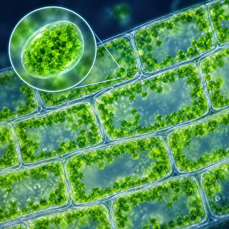
Chloroplasts under a microscope look nothing like the crisp, striped ovals in a biology textbook. At 400x on a standard light microscope, they appear as small green dots, roughly 2 to 7 microns across, packed in a ring just inside each plant cell’s wall. Watch a living Elodea leaf for a minute or two and those dots start to drift — a slow, continuous flow called cytoplasmic streaming.
What Chloroplasts Actually Look Like (Green Dots, Not Textbook Ovals)
A chloroplast measures about 2 to 7 micrometers across and only 1 to 2 micrometers thick — a flattened, lens-shaped disc, not the fat football-with-stripes shape most diagrams draw. Under a light microscope at high power, that shape resolves into a small, smooth green dot with no visible internal detail. A single mesophyll cell (the photosynthetic tissue inside a leaf) can hold 20 to 100 or more of them. They’re usually pressed in a loose ring against the inside of the cell wall, around a large central vacuole. The overall effect is closer to a scatter of green lentils pressed against a clear glass brick than a labeled diagram. OpenStax includes an Elodea micrograph showing chloroplasts as small green bodies inside leaf cells.
Chlorophyll a and b, the pigments packed inside each chloroplast, absorb red and blue light and reflect green — that’s the light-absorption chemistry behind photosynthesis, and it’s why the dots read as vivid green rather than any other color. What you won’t see, even at 400x, is the internal architecture: the thylakoid membranes, their stacked grana, and the surrounding stroma. Those structures are real — a chloroplast is a double-membrane plastid with its own DNA, most likely descended from a free-living cyanobacterium that a plant cell absorbed billions of years ago, as Britannica’s overview of chloroplast structure lays out. But they sit below the resolution of a hobbyist light microscope. Naming them is useful; expecting to see them is the mistake the textbook diagram sets you up for.
The Best Specimen: Why Elodea Leaf Wins
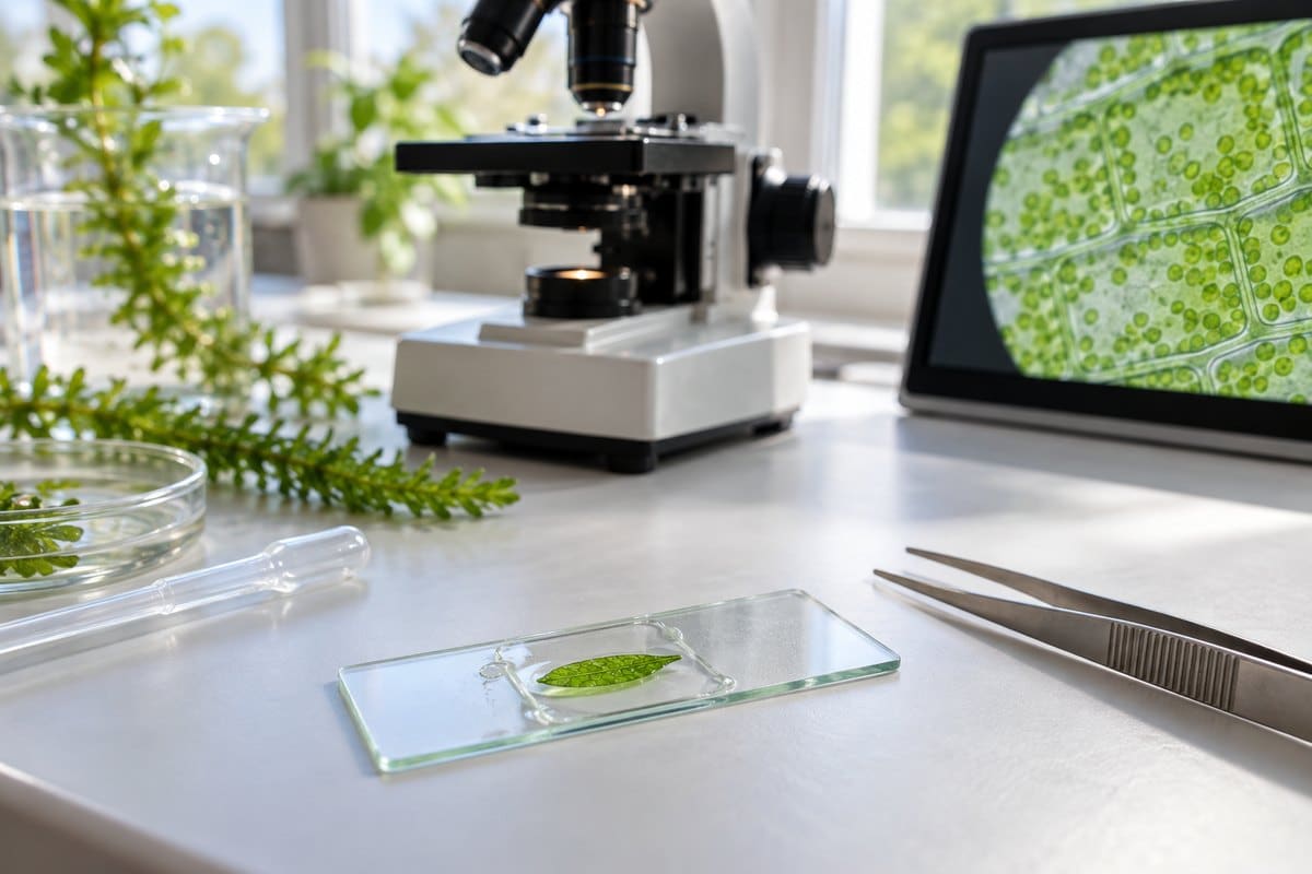
Elodea (also sold under the aquarium-trade name Anacharis) is the specimen that makes this whole observation easy. Its leaves are only two cell layers thick, so light passes straight through without any cutting, staining, or sectioning — drop a whole leaf tip in water, add a coverslip, and you’re looking at intact, living cells with their chloroplasts in place. A sprig from a pet store’s aquarium plant bin or a scoop from a drop of pond water is usually all you need. Moss leaves, such as those from Plagiomnium, are a solid backup for the same reason — they’re also only one or two cells thick.
Compare that to a typical land-plant leaf, where the chloroplast-dense mesophyll layer sits sandwiched between the epidermis and vein tissue — worth understanding if you want the bigger picture of the layers of a leaf under the microscope, but not something you can view live without slicing a thin cross-section first. Elodea skips that step entirely, which is why it’s the standard classroom and hobbyist choice for this exact question.
How to See Chloroplasts Under a Microscope (Step by Step)
This is a genuine wet-mount observation you can do with a basic student microscope. Follow these steps:
- Pull a fresh young leaf tip from an Elodea or Anacharis sprig — new growth at the stem tip is thinnest and most active.
- Place the leaf flat on a clean slide and add one drop of water. (If you need the full technique, see how to make a wet mount slide.)
- Lower a coverslip onto the drop at a shallow angle to avoid trapping air bubbles.
- Start at low power (40x or 100x) and scan for the thinnest, most translucent part of the leaf.
- Once you’ve centered a thin, well-lit area, switch to high power (400x).
- Dim the light source or close the diaphragm partway — full brightness washes the green out to pale gray.
- Give it two to three minutes under the lamp’s gentle warmth before deciding nothing is happening.
What You’ll See at Each Magnification (40x, 100x, 400x)
| Magnification | What you’ll actually see |
|---|---|
| 40x | The outline of the leaf and rows of rectangular cells; an overall green tint but no individual chloroplasts. |
| 100x | Cell walls become distinct; chloroplasts appear as a grainy green haze inside each cell, not yet separated. |
| 400x | Each chloroplast resolves into a separate green dot or lens shape lining the cell wall; streaming motion becomes visible. |
Watching Them Move: Cytoplasmic Streaming
Cytoplasmic streaming, also called cyclosis, is the slow, continuous circulation of cytoplasm around a plant cell’s central vacuole, and it’s what carries the chloroplasts along in a loop near the cell wall. The motion is driven by myosin motor proteins walking along tracks of actin filaments. It’s a mechanism covered in detail in open biology reference texts on plant cell motility. It’s temperature- and light-dependent, which is why streaming often looks still for the first minute or two and then picks up speed as the slide warms slightly under the microscope’s lamp.

The video below shows exactly this moment in a live Elodea leaf at hobbyist magnification — the same slow, conveyor-belt drift you’ll see on your own slide once the cell warms up.
Once you spot it, you can’t unsee it: dozens of green dots, all drifting the same direction around the cell edge, in near silence, on a slide you made yourself in under five minutes.
Chloroplasts in Other Cells (and Where You Won’t Find Them)
Chloroplast density is highest in leaf mesophyll cells, but they’re not exclusive to leaves. Green algae carry them too — Spirogyra is famous for a single spiral-ribbon chloroplast winding through each cell, a completely different shape from the scattered dots in Elodea. Euglena, which carries its own chloroplasts, is another common pond-water find worth comparing against a plant leaf sample. Its photosynthesis overview also explains the internal organization and role of chloroplasts.

Just as important is knowing where chloroplasts are absent. Onion cells, which have no chloroplasts at all, are a classic surprise for students — the onion bulb grows underground and never photosynthesizes, so it’s used as a cell-wall-and-nucleus specimen, not a chloroplast one. The same goes for animal cells such as human cheek cells: no cell wall, no chloroplasts, no chlorophyll. For a related but different pigmented organelle, diatoms and their golden-brown plastids are worth a look — same basic idea, different pigment, different color entirely.
Common Mistakes and How to Fix Them
- Focusing on the thick middle of the leaf. You’ll get a dark, featureless blur. Fix: hunt along the thin, translucent leaf edge or the youngest tip.
- Running the light at full brightness. It washes chloroplasts from vivid green to pale gray. Fix: dim the diaphragm or LED to a moderate setting.
- Letting the mount dry out. Streaming stops and the cells look frozen. Fix: add a fresh drop of water at the coverslip’s edge, or remount.
- Mistaking air bubbles for cells. Bubbles show up as thick, black-rimmed circles with no internal detail — don’t confuse them with real cells.
- Using an old or wilted leaf. Older tissue is thicker and less active. Fix: pick the newest, thinnest shoot tip available.
Frequently Asked Questions
Can you see chloroplasts in onion cells?
No. Onion bulb cells come from underground tissue that never photosynthesizes, so they contain no chloroplasts. Onion is the standard specimen for viewing cell walls and nuclei, not chloroplasts — use Elodea or moss for that.
Why are chloroplasts green?
They’re green because the chlorophyll pigments packed inside them absorb red and blue light and reflect green light back to your eye. Other pigments, like carotenoids, are present too but are masked by the much larger amount of chlorophyll.
Do you need to stain chloroplasts to see them?
No. Chloroplasts are naturally pigmented, so they’re visible in a plain water wet mount with no dye needed. Staining with something like iodine can actually kill the cell and stop cytoplasmic streaming, which removes the best part of the observation.
Can you see chloroplasts with a light microscope, or do you need an electron microscope?
A basic light microscope is enough to see chloroplasts as green dots and to watch them move. What a light microscope cannot resolve is their internal structure — the thylakoids, grana, and stroma require an electron microscope to actually see.
How many chloroplasts does a single plant cell have?
It varies by cell type, but a typical mesophyll cell in a leaf holds anywhere from 20 to over 100 chloroplasts, usually arranged in a loose ring near the cell wall to maximize light exposure.
Is a chloroplast the same thing as chlorophyll?
No — they’re often confused, but they’re not the same thing. A chloroplast is the organelle, the physical structure you can see as a green dot under a microscope. Chlorophyll is the pigment molecule packed inside it that actually does the light absorbing.
Can I reuse the same Elodea leaf for a second look later?
Not the same wet mount — once it dries out, the cells stop streaming and won’t recover. But you can pull a fresh leaf tip from the same sprig and make a new slide in a couple of minutes, so one plant will keep you supplied for many observations.
Conclusion
Chloroplasts under a microscope are simpler and stranger than any diagram suggests: small green dots, dozens to a cell, quietly drifting around the cell wall in a loop you can watch happen in real time. An Elodea leaf, a drop of water, and a basic light microscope are all it takes to see the gap between the textbook picture and the real thing for yourself.
Have you tried this one yet? Pull a sprig of Elodea, make a quick wet mount, and see how long it takes you to spot the streaming — then tell us what you noticed in the comments below.