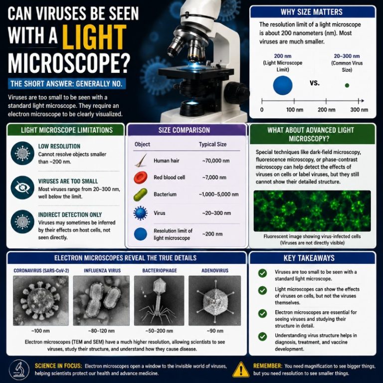
Viruses range from 20 to 300 nanometers in diameter, and a standard light microscope resolves no finer than about 200 nanometers — the Abbe diffraction limit — which means the optical microscope you find in a lab or school sits right at the edge of visibility for the very objects virologists most need to study. Whether a virus can be seen under a light microscope depends entirely on which light microscope, and what you mean by “see.”
Why the standard light microscope can’t see most viruses
The Abbe diffraction limit sets a hard ceiling on what any lens-and-visible-light system can resolve: roughly λ / (2 × NA), which works out to approximately 200 nm under typical laboratory conditions. Nobel Prize background, 2014 That ceiling is the reason conventional compound microscopes fail at virology for most species.
To put the scale in terms you can picture: if a typical bacterium (around 2 µm long) were the size of a car, a typical virus at 50 nm would be about the size of a coin sitting on the back seat. The light microscope can just resolve the car. The coin is invisible.
Bacteria typically measure 1–10 µm. Viruses range from roughly 20 nm (Parvovirus) up to about 300–400 nm (Poxvirus). Britannica That makes most viruses 10 to 100 times smaller than typical bacteria — not the 100–500× figure that circulates in older articles. The smallest bacteria (~400 nm) are about the same size as the largest common viruses; it is in that overlap zone that the comparison breaks down.
The compound light microscope, with a practical resolution of around 200 nm, can therefore only resolve a virus if it is unusually large — and as it happens, some viruses are.
Giant viruses you can actually see under a light microscope
Mimivirus has a capsid diameter of roughly 440 nm and an overall particle size of about 750 nm — well above the 200 nm diffraction limit. Mimivirus, Wikipedia When it was first discovered in a cooling tower in Bradford in 1992, researchers assumed it was a Gram-positive bacterium; they only realised their mistake a decade later when genomic analysis revealed it was a virus. Under a light microscope it appears as a small, slightly fuzzy dot — unremarkable to look at, but genuinely visible.
Pandoravirus is even larger: approximately 1 µm long and 0.5 µm wide. Both Mimivirus and Pandoravirus fall within the resolving power of a standard laboratory microscope. For the hobbyist reader: seeing these would require an actual culture and prepared slide, not something easily obtained — but the point stands that “light microscopes can’t see viruses” is an oversimplification.

Modified light microscopes: microsphere nanoscopy
For typical viruses, the tool that extends light microscopy into virus territory is microsphere-assisted optical nanoscopy (also called the white-light microsphere nanoscope). Developed by Wang et al. and published in Nature Communications in 2011, the technique places transparent silicon-dioxide microspheres (2–9 µm diameter) directly on the specimen. Wang et al., Nature Communications, 2011 The microspheres act as far-field superlenses, concentrating the evanescent near-field into a detectable propagating signal and achieving lateral resolution of approximately 50 nm — without a vacuum, without electron beams, and without cryogenic sample preparation.
A follow-on paper by Li et al. (2013) imaged adenoviruses (~75 nm) label-free using submerged microsphere optical nanoscopy, confirming individual viral particles as discrete dots. That is the system described in older accounts as “modified light microscopes with transparent microspheres” — now it has a proper name.

This comparison shows the same virus particles on a sample viewed through a scanning electron microscope versus a modified light microscope. The electron microscope image is sharper, but the virus particles are identifiable in both — the resolution gap is real, but not absolute.

One important caveat: microsphere nanoscopy, along with the other advanced optical techniques described below, are research laboratory tools, not clinical diagnostic instruments. Routine viral diagnosis relies on PCR, antigen tests, and serology — not on optically visualising the particle itself.
What is a virus?
A virus is an infectious agent that can only replicate inside the living cells of a host — bacteria, plants, animals, or humans. Unlike bacteria, viruses are not cells; they carry no ribosomes, produce no ATP, and cannot divide independently. Outside a host cell a virion is metabolically inert, closer to a sophisticated chemical package than to a living organism.

This microscope image shows virus particles distributed within host tissue. You can see various cells and tissue sections surrounding the virus particles — the density and distribution pattern varies with the stage of infection.
Structurally, most viruses are either helical (a coiled protein coat around a nucleic-acid strand, like Tobacco mosaic virus) or icosahedral (a 20-faced spherical shell, like adenovirus). Some, like Poxviruses, are brick-shaped and complex. The receptor-binding proteins on the capsid or envelope surface are what allow the virion to dock with and enter the correct host cell type.
Imaging methods compared: a practical guide
The question “can I see a virus under a microscope?” has a different answer depending on which microscope. This table maps the common methods to their resolution and practical capability:
| Method | Approximate resolution | Can it resolve a typical virus (20–300 nm)? |
|---|---|---|
| Standard light microscope (compound) | ~200 nm | No — only giant viruses ≥ ~400 nm |
| Microsphere nanoscope (white-light) | ~50 nm | Yes — research context only |
| Super-resolution fluorescence (STED / PALM / STORM / SIM) | ~20–50 nm | Yes — research context only |
| Transmission electron microscope (TEM) | <1 nm | Yes, including fine structure |
| Cryo-electron microscope (cryo-EM) | <1 nm (near-atomic) | Yes — gold standard for virus structure |
| Atomic force microscope (AFM) | ~1–10 nm | Yes — surface topography |
Super-resolution fluorescence microscopy (STED, PALM, STORM, SIM)
Super-resolution microscopy was awarded the 2014 Nobel Prize in Chemistry to Eric Betzig, Stefan Hell, and William Moerner. By exploiting clever fluorescence tricks — stimulated emission depletion (STED), photo-activated localization (PALM), or stochastic optical reconstruction (STORM) — these techniques push spatial resolution to 20–50 nm while still using visible light and standard sample preparation.
In practice, a researcher conjugates an antibody — one that binds specifically to a viral surface protein — to a fluorescent dye. The labeled virions then glow in the microscope, and the super-resolution algorithm reconstructs their positions at sub-diffraction precision. This is why STED or STORM can resolve the internal architecture of Herpes simplex virions or track individual HIV particles budding from a cell membrane.
Super-resolution fluorescence is now the dominant optical method for studying virus–cell interactions in real time. Unlike electron microscopy, it works on living cells and allows researchers to watch a virus enter, traffic, and replicate.
Electron microscopy and cryo-EM
Conventional electron microscopy — both transmission (TEM) and scanning (SEM) variants — uses electron beams rather than photons, bypassing the light-diffraction limit entirely and achieving sub-nanometre resolution. The advantages of electron microscopes for virology are well established: TEM was the primary tool for virus identification throughout the twentieth century.

This high-resolution TEM image of H1N1 influenza virus shows the characteristic rounded particles with surface-protein spikes clearly resolved. TEM images like this one are how virologists characterised influenza morphology long before sequence analysis was available.
Cryo-electron microscopy (cryo-EM) takes the technique further. The specimen is flash-frozen in vitreous ice — no fixatives, no staining, no dehydration — preserving it in a near-native hydrated state. Thousands of 2D projection images are then computationally assembled into a 3D structure at near-atomic resolution. Cryo-EM was awarded the 2017 Nobel Prize in Chemistry to Jacques Dubochet, Joachim Frank, and Richard Henderson. In 2020, the SARS-CoV-2 spike protein structure that featured in every vaccine report was determined by cryo-EM — not by light microscopy. The spike’s receptor-binding domain, ACE2 interactions, and prefusion conformation were all mapped at 3–4 Å resolution within weeks of the pandemic starting, enabling rapid vaccine design.

This is a three-dimensional reconstruction of the chikungunya virus particle, produced by cryo-EM. The icosahedral symmetry of the capsid is clearly visible, with surface glycoprotein spikes resolved at the periphery. Standard TEM would show the outline; cryo-EM reveals the architectural detail.
Fluorescence microscopy and labeling protocols
Fluorescence microscopy makes viral particles detectable by attaching a light-emitting molecule to the virus or to the host cell’s DNA being hijacked. In a typical labeling workflow a researcher either (a) conjugates an antibody targeting a specific viral surface protein to a fluorophore — the antibody binds the virion, the dye glows — or (b) labels the viral nucleic acid directly with an intercalating fluorescent dye that emits when the viral genome is expressed inside the host cell.
Once labeled, the sample is imaged on a confocal microscope, which uses a pinhole aperture to exclude out-of-focus light and build an optically sectioned, three-dimensional image of the labeled virions inside the cell. Confocal fluorescence is the standard tool for tracking viral trafficking — where the virus enters, how it travels toward the nucleus, and when it starts replicating.
Multiplexed fluorescence — using multiple fluorophores with different emission wavelengths simultaneously — allows a single experiment to image several different viral proteins at once, each appearing a different colour. This is what older descriptions of “identifying Ebola and Zika using different light wavelengths” are actually referring to: not simple colour filtering, but fluorescent labeling where each target emits at a distinct wavelength that spectral detectors separate out.
Total internal reflection dark-field microscopy (TIRDFM)
Total internal reflection dark-field microscopy is a label-free technique for detecting individual virus particles. Light strikes the glass–sample interface at an angle beyond the critical angle; it undergoes total internal reflection, generating an evanescent wave that extends only ~100 nm above the coverslip surface. Virus particles bound within that 100 nm zone scatter the evanescent field, appearing as bright spots against a dark background. PMC3499535
Objective-type TIRDFM replaces the dichroic mirror of a conventional TIRF setup with a perforated mirror. This modification improves the signal-to-background ratio for scattered light and allowed researchers to image single influenza virions in real time without any fluorescent label — a significant advantage when studying how unmodified native virions interact with cell surfaces. The downside is that sample preparation and optical alignment are demanding, and the evanescent field penetration depth (~100 nm) limits the technique to events happening at or very close to the coverslip surface. Dark-field microscopy in its conventional form is far simpler, though it lacks the evanescent-field specificity.
Atomic force microscopy
Atomic force microscopy (AFM) is a scanning probe technique that drags a nanometre-scale tip across the specimen surface, mapping topography by measuring the forces between tip and sample. AFM does not use light at all, and achieves 1–10 nm resolution on soft biological surfaces. Its advantage in virology is that it can image virions in near-physiological buffer conditions — seeing the native surface texture — and can even measure the mechanical stiffness of capsid walls. The disadvantage is throughput: AFM scans one small area slowly, making it impractical for counting large numbers of particles.
Can I see COVID-19 or flu on a home microscope?
No. SARS-CoV-2 is roughly 80–120 nm in diameter. The influenza virion is about 80–120 nm. The common cold rhinovirus is around 30 nm. All of these are well below the 200 nm resolution limit of any standard optical microscope. A hobby microscope at 400× or even 1000× magnification will not resolve any of these viruses as identifiable particles — only a blurry background or the cells they may be infecting.
The SARS-CoV-2 spike protein images that circulated widely in 2020 were produced by cryo-EM and computational reconstruction, not by anyone looking down a microscope eyepiece. The “coronavirus” rendered in popular media is a scientific model, not a direct optical photograph.
The only viruses a hobbyist could theoretically resolve on a well-maintained light microscope are the giant viruses — Mimivirus and Pandoravirus — and only if a culture were available, which it is not through normal consumer channels. For the types of microscopes that can genuinely resolve typical viruses, you are looking at research-grade instruments costing tens of thousands of dollars.
FAQ
What is the smallest thing a light microscope can see?
The Abbe diffraction limit sets the practical floor at approximately 200 nm for a standard optical microscope using visible light. The resolution limit of a light microscope is determined by wavelength: shorter wavelengths (UV, deep UV) push this toward 100 nm, which is why some UV microscopes can just resolve the largest common viruses. Super-resolution fluorescence systems break below 50 nm by using fluorophore photophysics to circumvent — not violate — the diffraction limit.
Can you see a virus with a regular microscope?
Only if the virus is a giant virus (Mimivirus, Pandoravirus) that exceeds ~400 nm. Standard viruses — influenza, HIV, coronaviruses, adenoviruses — are below 200 nm and invisible to any conventional light microscope. Electron microscopy, cryo-EM, or super-resolution fluorescence microscopy are required.
What microscope is used to see viruses in research?
Cryo-EM for structural characterisation; super-resolution fluorescence (STED, STORM, PALM) for live-cell trafficking studies; TEM for classical morphology and negative-stain imaging; TIRDFM for label-free single-particle detection. The choice depends on the question: structure, dynamics, or detection.
Why is cryo-EM preferred over standard electron microscopy for viruses?
Standard TEM requires fixing, dehydrating, and staining the specimen — all of which alter the native structure. Cryo-EM freezes the specimen in a glassy ice layer that preserves its hydrated, near-native conformation. This is why cryo-EM revealed the SARS-CoV-2 spike protein in its functionally relevant prefusion state, information that was directly used in mRNA vaccine design.
Can fluorescence microscopy identify the type of virus?
Yes, if appropriate antibodies or nucleic-acid probes are available. Multiplexed fluorescence labels different viral proteins or genomes with spectrally distinct fluorophores, so different virus species or strains can be distinguished by the colour and location of their signal. This requires prior knowledge of the virus (to design the probe) and is a research or clinical-lab technique, not a general diagnostic shortcut.
Conclusion
A light microscope cannot see most viruses — the Abbe diffraction limit of ~200 nm is a physical wall, and the majority of viruses sit well below it. The exceptions are the giant viruses (Mimivirus, Pandoravirus), which genuinely resolve under a standard optical microscope. For everything else, seeing viruses requires breaking the diffraction limit: microsphere nanoscopy reaches ~50 nm, super-resolution fluorescence (STED/PALM/STORM) reaches 20–50 nm, and cryo-EM goes to near-atomic resolution — the technique that mapped SARS-CoV-2’s spike protein and earned a Nobel Prize. If you want to explore this further, the comparison table above is the fastest way to match your question to the right tool: look up the virus diameter, check it against the method’s resolution, and that tells you what instrument you need.