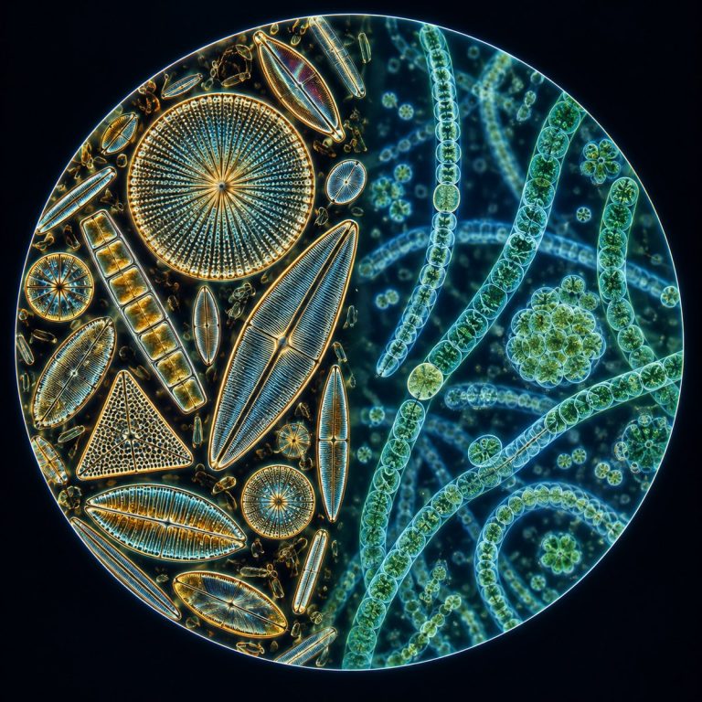
Diatoms vs cyanobacteria is ultimately a contrast between two different domains of life, not two flavors of the same thing: diatoms are eukaryotic algae encased in glass, while cyanobacteria are prokaryotic bacteria that only look like algae. A diatom carries a true nucleus and a silica shell called a frustule. A cyanobacterium has neither — no nucleus, no glass, just a soft peptidoglycan wall around a cell built more like an ordinary bacterium than a plant. That one split, eukaryote versus prokaryote, is the root of nearly every other difference below, from color and size to which one can fix nitrogen or turn toxic.
Diatoms vs Cyanobacteria at a Glance
| Feature | Diatoms | Cyanobacteria |
|---|---|---|
| Cell type | Eukaryote (alga) | Prokaryote (bacterium) |
| Nucleus | True, membrane-bound nucleus | None — DNA sits loose in a nucleoid |
| Organelles | Chloroplasts and mitochondria | None; free-floating thylakoids |
| Cell wall | Silica (glass) frustule | Peptidoglycan plus mucilage sheath |
| Size | ~2–200 µm | ~0.5–5 µm |
| Color | Golden-brown (fucoxanthin) | Blue-green (phycocyanin) |
| Pigments | Chlorophyll a & c, fucoxanthin | Chlorophyll a, phycobilins |
| Energy storage | Chrysolaminarin and oil | Glycogen |
| Nitrogen fixation | No | Some, via heterocysts |
| Reproduction | Binary fission, plus sexual auxospores | Binary fission (asexual) |
| Toxins | Essentially none | Some genera produce cyanotoxins |
| Example genera | Navicula, Pinnularia, Cyclotella | Anabaena, Oscillatoria, Microcystis |
| Under the scope | Ornate, symmetric glass shells | Tiny cells or beaded filaments |
The Core Difference — Eukaryote vs Prokaryote
Diatoms build their cells the way plants and animals do: a true, membrane-bound nucleus holds the DNA, and dedicated organelles — chloroplasts for photosynthesis, mitochondria for respiration — do the cell’s work in their own compartments. Cyanobacteria skip all of that. Their DNA sits loose in the cytoplasm in a region called the nucleoid, with no membrane around it, and they have no mitochondria or chloroplasts at all. Photosynthesis still happens, but on thylakoid membranes floating free inside the cell rather than tucked inside an organelle. This is the taxonomic line that actually separates the two: diatoms are eukaryotes, full stop, and cyanobacteria are bacteria, full stop. Everything else in this article — size, color, wall chemistry, what each one can and can’t do — traces back to that split. OpenStax distinguishes eukaryotic algae such as diatoms from prokaryotic cyanobacteria.
Cell Walls — Glass Silica vs Peptidoglycan
A diatom’s cell wall, called a frustule, is made of biogenic silica — hydrated silicon dioxide, essentially glass grown by a living cell. It comes in two interlocking halves, the larger epitheca fitting over the smaller hypotheca like the lid of a petri dish. The surface is often etched with rows of microscopic pores called striae, fine enough to test the resolving power of a lens. A cyanobacterium’s wall is built from peptidoglycan, the same Gram-negative-type material found in ordinary bacteria, usually wrapped in a slippery mucilage sheath. There’s no glass, no interlocking halves, and no fixed geometric symmetry — just a simple envelope around a much smaller cell. If you only remember one structural fact for telling them apart on a slide, make it this one: glass box versus soft bacterial wall.
What You See Under the Microscope — Size, Color & Shape
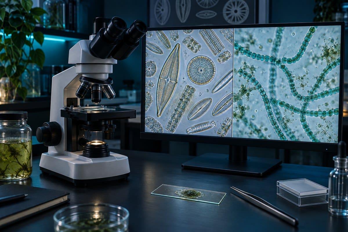
Diatoms read as little glass jewel boxes — sharply geometric, often tinted golden-brown or tan from fucoxanthin. Centric species look like ribbed drums or wheels, radially symmetric and often planktonic; pennate species look like tiny boats or canoes with a seam running down the middle. At 400x and up, the pinprick rows of striae pop in and out as you work the fine focus. That’s exactly why diatom test slides are a classic resolution check. You can get a closer look at that structure and symmetry in how diatoms look under a microscope.
Cyanobacteria are a different experience entirely: smaller, plainer, and usually seen as a group rather than as individuals. Oscillatoria looks like a straight blue-green hair that slowly glides across the field of view. Anabaena looks like a string of beads with the occasional larger, paler bead — that pale cell is a heterocyst, a specialized nitrogen-fixing structure diatoms simply don’t have. At low power a cyanobacterial mat is often just a blue-green smear; resolving individual cells usually takes 400x–1000x with oil immersion. The most common beginner mistake is expecting cyanobacteria to show the same fine detail as a diatom — they won’t, because there’s no ornate silica shell to resolve. Judge cyanobacteria by size and color rather than surface detail, and stop the condenser down if the light is washing out the faint blue-green tint. For the full walkthrough of what these look like up close, see cyanobacteria under a microscope.
How Each One Photosynthesizes
Diatoms photosynthesize inside true chloroplasts, using chlorophyll a and c along with the carotenoid fucoxanthin — the pigment that masks their chlorophyll and gives them their golden-brown color. The sugars they make are stored as chrysolaminarin, a polysaccharide, along with lipid droplets, not starch. As a group, diatoms are estimated to account for roughly a fifth to a quarter of global photosynthesis. That estimate comes from a diatom overview from Carleton College’s Microbial Life Educational Resources.
Cyanobacteria photosynthesize without a chloroplast at all, using chlorophyll a plus accessory pigments called phycobilins. The main one is the blue phycocyanin that gives them their color, and in some species the red-tinted phycoerythrin. These pigments sit in phycobilisomes studding the free-floating thylakoid membranes. Cyanobacteria store their sugars as glycogen instead of chrysolaminarin. They also hold a unique distinction: they were the first organisms on Earth to perform oxygenic photosynthesis. Every chloroplast in every plant and alga alive today — including a diatom’s — descends from an ancient cyanobacterium. That cyanobacterium was engulfed and retained by a larger cell, in a process called endosymbiosis. In a real sense, a diatom’s chloroplast is a captured cyanobacterium, several billion years removed.

Nitrogen Fixation and Toxins — Where Cyanobacteria Are Different
Cyanobacteria can do something diatoms cannot: pull nitrogen straight out of the air. Some filamentous genera, including Anabaena and Nostoc, build thick-walled cells called heterocysts that house the enzyme nitrogenase and convert atmospheric nitrogen into ammonia the cell can use. Diatoms have no equivalent structure and depend entirely on dissolved nitrogen compounds in the water around them. The EPA explains which cyanobacteria can form blooms and produce cyanotoxins.
Cyanobacteria are also the one with a real safety concern. Genera like Microcystis and Anabaena can form dense harmful algal blooms and produce cyanotoxins — including microcystin and anatoxin-a. These compounds are dangerous to people, pets, and livestock, according to the EPA. Diatoms carry essentially no comparable everyday risk. If you come across a blue-green bloom or scum in a pond, lake, or aquarium, treat it as potentially toxic. Keep people and pets out of the water, don’t taste or drink it, and wash your hands afterward. This is educational information, not water-safety or medical advice — a microscope can’t tell you whether a specific bloom is toxic, only laboratory testing can.
How to Tell Them Apart Under Your Own Microscope
On a real slide, four quick checks separate diatoms from cyanobacteria almost every time:

- Look for a nucleus. A distinct round body inside the cell means diatom; its absence means cyanobacterium.
- Check the size. Diatoms usually run 10–80 µm and are easy to spot at 100x–400x; cyanobacteria are often under 5 µm and need 400x–1000x to resolve individually.
- Check the color. Golden-brown means diatom; blue-green means cyanobacterium.
- Check the shape. Rigid, symmetric glass boxes (drums or boats) mean diatom; plain threads, beaded filaments, or slimy colonies mean cyanobacterium.
Diatoms are a major component of freshwater and marine plankton, so a plankton tow or a scoop of pond water will usually turn up both groups side by side. Prepare a standard wet mount slide for either one — no stain is required to see the basic shapes and colors described above. If you want a size and structure comparison against a different prokaryote entirely, what bacteria look like under a microscope is a useful reference point. Ordinary bacteria share the cyanobacterium’s lack of a nucleus but not its photosynthetic pigments.
Why Cyanobacteria Were Called “Blue-Green Algae”
Cyanobacteria earned the name “blue-green algae” the old-fashioned way: early naturalists sorted organisms by where they lived and what color they were, not by cell structure. Anything blue-green, aquatic, and photosynthetic got filed under “algae,” and cyanobacteria fit that description perfectly from the outside. It wasn’t until microscopy and cell biology matured enough to reveal the absence of a nucleus and membrane-bound organelles that scientists reclassified them as bacteria. According to an overview from the UC Museum of Paleontology, cyanobacteria are credited with the Great Oxidation Event roughly 2.4 billion years ago. Their photosynthesis first filled Earth’s atmosphere with oxygen, long before anything resembling a true alga existed. The old name persists in casual use, but taxonomically, “blue-green algae” is a misnomer: they are bacteria that happen to photosynthesize, not algae at all.
It’s also worth flagging the reverse trap: not every blue-green-looking filament in a pond sample is a cyanobacterium. Spirogyra, a common filamentous pond organism, is a true green alga with visible ribbon-shaped chloroplasts — a real eukaryote, despite sometimes getting lumped in with “pond scum” alongside cyanobacteria. The giveaway is the same nucleus-and-chloroplast check described above.
Frequently Asked Questions
Do both diatoms and cyanobacteria photosynthesize?
Yes. Both are photosynthetic, which is exactly why they get confused for each other. But diatoms do it inside a true chloroplast, while cyanobacteria do it on free-floating thylakoid membranes with no chloroplast at all.
Which is bigger, a diatom or a cyanobacterium?
Diatoms are almost always bigger. Most diatoms measure 10–80 µm, occasionally up to 200 µm, while individual cyanobacterial cells typically run just 0.5–5 µm. Cyanobacteria are visible mainly because they form long filaments or colonies, not because any single cell is large.
Can you see diatoms and cyanobacteria with a home microscope?
Yes. Diatoms are easy to spot at 100x–400x thanks to their size and glass shells. Cyanobacteria are trickier — you’ll usually need 400x–1000x with oil immersion to resolve individual cells and structures like heterocysts.
Are diatoms good or bad for a fish tank or pond?
Generally good, or at least neutral. Diatoms are a normal, non-toxic part of the food web and often show up as a harmless brown film early in a new tank’s cycle. Cyanobacteria blooms are the ones to watch, since some produce toxins.
Did cyanobacteria really create Earth’s oxygen?
Largely, yes. Cyanobacteria were the first organisms to evolve oxygenic photosynthesis. Their activity over hundreds of millions of years drove the Great Oxidation Event, which filled Earth’s atmosphere with free oxygen for the first time.
Conclusion
Diatoms and cyanobacteria both live in water, both photosynthesize, and both get casually lumped under “algae” — but they sit on opposite sides of one of biology’s deepest divides. A diatom is a eukaryote with a nucleus and a glass shell; a cyanobacterium is a bacterium with neither. Once you know to check for a nucleus, judge the size, and read the color, telling them apart at the eyepiece becomes second nature rather than a guessing game.
Have you spotted both in the same drop of pond water? Tell us what you found — and whether it was the golden glass boxes or the blue-green threads that gave it away — in the comments below.