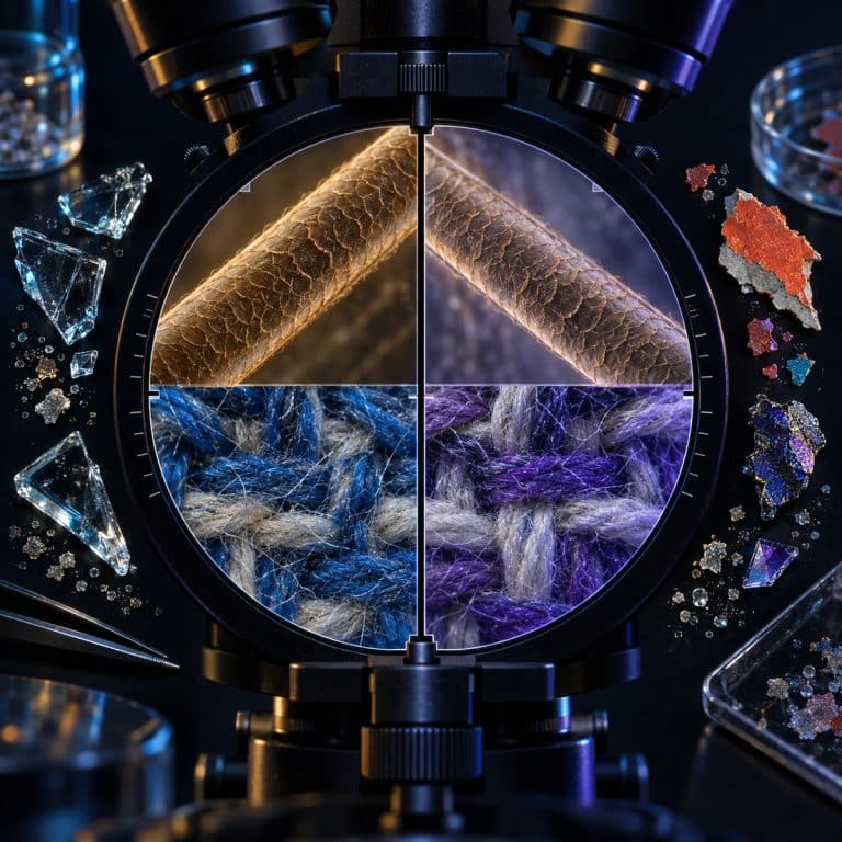
Forensic microscopy is the use of microscopes to examine, identify, and compare physical evidence — hairs, fibers, glass, paint, and more — recovered from a crime scene. Crime labs rely on it to answer one narrow but crucial question: does this trace of evidence match, or fail to match, a known sample? This overview covers the instruments forensic scientists actually use, the evidence types they examine, and where microscopy’s conclusions end and DNA testing’s begin.
What Forensic Scientists Actually Do With a Microscope
Trace evidence is the small, often microscopic material transferred during a crime — fibers, hairs, glass fragments, paint chips, soil, and gunshot-residue particles. It is the raw material forensic microscopy exists to examine. A forensic microscopist spends far less time chasing dramatic revelations than television suggests. Most of the job is patient screening: sorting debris recovered from a victim’s clothing, a suspect’s vehicle, or a crime scene into categories — fiber, hair, mineral, plant material — before any comparison work even begins. Only after that sorting does a sample reach a comparison microscope. NIST describes how hair, fibers, paint and glass function as trace evidence in forensic examinations.
Locard’s Exchange Principle: Every Contact Leaves a Trace
Locard’s exchange principle underpins the entire discipline. French criminologist Edmond Locard proposed that any contact between two objects or people results in a mutual transfer of material — fibers from a sweater snag on a car seat, carpet fibers cling to a shoe sole, a strand of hair drops onto a doorframe. Locard’s exchange principle, the idea that every contact leaves a trace, is why crime scenes and suspects alike get searched for microscopic material in the first place: whatever a perpetrator touched, they likely also carried something away from.
Identifying Evidence vs. Comparing Evidence
Forensic microscopy does two distinct jobs, and mixing them up causes real confusion about what a lab report actually proves. Identification asks what a material is — is this powder cornstarch or cocaine, is this fiber cotton or polyester. Comparison asks whether two samples could share a common source — does this carpet fiber recovered from a trunk match the carpet fiber standard cut from a specific vehicle. Identification produces a fact; comparison produces an opinion about consistency, which is a meaningfully weaker claim and one the later sections of this page return to.
The Microscopes Used in Forensic Science
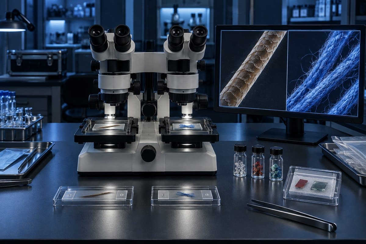
No single microscope handles every kind of evidence. Crime labs keep several types on hand, each suited to a different scale of detail and a different kind of question. Its forensic lexicon defines a comparison microscope as two microscopes joined optically for simultaneous viewing.
| Instrument | What It Does | Typical Forensic Use |
|---|---|---|
| Stereo (dissecting) microscope | Low magnification (~10–50x), three-dimensional view, top lighting | First-look screening, sorting trace evidence, examining tool marks and documents |
| Compound light microscope | Higher magnification (40–1000x), light passes through the sample, flat two-dimensional view | Examining mounted hairs, fibers, and other thin, transparent specimens |
| Comparison microscope | Two compound scopes joined by an optical bridge; two samples viewed side by side in one split field | Bullet and cartridge-case striations, hair comparison, fiber comparison |
| Polarized light microscope (PLM) | Uses polarized light to reveal optical properties like birefringence and refractive index | Identifying synthetic vs. natural fibers, minerals in soil, and some drug crystals |
| Scanning electron microscope (SEM) | Electron beam imaging at very high magnification, often paired with elemental analysis | Gunshot-residue particle confirmation, paint-layer analysis, fine surface detail beyond a light microscope’s limits |
The Comparison Microscope: Forensic Science’s Signature Instrument
The comparison microscope is the instrument most associated with forensic work, and for good reason: it is purpose-built for the job of comparison. Two compound microscopes are joined by an optical bridge so an examiner looks into a single eyepiece and sees a circular field split down the middle by a fine vertical line — a known sample fills one half, a questioned sample fills the other. The examiner nudges each stage until striations on a bullet, or scale patterns on a hair, line up across that divider like two halves of one continuous image. The instrument itself was adapted for forensic use in 1925 by firearms examiners Calvin Goddard and Philip Gravelle, who built it specifically to match striations on fired bullets and cartridge cases — work that remains its core application today.
Stereo and Compound Microscopes: Screening vs. Detailed Examination
Stereo and compound microscopes get confused constantly by beginners, and the mix-up matters: reaching for high magnification when a job calls for a low-power surface view (or the reverse) wastes time and can damage delicate evidence. A stereo microscope gives a three-dimensional view at low power using reflected light bouncing off the surface of an object — the right tool for the first pass over a garment or a piece of debris. A compound microscope pushes to much higher magnification using light transmitted through a thin, mounted sample, which is what a hair or fiber needs once it has been picked out and prepared on a slide.

Polarized Light and Electron Microscopes
A polarizing microscope passes light through two filters set at right angles, and the way a material bends that light — its birefringence — identifies what the material is. This makes it the workhorse for telling a synthetic fiber from a natural one, spotting mineral grains in soil, and screening some drug crystals, all without destroying the sample. For evidence too fine for any light microscope to resolve, labs step up to an electron microscope, which scans a surface with a focused electron beam instead of light. Paired with elemental analysis, it can confirm the specific metallic composition of a gunshot-residue particle or reveal the individual layers inside a chip of automotive paint.
What Evidence Looks Like Under the Microscope
Fibers, hair, fingerprints, and other trace materials each have their own deep-dive guide on this site; this section is the quick tour that sends you to the right one.
Fibers: Natural vs. Synthetic
Under a compound or polarized light microscope, synthetic fibers tend to look like smooth, uniform, glass-clear or evenly dyed rods, often with tiny black delustrant speckles baked in to cut down shine. Natural cotton, by contrast, looks like a flat, twisted ribbon — not unlike a deflated garden hose — while wool shows a scaly, overlapping surface closer to what you’d expect from an animal fiber. A fiber examiner sorts by cross-sectional shape, diameter, dye, and (under polarized light) refractive index before ever calling two fibers a possible match. The full breakdown of what these differences look like lives in our guide to textile fibers under a microscope.

Hair: Cuticle, Cortex, and Medulla
A hair mounted on a slide reveals three layers to a trained eye: the cuticle, cortex, and medulla. The cuticle scales overlap along the shaft like roof shingles or fish scales, and their pattern differs by species. The cortex carries the pigment granules that give hair its color and shows the internal structure examiners use for comparison. The medulla — the central core running down the shaft — is often a bold, continuous “ladder” in many animal hairs, while in human head hair it’s typically thin, fragmentary, or absent altogether, which is one of the fastest ways to sort human from animal hair at a glance. See this contrast directly in our comparison of animal fur next to human hair under a microscope, or go deeper into what human hair looks like under a microscope on its own.
Fingerprints and Ridge Detail
Friction-ridge skin on fingertips forms patterns — loops, whorls, and arches — built from fine minutiae like ridge endings and bifurcations. Magnification through a stereo microscope, a loupe, or high-resolution imaging helps an examiner resolve that fine ridge detail, and in some cases even third-level detail like individual sweat pores along a ridge edge. Fingerprint examination is a large enough topic that it gets its own dedicated deep dive on this site, covering how ridge detail is captured, compared, and interpreted.
Other Trace Evidence: Glass, Paint, Soil, and Dust
Trace evidence extends well beyond fibers and hair. Glass fragments recovered from a broken window or a hit-and-run scene get compared by refractive index and thickness — you can see the fracture patterns and layering yourself in our look at glass fragments under a microscope. Paint chips reveal a stack of distinct layers under magnification, each layer a potential match point to a specific vehicle or surface. Soil and household dust carry their own mixed signature of fibers, mineral grains, pollen, and skin cells, which is exactly what shows up in our piece on the fibers and debris hiding in household dust.
The Limits of Forensic Microscopy
This article is an educational overview, not legal or professional forensic guidance — real casework is performed by trained examiners working in accredited labs under strict protocols. With that framing in mind, forensic microscopy’s biggest limitation deserves a plain, honest statement: microscopic hair comparison establishes that two hairs are consistent with each other, not that they came from the same person. It is an associative method, not a positive identification, and history shows examiners have not always drawn that line as carefully as they should have. A 2015 FBI review of microscopic hair comparison testimony found erroneous or overstated statements in the vast majority of cases it examined, prompting a lasting shift toward describing hair comparison results in cautious, measured language — “is consistent with” or “cannot be excluded,” never “matches” or “is identical to.”
That doesn’t mean microscopy has been pushed aside by DNA testing. The two work alongside each other: microscopy screens, sorts, and characterizes trace evidence quickly and non-destructively, narrowing down which samples are even worth the time and cost of DNA analysis. The National Institute of Justice’s overview of trace evidence frames microscopy as a foundational first step in that pipeline rather than a method DNA has replaced.
Frequently Asked Questions
Can hair be used to identify a person under a microscope?
Not on its own. Microscopic hair comparison can show that a hair is consistent with a known sample and rule out hairs that clearly don’t match, but it cannot uniquely identify one specific person the way DNA testing can. Treat a microscopic hair “match” as investigative support, not proof of identity.
Is forensic microscopy still used now that DNA testing is available?
Yes. Microscopy is faster and cheaper than DNA analysis, so labs use it to screen and sort trace evidence before deciding what’s worth sending for DNA testing. The two methods work together rather than one replacing the other.
Can trace evidence be contaminated during microscopic examination?
Yes, which is why labs work under strict protocols. Locard’s exchange principle cuts both ways: handling a sample without gloves, a clean workspace, and a known control can transfer new material onto the evidence or lose material that was already there, undermining a comparison before it even starts.
Do you need a special microscope to see fingerprints?
A standard stereo microscope or even a hand loupe is often enough to resolve ridge detail on a fingerprint. Higher-resolution imaging comes into play for finer third-level detail like individual pores, but no forensic-specific microscope model is required just to view ridge patterns.
Do all crime labs have the same microscopy equipment?
No. Equipment varies by lab size, budget, and accreditation level — a small regional lab may rely on a handful of stereo and compound microscopes, while a larger state or federal lab is more likely to have a comparison microscope, polarized light microscope, and access to an SEM in-house or through a partner facility.
Conclusion
Forensic microscopy gives crime labs a fast, non-destructive first look at the smallest physical evidence a case has to offer — fibers, hair, glass, paint, and more — using tools purpose-built for identifying what a material is and comparing it against a known sample. The honest caveat that runs through all of it is that comparison work establishes consistency, not certainty, which is why microscopy today operates alongside DNA and chemical testing rather than standing in for them.
Have you looked at any of these evidence types — fiber, hair, or glass — under your own microscope at home? Tell us what you noticed in the comments below.