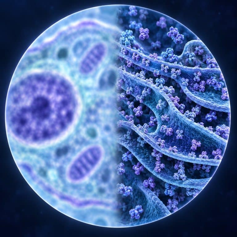
To see ribosomes under a microscope is impossible — not with a budget student scope, not with a research-grade one. A single ribosome measures roughly 25 nanometers, about eight times smaller than the ~200-nanometer resolution limit of visible light. The same physics hides the endoplasmic reticulum, the Golgi apparatus, lysosomes, and the individual filaments of the cytoskeleton. None of that is a hardware problem you can fix by spending more money. It is the wavelength of light itself drawing a hard line under what any light microscope can ever resolve.
| Structure | Real size | Below the 200 nm limit? | What reveals it | What you see instead |
|---|---|---|---|---|
| Ribosome | ~20–30 nm | Yes — ~8x too small | TEM, cryo-EM | Fine grain in the cytoplasm |
| Endoplasmic reticulum | membrane ~4–10 nm thick | Yes | TEM | A faint, denser patch near the nucleus |
| Golgi apparatus | stack ~1–2 µm, cisternae ~15–30 nm apart | Yes (stack architecture) | TEM | Nothing distinct in a live prep |
| Lysosome | ~0.1–1.2 µm | Borderline — visible but not identifiable | Fluorescence (LysoTracker), TEM | Tiny jiggling specks with no label |
| Cytoskeleton filaments | 7–25 nm diameter | Yes | Fluorescence, super-resolution (STED/STORM) | Nothing in bright field — the cell just holds its shape |
The 200-Nanometer Wall: The Smallest Thing Light Can Show You
A light microscope’s resolution tops out at around 200 nanometers (0.2 µm) under the best conditions, and that number comes from the Abbe diffraction limit, not from lens quality or price. Ernst Abbe worked out in 1873 that resolution d equals wavelength λ divided by twice the numerical aperture (NA): d = λ / (2 × NA). Plug in green light at 550 nm and a top oil-immersion objective at NA 1.4, and you get d = 550 / (2 × 1.4) ≈ 196 nm. That is the physical floor — no amount of extra magnification pushes past it. OpenStax explains why wavelength and numerical aperture determine microscope resolution.
For scale, the unaided human eye resolves down to about 0.1 mm (100 µm) and a light microscope reaches roughly 200 nm. A transmission electron microscope (TEM), by comparison, can resolve down to about 0.1–0.2 nm. A light microscope is genuinely useful up to about 1,000–1,500x magnification; past that, you are not seeing more detail, you are seeing a bigger blur.
Magnification Isn’t the Problem — Resolution Is
Magnification enlarges an image; resolution is the ability to tell two close points apart as separate. Crank a light microscope’s magnification past its resolution limit and you get “empty magnification” — a larger, dimmer, softer image with zero new detail. The information was never captured by the lens in the first place, so there’s nothing extra to reveal. This is why resolution, not magnification, sets the limit on what any light microscope can ever show you. It’s the single most useful thing to understand before you go hunting for organelles.
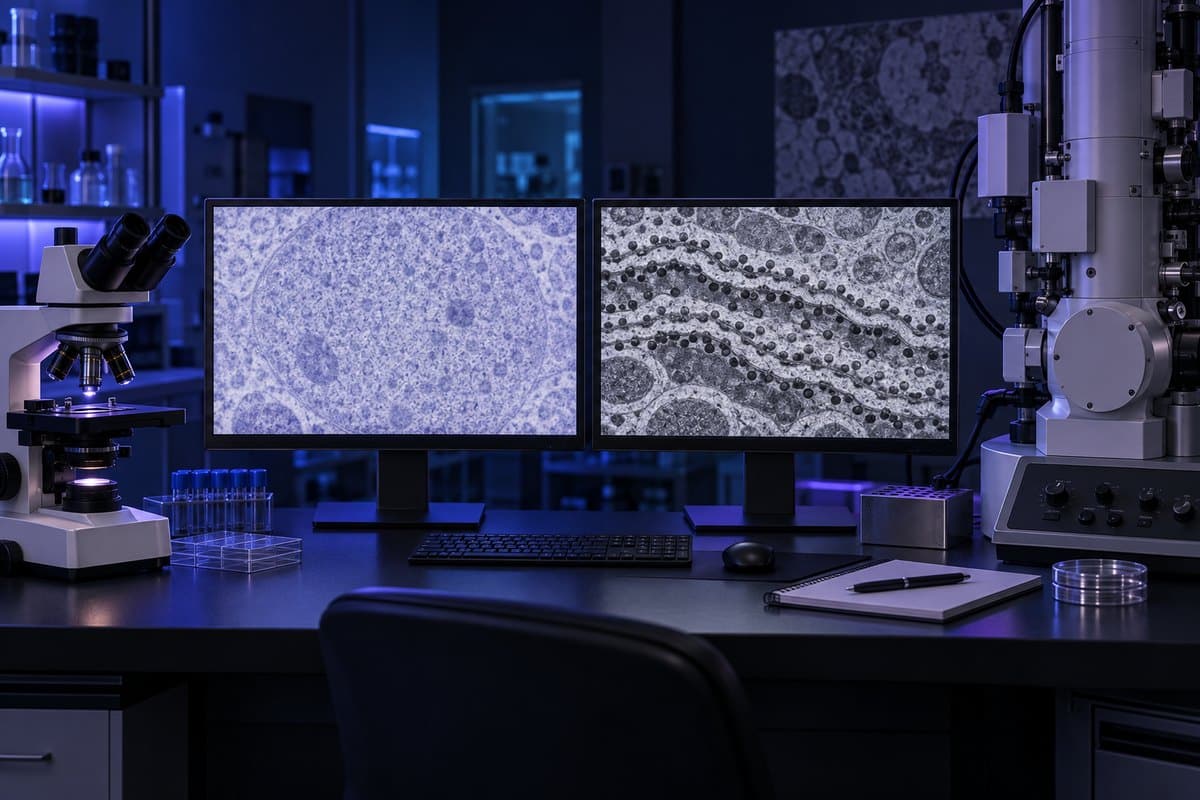
Instruments that resolve finer than light ever can rely on a shorter “probe” than a light wave. An electron microscope resolves what light can’t because electron wavelengths are thousands of times shorter than visible light. You can browse real electron microscope images of these structures to see what that actually looks like.
Ribosomes: About Eight Times Too Small to Resolve
A ribosome is a compact machine of ribosomal RNA and protein — in eukaryotic cells, an 80S particle built from a 60S and a 40S subunit. It measures only about 20–30 nm across. Set beside the ~200 nm resolution limit, that puts a single ribosome roughly eight times below what a light microscope can distinguish. That figure comes from the NCBI Bookshelf’s structural overview of the ribosome. Under TEM, ribosomes show up as small dark dots, often studding the rough endoplasmic reticulum like grains on sandpaper. Under a light microscope, you never see one individual ribosome resolve into a dot. What you see instead is the cytoplasm’s overall grainy, faintly sandy texture — thousands of ribosomes and small vesicles packed together, blurred into a single haze by the diffraction limit. Its biology guide describes ribosome structure and its role in protein synthesis.
The Endoplasmic Reticulum: Membranes Below the Line
The endoplasmic reticulum (ER) is a folded network of membrane that can span microns across a cell. The membrane itself — the part that actually needs resolving — is only about 4–10 nm thick, according to Britannica’s overview of the endoplasmic reticulum. It also has very low inherent contrast against the surrounding cytoplasm. Rough ER, studded with ribosomes, and smooth ER, involved in lipid synthesis, are both structures that TEM defined; a light microscope was never going to trace their folded sheets. At the eyepiece, the most an experienced observer can point to is a slightly denser, more refractile patch tucked beside the nucleus. It’s a hint that something busy is happening there, never a labeled stack of membranes.

The Golgi Apparatus: Discovered, But Not by a Home Microscope
Camillo Golgi found this organelle in 1898 using a silver-nitrate impregnation technique he called “la reazione nera” — the black reaction. The work later earned him a share of the 1906 Nobel Prize, detailed in his own Nobel biographical account. But that stain only produced a dark blob under a light microscope, not the stacked cisternae shown in textbooks. Some scientists argued for decades that the Golgi apparatus was just a staining artifact, until TEM confirmed its true structure in the 1950s. The real Golgi apparatus is a stack of four to eight flattened membrane sacs, each cisterna only 15–30 nm from the next, with membranes around 6–8 nm thick. That internal architecture sits well below the light microscope’s resolving power. Seeing it needs the heavy-metal staining and vacuum optics of TEM — not an ordinary live prep on a hobbyist’s scope.
Lysosomes: Big Enough to See, Impossible to Identify
Lysosomes are the one structure on this list that isn’t automatically ruled out by size alone. At roughly 0.1–1.2 µm, the larger ones sit right around or just above the resolution limit. The problem is identification, not visibility. In a live cheek cell at high power you may genuinely catch tiny dark specks jiggling with Brownian motion. But nothing in a plain bright-field view tells you which speck is a lysosome versus any other small vesicle or granule. Distinguishing them takes a specific tool: fluorescent dyes like LysoTracker that bind selectively to lysosomes, or acid-phosphatase histochemistry that stains for a lysosomal enzyme. Without one of those, a lysosome under your microscope is just an anonymous dot in the crowd.
The Cytoskeleton: Invisible Until You Make It Glow
The cytoskeleton is a mesh of protein filaments that gives a cell its shape, and every strand in it is far too thin for light to resolve. Microtubules run about 25 nm across (and are hollow), actin filaments about 7 nm, and intermediate filaments about 10 nm. In plain bright-field microscopy you see nothing of this scaffolding at all — the cell simply holds its shape, with no visible wires. What makes the cytoskeleton visible is fluorescence: antibodies tagged with fluorescent labels bind to specific filament proteins, or a dye like phalloidin binds actin directly. The glow traces out a web the eye could never otherwise detect. For individual filaments rather than a general glow, researchers reach for super-resolution methods like STED and STORM, which sidestep the diffraction limit using clever optical tricks rather than shorter wavelengths.

So What CAN You See? (And What That Grainy Texture Really Is)
None of this makes a light microscope useless for cell biology — it just means the target list is different from the textbook diagram. With a well-prepared slide and decent contrast, you can genuinely see the nucleus, often the nucleolus inside it, chloroplasts in plant cells, and the central vacuole. Sometimes mitochondria show up too, as small moving specks. Reviewing cheek cells under a microscope is a good way to see this contrast firsthand: a clear nucleus against a cytoplasm that looks softly grainy rather than empty. That graininess is not background noise — it is ribosomes, small vesicles, and fragments of ER, all packed together and blurred by diffraction into one texture. You are seeing the crowd, not the individuals, and once you know that, the texture actually makes sense instead of looking like a disappointing blur.
If you’re chasing detail rather than identification, it also helps to know the highest magnification a microscope can actually reach before it stops adding anything useful. Pushing past that point just enlarges the same grainy blur, not the organelles hiding inside it.
Frequently Asked Questions
I saw a tiny dot moving in the cytoplasm — was that a ribosome?
Probably not. A single ribosome is far too small to resolve as an individual moving dot under a light microscope — what you most likely saw was a small vesicle, a lipid droplet, or, in a well-focused live prep, a mitochondrion, all of which sit closer to or above the 200 nm resolution limit. A genuine single-ribosome sighting isn’t possible with any light microscope, regardless of price or magnification.
Would phase-contrast or dark-field microscopy reveal ribosomes that a standard light microscope misses?
No. Phase-contrast and dark-field techniques improve contrast so transparent structures stand out more clearly, but both still use visible light, so they’re bound by the same ~200 nm diffraction limit. They can make the cytoplasm’s grainy texture easier to see, not resolve the individual ribosomes producing it.
Are the colors in electron microscope images of organelles real?
No. TEM images are grayscale, formed by how densely electrons pass through the sample. The blues, purples, and greens you see in textbook or magazine images are added afterward — a technique called false coloring — to make different structures easier to tell apart. It isn’t a record of the organelle’s actual appearance.
Would a more expensive light microscope let me see ribosomes?
No. A pricier light microscope can improve contrast, optics, and ease of use, but it cannot beat the ~200 nm diffraction limit set by the wavelength of visible light itself. Seeing a ribosome requires a fundamentally different tool, such as a transmission electron microscope, not a better version of the same tool.
Can staining make organelles like the Golgi visible in a home lab?
Basic stains used in home or classroom labs, like methylene blue or iodine, boost contrast but don’t change what the optics can resolve — they won’t reveal the Golgi’s stacked structure. The silver-nitrate staining that first revealed the Golgi apparatus was a specialized 19th-century histology technique. Even it only showed a dark blob, not the true stack that TEM later confirmed.
Can a hobbyist access an electron microscope?
Rarely, and not casually. TEM instruments cost hundreds of thousands of dollars, need a trained operator, and require samples to be dehydrated and placed in a vacuum, which rules out live specimens. Some university outreach programs and core imaging facilities occasionally offer public access or tours, but it isn’t a weekend-hobbyist tool the way a light microscope is.
Conclusion
Ribosomes, the endoplasmic reticulum, the Golgi apparatus, lysosomes, and the cytoskeleton all sit below — or right at the edge of — the ~200 nanometer wall that visible light can never resolve. No amount of spending on the microscope doing the looking changes that. That grainy, sandy texture in the cytoplasm you’ve probably already seen isn’t a disappointing blur. It’s the crowd-level signature of exactly the structures the textbook diagram draws individually. Knowing that turns a “why can’t I see this?” moment into a genuine piece of understanding about how light itself behaves.
Have you looked closely at cheek cells or onion skin and noticed that grainy cytoplasm yourself? Tell us what you saw — and whether it matched what you expected from the diagrams — in the comments below.