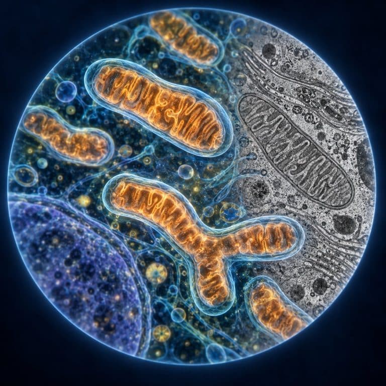
Mitochondria under the microscope appear as little more than faint, dust-like specks scattered through the cytoplasm — not the crisp, bean-shaped organelles from the textbook diagram. A standard light microscope, even at 1000x with oil immersion, can only tell you that mitochondria exist and roughly where they cluster. The double membrane and folded cristae that make them recognizable are strictly an electron-microscope view. Functionally, they’re the cell’s main site of aerobic respiration, turning nutrients and oxygen into ATP — though that’s only part of the job.
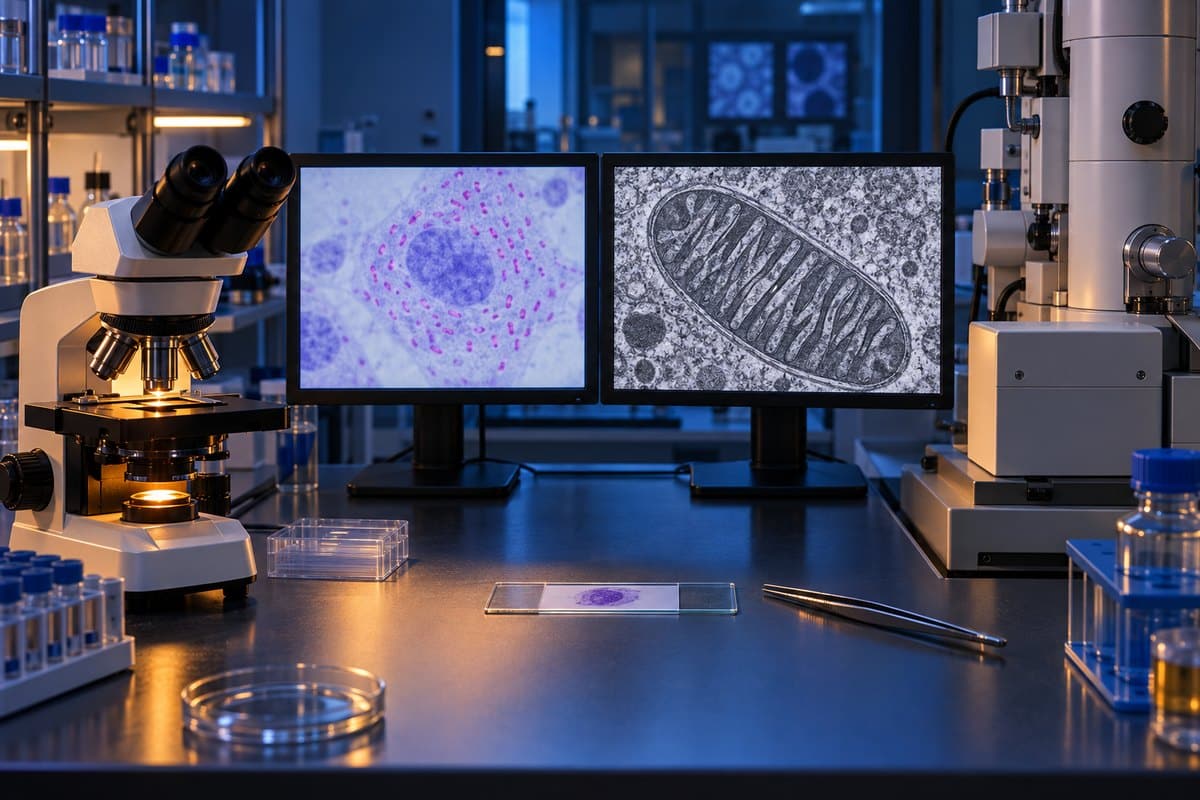
Can You Actually See Mitochondria Under a Light Microscope?
The honest answer is barely, and only with help. A typical mitochondrion measures just 0.75–3 µm long and 0.5–1 µm wide — around the size of a bacterium. A standard light microscope, even a good compound scope with an oil-immersion objective, tops out at a practical resolving power of about 200 nanometers (0.2 µm). That’s close enough to the size of a mitochondrion that you can detect its presence as a dot, but nowhere near sharp enough to resolve its internal architecture. Want to understand why that ceiling exists at all? The resolution limit of light microscopes comes down to the wavelength of visible light itself, not the quality of your lenses. OpenStax reviews the structure of mitochondria and other eukaryotic organelles.
Unstained, mitochondria are essentially invisible. They’re nearly transparent and blend into the surrounding cytoplasm, which is why beginners staring at an unstained wet mount usually see nothing distinct — just a grainy, featureless cytoplasm. That disappointment is normal; it isn’t a sign you’re doing something wrong.
What Staining Reveals
Contrast is the fix. The classic tool is Janus Green B, a vital dye that’s selectively taken up by active mitochondria and turned into a stable blue-green color by their internal enzymes — cells that are alive and respiring hold the color; dead ones don’t. Under a light microscope, stained mitochondria show up as tiny blue-green granules and short rods, densest in the cytoplasm around the nucleus, like a light dusting of blue-green pepper. Onion epidermal cells and human cheek cells are the two classic subjects for this prep because they’re thin, easy to obtain, and readily available in a classroom or home setup.
Modern labs more often reach for fluorescent probes like MitoTracker dyes, viewed with fluorescence microscopy instead of plain brightfield. Under fluorescence, mitochondria don’t look like isolated beans at all — they glow as a thread-like, branching green network, because in a living cell they constantly fuse and divide rather than sitting still as separate units.
What You See vs What You Don’t
With staining and a light microscope, you get location and rough shape: scattered granules or short rods, or a glowing network under fluorescence. What you do not get, at any light-microscope magnification, is the double membrane, the cristae, or the matrix — those require an electron microscope, full stop. Treat a light-microscope view as a presence-and-distribution map, not a structural one.
What Mitochondria Look Like Under an Electron Microscope
A transmission electron microscope (TEM) is what actually resolves mitochondrial architecture, because it uses electron beams with wavelengths thousands of times shorter than visible light. In published electron microscope images, a mitochondrion looks like a stadium-shaped or oval capsule, its inner membrane crammed into parallel, shelf-like folds that resemble a stack of ridges or a maze. A darker, granular matrix fills the space between them. This is the image every biology textbook diagram is trying to simplify.

The Parts, Labeled
- Outer membrane — smooth and permeable to small molecules via embedded porin proteins; it’s the mitochondrion’s outer boundary.
- Inner membrane — folded and selectively permeable; it holds the electron transport chain and ATP synthase, the machinery that actually generates ATP.
- Cristae — the folds of the inner membrane; they exist purely to increase surface area for the electron transport chain, packing more ATP-producing capacity into a tiny space.
- Matrix — the gel-like interior enclosed by the inner membrane; it holds the Krebs cycle enzymes, the mitochondrion’s own circular DNA, and its ribosomes.
- Intermembrane space — the narrow gap between the outer and inner membranes, where protons accumulate to drive ATP synthase.
Light vs Electron Microscope: What Each Reveals
| Factor | Light Microscope | Electron Microscope (TEM) |
|---|---|---|
| What’s visible | Presence, rough shape, distribution as stained dots or a glowing network | Double membrane, cristae, matrix, full internal architecture |
| Practical resolution | ~200 nm (0.2 µm) | Below 1 nm |
| Typical magnification | Up to ~1,000x–1,500x | Up to several hundred thousand times |
| Stain/prep needed | Janus Green B or MitoTracker on living or freshly fixed cells | Heavy metal stains, resin embedding, ultra-thin sectioning |
| Typical access/cost | Classroom or hobbyist scope, low cost | University or research-lab instrument, high cost |
What Do Mitochondria Do? (Function)
A mitochondrion runs the cell’s aerobic respiration — the multi-step process that converts nutrients and oxygen into ATP, the molecule cells spend to power almost everything they do. The Krebs cycle (also called the citric acid cycle) takes place in the matrix, breaking down fuel molecules and feeding electrons into the electron transport chain embedded in the cristae. That chain pumps protons across the inner membrane into the intermembrane space, building up a gradient that ATP synthase then uses to manufacture ATP — a process called chemiosmosis. Its energy guide also explains how mitochondrial membranes and cristae support ATP production.
Cells vary enormously in how many mitochondria they carry, from a handful to several thousand, and the count tracks energy demand closely. Muscle tissue, especially cardiac and skeletal muscle, is packed with mitochondria, and so are liver cells and neurons — all high-energy-demand tissue. Beyond ATP, mitochondria also buffer intracellular calcium, trigger apoptosis (programmed cell death) when a cell is damaged beyond repair, and generate heat in specialized tissue. For a deeper walkthrough of the biochemistry, Khan Academy’s cellular respiration unit covers the Krebs cycle and electron transport chain step by step.
Why “The Powerhouse of the Cell” Is Only Half the Story
Biologist Philip Siekevitz coined “the powerhouse of the cell” in a 1957 Scientific American article, and as a summary of the ATP story, it holds up — mitochondria genuinely are the cell’s dominant energy generator. The problem is that the slogan is where most explanations stop, and mitochondria do considerably more than generate power. They regulate calcium signaling between organelles, they initiate apoptosis when a cell needs to die on schedule (during development, or after DNA damage a cell can’t fix), and they produce heat directly in tissues like brown fat. They’re also historically strange: mitochondria carry their own small, circular DNA and their own ribosomes, distinct from the cell’s nuclear genome. That’s strong evidence for the endosymbiotic theory, which holds that mitochondria descend from a free-living aerobic bacterium engulfed by an ancestral cell roughly two billion years ago. None of that fits inside a four-word slogan, which is exactly why it’s worth knowing the fuller picture. The Britannica entry on the mitochondrion and the NCBI Bookshelf chapter on mitochondrial structure both cover this fuller functional picture in more depth.

How to Try Observing Mitochondria Yourself
This is a genuinely doable home or classroom setup, with modest expectations.
- Gather a fresh sample of live cells — a cheek-cell swab or a thin peel of onion epidermis both work well.
- Make a wet mount slide, keeping the sample as thin as a single cell layer wherever possible.
- Add a drop of Janus Green B stain and let it sit briefly — work quickly, since the dye fades as cells die and stop respiring.
- View at your highest dry objective (40x) or with oil immersion (100x); mitochondria are invisible at low power.
- Close down the condenser aperture slightly to boost contrast on the faint granules.
- Focus on a thin edge of the sample, not the thick center, and watch for small blue-green specks or short rods clustered near the nucleus.
Common snags: the slide drying out under the objective and oil, too much illumination washing out the faint color, and mistaking air bubbles or dye crystals for organelles. None of that means you’ve failed — it means you’re seeing exactly what a light microscope is capable of showing.
Frequently Asked Questions
How big are mitochondria?
Most mitochondria run about 0.75–3 µm long and 0.5–1 µm wide, which puts them in roughly the same size range as a typical bacterium.
Do mitochondria have their own DNA?
Yes. Mitochondria carry their own small, circular DNA (mtDNA) and ribosomes, separate from the cell’s nuclear DNA — a legacy of their bacterial origin under the endosymbiotic theory.
What cells have the most mitochondria?
High-energy-demand cells carry the most: cardiac and skeletal muscle cells, liver hepatocytes, and neurons can each hold from hundreds to several thousand mitochondria.
Are mitochondria only in animal cells?
No. Mitochondria are present in nearly all eukaryotic cells, including plant cells, fungi, and protists — not just animal cells.
What is the difference between mitochondria and chloroplasts?
Both are double-membrane organelles with their own DNA, but they do opposite jobs: mitochondria release energy by breaking fuel down into ATP through respiration, while chloroplasts (found only in plants and algae) capture light energy to build sugars through photosynthesis. Plant cells contain both.
Conclusion
Mitochondria under the microscope are a study in managed expectations: a light microscope, even at its best, only ever shows you presence and rough location as stained specks or a glowing network, while the double membrane and cristae that define mitochondrial structure belong entirely to the electron microscope. Knowing that gap up front — and understanding what mitochondria actually do beyond the “powerhouse” tagline — turns a potentially disappointing look through the eyepiece into a genuinely informative one.
Have you tried staining your own cheek or onion cells to spot mitochondria? Tell us what you saw — or didn’t see — in the comments below.