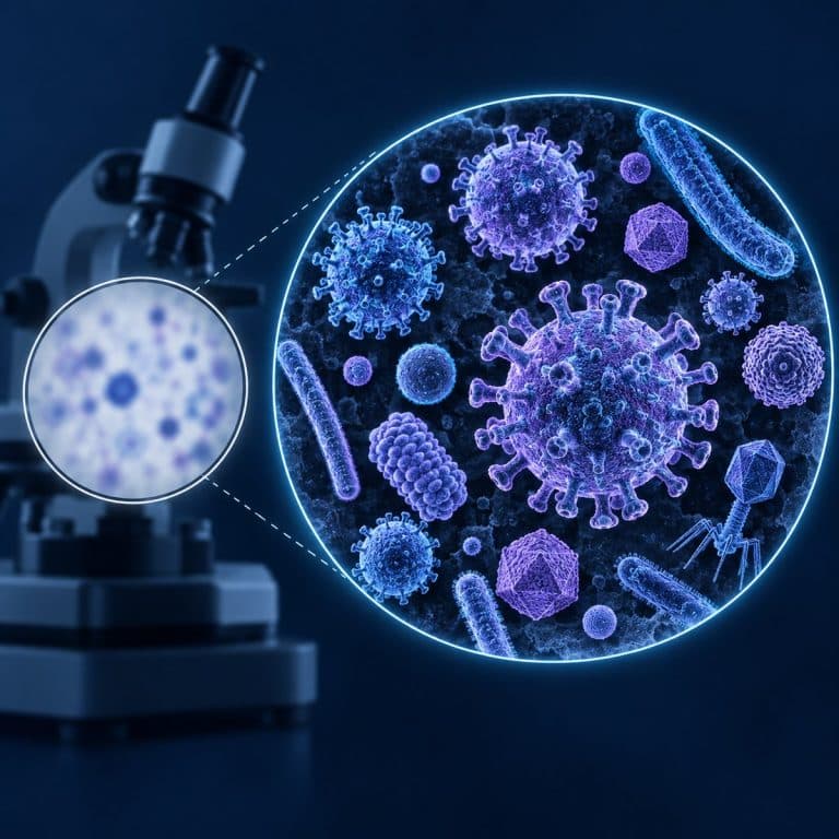
A virus under a microscope stays invisible on any standard light scope, no matter how far you crank the magnification dial — the problem is resolution, not zoom. Viruses measure just 20–300 nanometers, well below the roughly 200-nanometer limit that light itself imposes on any optical instrument. That doesn’t mean your microscope is useless around infection, though: it just means you’re looking for the wrong thing.
How Big Is a Virus — And Why Size Is the Whole Problem
A typical virus measures somewhere between 20 and 300 nanometers across, depending on the species. Poliovirus is one of the smallest at roughly 30 nm, influenza runs 80–120 nm, SARS-CoV-2 sits around 60–140 nm, HIV is about 120 nm, and the bulkier poxviruses reach 200–300 nm. Every one of those numbers is a fraction of a micrometer — and a micrometer is already a thousand times smaller than a millimeter. OpenStax compares virus sizes with bacterial and eukaryotic cells.
Context makes the gap obvious. A typical bacterium runs 0.5–5 micrometers (500–5,000 nm), which puts it 10 to 100 times larger than a virus and comfortably within reach of a home light microscope. A human red blood cell, for comparison, is about 7 micrometers across. So when someone insists they “saw a virus” wriggling across their field of view, what they almost certainly saw was a bacterium, a protist, or plain debris. All of those dwarf an actual virus.
| Entity | Typical Size | Visible With a Light Microscope? |
|---|---|---|
| Virus (poliovirus, influenza, SARS-CoV-2, HIV) | 20–300 nm | No — below the ~200 nm resolution limit |
| Bacterium | 0.5–5 µm (500–5,000 nm) | Yes — 10 to 100× larger than a virus |
| Red blood cell | ~7 µm | Yes — easily resolved |
| Light microscope resolution limit | ~200 nm | — |
For a closer look at what those larger organisms actually look like under glass, see what bacteria actually look like under a microscope — it’s the thing most people mistake for a virus.
Why a Light Microscope Can’t Resolve a Virus
Resolution — not magnification — is what decides whether a virus can appear on a standard bright-field microscope. Resolution is the ability to see two points as distinct rather than as one blurred smear. It’s fundamentally limited by the wavelength of visible light, which runs about 400–700 nm. The relationship is described by the Abbe diffraction limit: resolution equals wavelength divided by twice the numerical aperture (d = λ / 2NA). Plug in green light and a top-tier oil-immersion objective and the best-case result is about 200 nanometers — a hard physical wall, not an engineering shortfall.
Most viruses sit at or below that 200 nm line. According to microscopy educators, that limit applies to every light-based optical system regardless of brand or price. That’s why the National Institutes of Health and university virology programs consistently describe individual virus particles as invisible to light microscopy (NCBI’s overview of virus structure and size). Light simply can’t “see” something smaller than its own wavelength allows it to distinguish.
Magnification vs. Resolution — The Mistake Almost Everyone Makes
Magnification is the single most misunderstood word in amateur microscopy. Magnification just enlarges an image; resolution determines whether enlarging it reveals any new detail. Push magnification past what your optics can actually resolve and you get “empty magnification” — a bigger, blurrier blob with zero new information, not a sharper picture of something smaller. A toy microscope boasting “2000x zoom” cannot out-muscle a 200 nm physics wall any more than stretching a low-resolution photo makes the pixels turn into a face.

Nikon’s own microscopy education team makes this same distinction central to how optical systems are designed. Past the resolution limit, adding magnification only makes the blur bigger (Nikon MicroscopyU’s guide to resolution). It’s the single most important correction to make before buying — or blaming — a microscope.
So What Magnification Would You Actually Need?
There isn’t one — and that’s the trick in the question. No magnification number fixes a resolution problem, because resolution is set by the wavelength of light itself, not by how many times an eyepiece and objective multiply the image. Doubling, tripling, or maxing out magnification on a 200 nm-limited system still caps out at 200 nm of real detail. The only way to break that wall is to stop using light altogether, which is exactly what electron microscopes do.
The Exceptions — Giant Viruses You Can Barely See
A small handful of giant viruses push right up against the resolution wall and can appear as faint dots under a high-power light microscope. Mimivirus measures roughly 400–750 nm including its surface fibers, Pandoravirus stretches to about 1,000 nm (a full micrometer), and Pithovirus reaches nearly 1,500 nm. All three are large enough to just clear the ~200 nm threshold, which is why researchers first mistook Mimivirus for a bacterium when it was discovered. These are laboratory curiosities isolated from amoebae, though — not something a hobbyist will ever encounter in a backyard pond sample.

What Scientists Use to Actually See Viruses
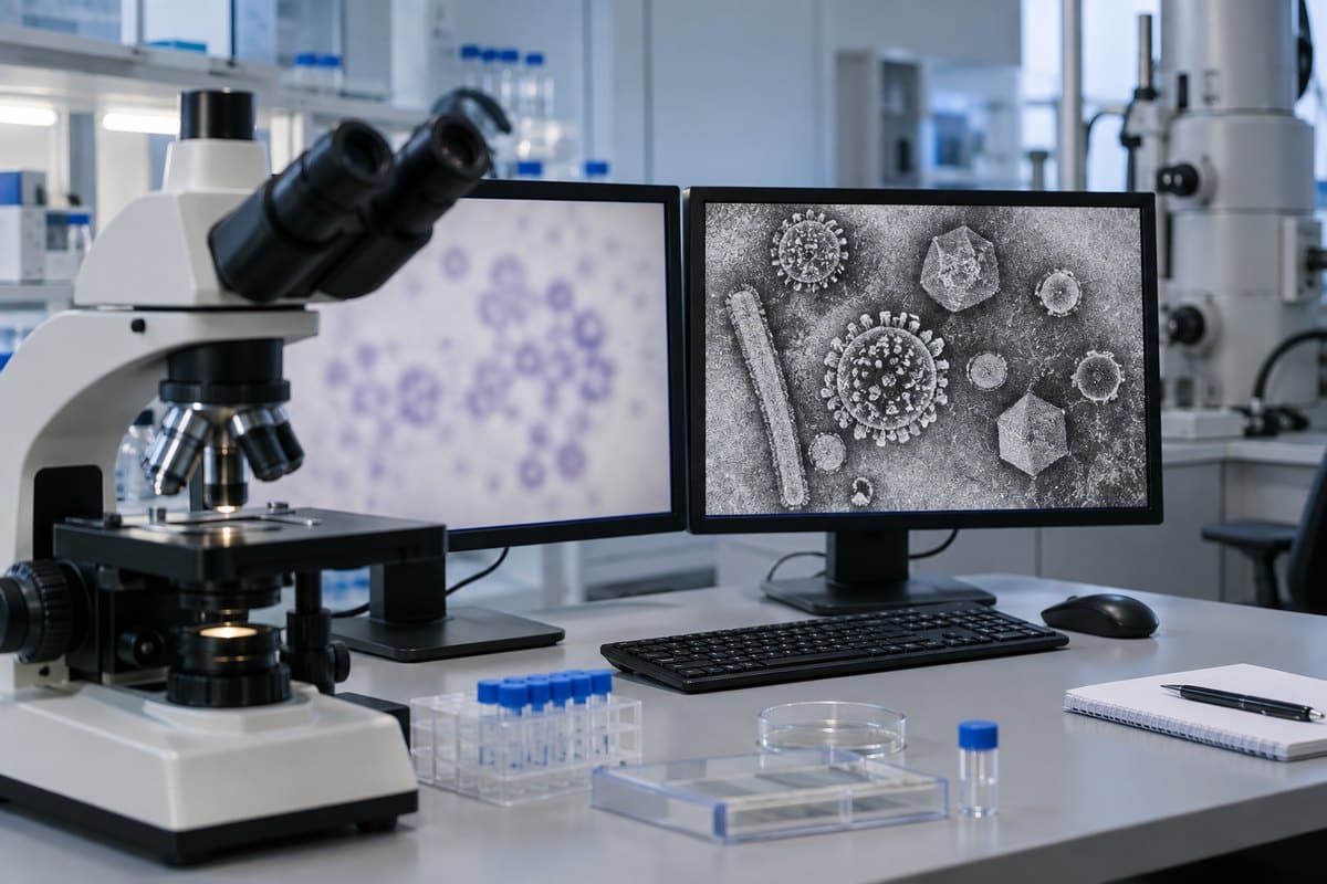
Electron microscopes reveal individual virus particles because electron beams have a wavelength thousands of times shorter than visible light, pushing resolution down into the sub-nanometer range. Transmission electron microscopy (TEM) is the workhorse for capturing a virus’s actual shape and surface structure. Cryo-EM freezes samples in near-native ice to build detailed 3D models without staining artifacts. Super-resolution fluorescence techniques — STED, STORM, PALM, and SIM — take a different approach, tagging viral proteins with fluorescent labels and localizing those tags below the normal diffraction limit. That approach reveals a glowing marker rather than the particle’s true shape, though — not the virus imaged directly. Its microscopy guide explains why electron microscopes achieve much higher resolution.
| Imaging Method | What It Can Resolve |
|---|---|
| Light microscope (bright-field) | Down to ~200 nm — bacteria, cells, organelles. Not individual viruses. |
| Transmission electron microscope (TEM) | Sub-nanometer detail — full virus particle shape and surface structure. |
| Cryo-EM / super-resolution fluorescence | Near-native 3D structure, or labeled viral proteins below the diffraction limit. |
For a full breakdown of how each of these instruments actually images a virus, see our deeper breakdown of which microscopes can image viruses. It’s also worth browsing real electron microscope images of viruses to see what these particles genuinely look like once resolution stops being the obstacle. And for readers curious how far resolution can be pushed, super-resolution microscopy techniques and atomic force microscopy both go beyond what light alone can do.
What You CAN Observe at Home Instead
A home light microscope can still show you plenty about viral infection — just not the virus itself. The clearest example is the cytopathic effect (CPE): the visible cell damage a virus causes as it replicates. Healthy cells in a culture sit flat and evenly spaced, almost like paving stones. Infected cells round up, pull away from their neighbors, and clump together. Some fuse into bloated, multinucleated giant cells called syncytia, while nuclear or cytoplasmic inclusion bodies appear inside damaged cells. Eventually, sheets of dying cells lift off and float free. You’re watching the aftermath of infection play out in real time — not the attacker itself, but unmistakable evidence it was there. The Nature Scitable glossary lays out the same staged progression that virologists use to score infections in the lab (Nature Scitable’s definition of cytopathic effect).
Bacteriophages — viruses that infect bacteria — offer another indirect but genuine observation. Look for plaques, the clear circular zones that appear in a bacterial lawn where phages have killed off every cell in their path. Realistically, though, most of what a hobbyist scope turns up around a “virus hunt” is the everyday cast of pond and saliva samples. Expect rod- and dot-shaped bacteria, drifting protists, jittering debris caught in Brownian motion, and the occasional squared-off salt or dust crystal. None of it is a virus, but all of it is genuinely worth learning to identify. Start with a beginner mistake or two so you stop mislabeling what’s actually in view.
Two habits cause most of the confusion. First, cranking magnification to “find” the virus only produces a bigger blur — remember, it’s a resolution wall, not a zoom problem. Second, assuming the smallest, fastest-moving dots in a sample must be viruses. They’re almost always bacteria or debris jittering under Brownian motion, since a true virus sits 10 to 100 times smaller than the smallest thing a light microscope can resolve at all. It also helps to know that the vivid, colorful “virus” images circulating online are colorized electron micrographs. That’s a completely different instrument working at a completely different scale, not a preview of what any home scope will ever show. In practice, samples that dry out mid-observation, too much light washing out low-contrast cells, and air bubbles or oil droplets mistaken for cells are the three most common ways a session goes sideways. Reducing the condenser aperture — or switching to phase contrast, if your scope supports it — helps low-contrast living cells stand out, though no amount of technique crosses the 200 nm wall.
Educational note, not medical advice: nothing in this article is a substitute for clinical testing. A home microscope cannot diagnose an infection or detect a virus in a person or sample. Attempting to culture or concentrate pathogens outside a proper clinical or BSL-rated lab setting is not safe. If illness is a concern, that’s a question for a healthcare provider and an approved diagnostic test, not a hobby scope. See the CDC’s guidance on how respiratory viruses are actually confirmed (CDC on flu diagnostic testing).
Frequently Asked Questions
Can you see COVID-19 or the flu virus under a home microscope?
No. Both are well within the 60–140 nm size range that sits below the ~200 nm resolution limit of any light microscope, home or professional. Diagnosis requires PCR or antigen testing, never a microscope.
Is it a virus or bacteria I’m looking at?
If you can see it moving or resolve any shape at all on a light microscope, it is almost certainly not a virus — it’s bacteria, a protist, or debris. A genuine virus particle is too small to resolve as a shape under visible light.
What’s the smallest thing a light microscope can actually see?
Around 200 nanometers, under ideal conditions with a high-numerical-aperture objective and short-wavelength light. That’s enough to resolve most bacteria and cell structures, but not an individual virus particle.
Can you see a virus with an electron microscope?
Yes. Electron beams have a far shorter effective wavelength than visible light, so transmission electron microscopes can resolve sub-nanometer detail. That’s easily sharp enough to reveal a virus’s shape and surface structure.
Would a stain or dye help you see a virus under a light microscope?
No. Stains like iodine or methylene blue improve contrast, not resolution — they make already-visible structures easier to tell apart, but they can’t shrink the ~200 nm diffraction limit or bring a virus into view. Only a shorter-wavelength probe, like an electron beam, or fluorescent tagging combined with super-resolution imaging can get around that wall.
Can I buy an electron microscope for home use?
Technically yes, but they cost tens of thousands of dollars upward and need specialized vacuum systems and sample preparation. That makes them impractical outside a lab or university setting — not a realistic hobbyist purchase.
Conclusion
A virus under a microscope will always be invisible on standard light equipment, and that comes down to physics rather than a cheap lens or low magnification. Viruses are simply smaller than light’s own resolution limit allows anything to appear. What a home scope can show you instead is just as fascinating once you know where to look. Consider the size contrast against bacteria and cells, the rare giant-virus exceptions, and above all the cytopathic effect that reveals a virus’s fingerprints on the cells it infects.
Have you ever spotted cell damage, a plaque, or something you first mistook for a virus under your own scope? Tell us what you found in the comments below — it’s exactly the kind of “what am I looking at” question this site was built to help answer.