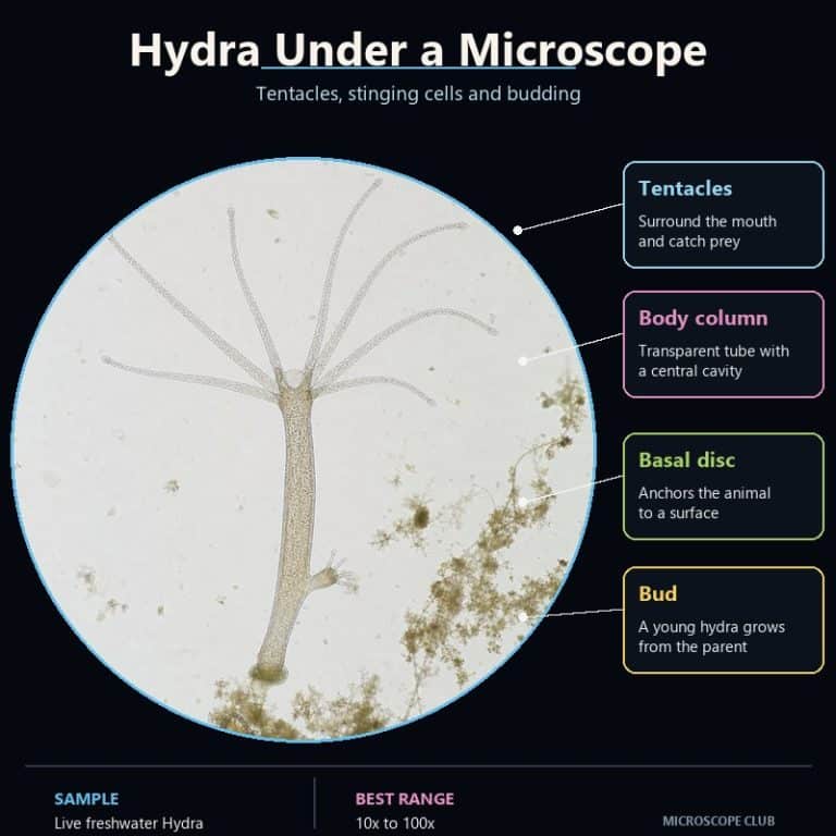
A hydra under a microscope looks like a translucent thread glued to the glass, crowned with a starburst of tentacles armed with stinging cells. This freshwater cnidarian, usually just 1–20 mm long, is one of the best live specimens a hobbyist can view: relax it under the right conditions and you can watch it hunt, sting, and even sprout a baby hydra from its own side.
How to Find and Collect Hydra
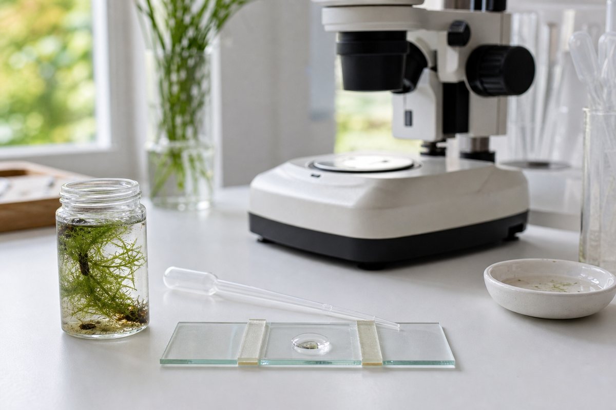
Hydra live attached to the undersides of submerged leaves, sticks, and stones in clean, slow-moving water — ponds, lake edges, and quiet streams are the best bets. Elodea and duckweed are favorite anchor points because hydra feed on the tiny crustaceans that cluster there. Scoop a handful of pond water along with a few submerged plants and stones into a clear jar, then let it sit undisturbed overnight near a window. Hydra crawl toward light, so check the glass wall on the sunny side of the jar the next morning. You’ll often spot several stuck to the glass like pale threads before you ever find one on a leaf. If collecting isn’t practical, live hydra cultures are available from biological supply companies and arrive ready to view.
Keeping Hydra Alive to Observe
Hydra tolerate a narrow set of conditions well. Use pond water, spring water, or dechlorinated tap water — never water straight from the tap, since chlorine kills them quickly. Keep the culture at 65–72°F (18–22°C) in dim, indirect light, and feed live Daphnia (water fleas) or newly hatched brine shrimp (Artemia) every few days. Overfeeding fouls the water faster than the hydra can use the food, so offer only a few prey items at a time and do a partial water change afterward.
How to View a Hydra Under the Microscope
Getting a hydra to sit still and extend is less about the scope and more about patience. Follow these steps:
- Transfer one hydra with a wide-mouth pipette into a depression (cavity) slide, not a flat wet mount — a standard coverslip pressed flat will crush it.
- Add a few drops of the same pond or culture water the hydra came from, so it doesn’t experience a temperature or chemistry shock.
- Set up first on a stereo (dissecting) microscope at its lowest power, where you can see the whole animal at once.
- Dim the room lights and back away from the slide. Hydra contract instantly when disturbed by light, heat, or vibration.
- Wait at least 10–15 minutes without touching the stage. The tentacles will slowly unfurl as the animal relaxes.
- Once it’s extended, move to a compound microscope with a well slide if you want cell-level detail on the tentacles and body wall.
- Optional: pipette in a single live Daphnia near the tentacles to trigger a feeding response.
Stereo vs. Compound Microscope for Hydra
A stereo microscope is the right first tool because hydra are a 3D, whole-animal subject — you need working distance and depth to watch it move, extend, and feed without cooking it under a tight compound-scope light. Once you’ve located a good specimen and want to resolve individual stinging cells, switching to a compound microscope at higher power reveals detail the stereo scope can’t.
What You See at Each Magnification
Total magnification changes what a hydra reveals, from whole-body behavior at low power down to individual stinging capsules at high power.
| Magnification | What’s Visible |
|---|---|
| 10–20x | Whole body, tentacle waving, contraction response, gliding movement |
| 40x | Individual tentacles, hypostome, a bud forming on the body column |
| 100x | Batteries (clusters) of cnidocytes studding the tentacle surface |
| 400x | Individual nematocysts, the two tissue layers, and mesoglea between them |
Hydra Anatomy: Every Part You Can See
A hydra’s body is a simple tube, and every visible part has a job. From foot to crown:

- Basal disc (pedal disc) — the sticky “foot” that anchors the animal to glass, stone, or a plant leaf.
- Body column — the tubular stalk; its hollow interior is the gastrovascular cavity, where digestion happens.
- Hypostome — a raised cone at the top of the column that carries the mouth, the animal’s only opening.
- Tentacles — usually 4 to 12, ringing the hypostome, capable of stretching to several times the body’s length when relaxed.
The body wall itself is only two cell layers thick — an outer epidermis and an inner gastrodermis — separated by a jelly-like layer called the mesoglea. That two-layer arrangement is a diploblastic body plan shared with jellyfish and sea anemones in the phylum Cnidaria. That thinness is exactly why a compound scope at 400x can show you both layers at once through the translucent column.
Nematocysts: The Stinging Cells
The beaded, warty texture along a hydra’s tentacles is made of cnidocytes — stinging cells, each loaded with a coiled capsule called a nematocyst. A trigger hair called the cnidocil sets it off, and the discharge is one of the fastest processes known in biology: the capsule turns inside out and fires a barbed thread in microseconds. Nudge a tentacle gently with a fine bristle, or prepare a squash slide with a drop of dilute acetic acid and a touch of methylene blue. Either method lets you trigger and actually watch nematocysts fire under the lens — a moment captured well in microscopy video galleries if your own timing is off.
| Nematocyst Type | Function |
|---|---|
| Stenotele (penetrant) | Largest type; harpoons prey and injects a paralyzing toxin |
| Desmoneme (volvent) | Fires a coiled thread that wraps around prey bristles |
| Holotrichous isorhiza | Sticky thread used for defense and somersaulting locomotion |
| Atrichous isorhiza | Sticky thread used mainly for adhesion and locomotion |
Feeding shows the whole system working together. A tentacle brushes a Daphnia, nematocysts fire, and paralyze it. The amino acid glutathione leaking from the wounded prey then triggers the hypostome to balloon open — wide enough to swallow prey noticeably larger than the hydra’s own column.

Budding and Regeneration
Under good conditions, a hydra reproduces asexually by budding. A small outgrowth partway down the body column grows its own tentacle crown and mouth, then pinches free as an independent animal. Watch for a lump on the column that slowly sprouts tiny tentacles — it’s a hydra growing a second hydra off its own side. Under stress or cold, hydra instead reproduce sexually, with testes appearing as small bumps high on the column and an ovary as a bulge lower down.
Hydra are also famous regenerators. A cut fragment can rebuild an entire animal, powered by interstitial stem cells that continuously renew its tissues. That continuous renewal is linked to what biologists call negligible senescence — a hydra shows no measurable aging. The genus takes its name from the many-headed Hydra of Greek myth, and naturalist Abraham Trembley first documented this regeneration in the 1740s, a discovery that helped launch experimental biology.
Green Hydra vs. Brown Hydra
Green hydra (Hydra viridissima) get their unmistakable grass-green color from symbiotic Chlorella algae living inside their gastrodermis. It’s a mutualism: the algae get shelter and carbon dioxide, and the hydra gets a share of the sugars the algae photosynthesize. Brown hydra, such as Hydra vulgaris and Hydra oligactis, lack this partnership and are tan to brown, their color coming mostly from gut contents rather than pigment. Beginners sometimes mistake green hydra for a plant or alga at first glance; watching one contract, glide, or catch prey quickly confirms it’s an animal.
Frequently Asked Questions
Are hydra harmful to humans?
No. Hydra nematocysts can’t penetrate human skin, so they’re completely harmless to handle. Wash your hands after working with pond water as general hygiene, since ponds can carry other microorganisms.
Can you see a hydra with the naked eye?
Yes, just barely. At 1–20 mm, a hydra looks like a short, whitish or greenish thread against glass or a leaf — visible without magnification if you know what to look for, though the tentacles and detail only resolve under a scope.
Why is my hydra shrunk into a tiny ball instead of extended?
It’s contracted in response to disturbance — light, heat, vibration, or being recently moved. Leave the slide undisturbed for 10 to 15 minutes in dim light and it will usually re-extend on its own.
Do hydra have a brain?
No. Hydra have no brain or centralized nervous system, only a diffuse nerve net spread through the body wall that coordinates contraction and feeding responses.
How long does a single hydra live?
Individual hydra can survive for years in stable lab or aquarium conditions, thanks to continuous renewal by interstitial stem cells. In the wild, predation, disease, or poor conditions typically end a hydra’s life well before that biological limit is reached.
Can I feed my hydra something other than Daphnia or brine shrimp?
Those two are the easiest to culture and are the standard live foods, but hydra will take other small live crustaceans of a similar size. Avoid dead or frozen food — hydra respond to movement and to chemical cues from live, struggling prey, and largely ignore anything that isn’t moving.
Conclusion
A hydra under a microscope rewards patience more than equipment. A depression slide, dim light, and ten quiet minutes turn a contracted blob into a fully extended animal with a working tentacle crown and visible stinging cells. Sometimes you’ll even catch a bud forming or a feeding in progress. Once you know what you’re looking at — the basal disc, the hypostome, the batteries of nematocysts — the anatomy stops being an abstract diagram and becomes something you watched happen live.
Have you managed to catch a hydra mid-feed, or spot a bud forming on yours? Tell us what you found in the comments below. And if you’re hunting for other pond-water finds, other life in a drop of pond water like Paramecium is often crawling on the same leaf.
Related Specimen
Sharing the same freshwater habitat, see our full guide to rotifers under a microscope.